IJCRR - 6(11), June, 2014
Pages: 30-35
Date of Publication: 13-Jun-2014
Print Article
Download XML Download PDF
SEX DETERMINATION IN FEMUR: PRESENCE OR PROMINENCE OF ITS FEATURES
Author: Sayee Rajangam, Vidhya R, Siva Charan, Flossie Jayakaran
Category: Healthcare
Abstract:Objectives: The present study was undertaken to find out whether the 'general features' in the femur by their presence or prominence could aid in its sex determination. Material and Method: 63 femur available at the International Medical School Bangalore were subjectively grouped into 34 male and 29 female femur. The total of the studied features becomes 504 (63x8). The features were graded as single '+' and double '++' for their presence or prominence. Results: The presence or prominence of the features in male and female femur was observed to be around 50%. Between the right and left sides, the total was more for the right side for the presence (141/251) or prominence (147/253) of the features. Within the right side, the prominence of the features was more (147/288) and within the left side the presence of the features was more (110/216). For the male femur, the presence (54%) or prominence (58%) of the features was found to be high for right side; within the right it was prominence (50.2%) and within the left (53.3%) it was presence of the features. For the female femur too, the presence (59%) or prominence (58.3%) were found to be high for the right; but, within the right (51.5%) and left (52%) it was the prominence of the features. For male femur, the high scores (22/34) were found for the presence of the medial epicondyle and depth of intercondylar fossa and for female femur, it was the prominence of nutrient foramen (25/29,86.2%). Conclusion: From the present study, it is seen, that the nutrient foramen could be 'the' constant feature in the sex determination of female femur followed by 'prominence of the features.'
Keywords: femur, features, male, female, right, left.
Full Text:
INTRODUCTION
The process of sex determination in unknown skeletal materials, in case it has to be accurate, usually depends on the (i) available fragmented or isolated remains; (ii) aging (iii) non-availability of ‘standards’. That is why, it is reported that the problems exist between the objective (‘descriptive measures, experience’) and subjective (measurements, statistical methods) sexing methods. (Krogman 1962)1 For the long bones of the adult, the ‘size alone’ could be the key factor in sex determination. (Stewart 1951-cited in Krogman 1962)1 Moreover, from the standard text book in Anatomy, it is seen, that the typical male long bones are ‘large, long, rough and massive’ than the typical female long bones.(Standring 2008)2 It is well known, that among long bones, it is the femur, which is the most ‘studied’ bone. (Krogman 1962)1 During osteology lesson in femur, the general and specific features are described. At that time, it was observed that there are some general features, which seemed to be ‘prominent’. Hence, the present study was undertaken to find out whether ’just the presence or the prominence of the features’ could contribute to the subjective method of sex determination in femur.
MATERIAL AND METHOD
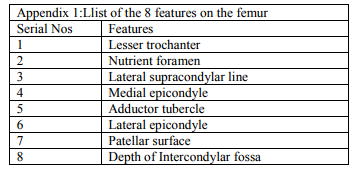
A total of 63 femur available at the International Medical School, Bangalore were subjectively grouped into 34 male and 29 female femur. There were 36 right and 27 left femur. Further grouping of the femur were done as per the subjective sexing and the sides: Male: 19 right and 15 left; Female: 17 right and 12 left. A total of 8 features were studied for their presence or prominence in the femur. On multiplication, it is seen that the total features were 504 (63x8=504); out of which for the male femur, it was 272 (34x8) and for the female femur, it was 232 (29x8). The grading given to the presence of the features was single plus (+) and for the prominence was double plus (++). In Appendix 1 is listed the 8 features. The percentage analysis and the X2 test were the applied statistical measures to the obtained values of the 8 features.
RESULTS
The values gathered for the 8 features from femur were studied under 6 categories for their ‘presence or prominence’: total for the male and female (1.1) and the right and left sides (1.2); total for the right and left sides of male and female femur (1.3 and 1.4); total between the right and left of male and female femur (1.5 and 1.6).
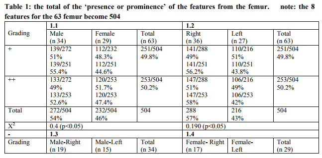
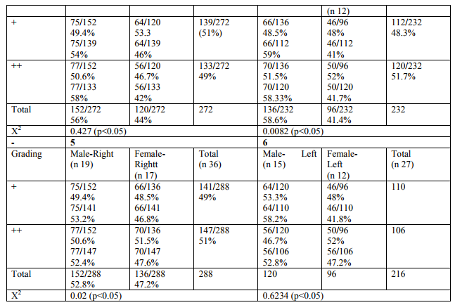
1.1: The total of the ‘presence or prominence’ of the features in the femur was observed to be more or less of equal percentage i.e. around 50%. 1.2: Between right and left sides, the total was more for the right for both the presence (141/251) as well as the prominence (147/253) of the features. When only the sides were considered, within the right, ‘prominence’ of the features was more (147/288) and within the left, ‘presence’ of the features was more (110/2161). 1.3: For the male femur, between right and left, ‘presence (54%) and prominence (58%)’ of the features were found to be of high percentages for the right; within the right it was ‘prominence (50.2%)’ and within the left (53.3%) it was ‘presence’ of the features. ‘presence (59%) and prominence (58.3%)’ of the features were found to be of high percentages for right; but, within the right (51.5%) and left (52%), it was ‘prominence’ of the features. 1.5: Between male and female femur of right side, male femur showed high percentages both for ‘presence (53.2%) and prominence (52.4%)’ of the features. 1.6: Between male and female femur of left side, male femur showed high percentages both for ‘presence (58.2%) and prominence (52.8%)’ of the features. The X2 test showed significant values for sex and side determination of the femur.
It was seen that in male femur, the high scores (22/34) were found for the presence (+) of the features numbered 4 and 8 (medial epicondyle, depth of intercondylar fossa); whereas for the female femur, it was for the feature number 1(21/29,72.4%) (lesser trochanter). It is to be noted, that, among the 8 features, the feature that stood out was nutrient foramen number 2;86.2%) in the female femur.
DISCUSSION
From literature review, it was seen, that the long bone that has been studied in detail in the human skeleton is the femur. In 1985, Meindel et al analysed the application of non-metric (subjective) versus metric (objective) parameters for sex determination in the skeletal material and found ‘no significant difference in the accuracy; so long care and skill are employed’. (Meindel et al 1985)3 Studies in femur have reported subjective and or objective methods of sex determination. (Soni et al 2010, Bhosale and Zambare 2013)4,5 In the present study, from the regular description of the general features of the femur, 8 were selected for their ‘presence or prominence’ and its application to sex determination.
Present study
Interpretations
=The total occurrence of the ‘presence or prominence’ of the 8 features in the male and female femur was observed to be more or less the same.
= Both ‘presence or prominence’ of the features have occurred more on right femur.
= Both male and female right femur showed more number of ‘presence as well as prominence’ of the features.
= Male femur for the occurrence of the features within the right or left sides showed for the right side; ‘prominence’ and for the left side ‘presence’ of the features.
= Female femur when considered for the occurrence of the features within the sides showed ‘prominence’ of the features on both sides. The X2 test showed that the features selected could contribute significantly in the sex determination of femur.
The ‘prominence of the features’ in the female femur may be correlated to them performing more tasks; especially pertaining to their ‘daily chore activities’.
Krogman1 in 1962 has summarized on 3 issues on the sexing of the skeletal remains and they are given below more or less in verbatim. i) sexing of unknown skeletal material could depend relatively on the availability of the complete skeletal material. The proposed percentage of the accuracy for the adult long bones alone is around 80%. In the present study, the subjective sexing was carried out on the available known and complete skeletal material i.e. femur. The feature which showed 86.2% is the nutrient foramen in the female femur. ii) The estimates are usually based on description, dimensions, proportions which are the morphology and morphometry methods. It seems that the ‘elaborate statistical analysis does not raise appreciably the average’. Hence, it is paraphrased, that with statistics ‘one can be sure or at least more sure in an individual case’. In the present study, the X2 test did made it sure that the 8 features could be utilized in sex determination of the femur and has identified the ‘nutrient foramen as the feature in female femur.’ iii) The standards of the ‘morphological and morphometric sex differences’ in the skeletal material may differ depending on the population of the samples. This notion is considered to be true with reference to ‘dimensions and indices’. As a general rule, the standards should be used with reference to the group from which they were drawn and upon which they are based and they are not ‘ordinarily interchangeable’. In the present study, the observed differences could be due to the sample size and the selected features. Moreover, as mentioned by Krogman1 the femur were available; but there was lack of information about their ‘population sample. In view of the non-availability of any publications similar to the present study, the study could not be discussed further. The ‘presence or prominence of the features’ could be because of the genetic, hormonal, environmental conditions, such as nutrition and bio-mechanic forces on the bones/joints/movements. It may be noted, that a paper on ‘Variations in the presence and prominence of the features in the long bones of the limbs’ has been published (Rajangam et al 2014)6 . In that study, for femur, 16 features were analysed. As a continuation, for the present study 8 were selected in tracing their contribution towards the subjective way of determining the sex in femur.
CONCLUSION
It may be concluded, that for subjective sex determination of the femur, in addition to the thickness, size, length, robustness and massiveness, the features and their findings from the present study could also be applied.
ACKNOWLEDGEMENT
Authors acknowledge the immense help received from the scholars whose articles are cited and included in references of this manuscript. The authors are also grateful to authors/editors/ publishers of all those articles, journals and books from where the literature for this article has been reviewed and discussed.
References:
1. Krogman WM. The Human Skeleton in Forensic Medicine. Illinois: Charles C Thomas Pub Ltd; 1969.
2. Standring S. Gray’s Anatomy. The Anatomical Basis of Clinical Practice. 40th edition. London: Churchill Livingstone Elsevier; 2008.
3. Meindl RS, Lovejoy CO, Meneforth RP, Don Carlos L. Accuracy and Direction of Error in the Sexing of the Skeleton: Implications. AJPA 1985; 68, 79-85.
4. Soni G, Dhall U, Chhabra S. Determination of Sex from Femur: Discriminant Analysis. Journal of Anatomical Society of India 2010; 59(2): 216-221.
5. Bhosale RS, Zambare BR. Sex Determination from Femur using Length of Femur in Maharashtra. J of Dental and Medical Sciences 2013; 3(4): 1-3.
6. Sayee Rajangam, Vidhya R, Shiva Charan, Flossie Jayakaran. Variations in the presence and prominence of the features in the long bones of limbs. Int J Cur Rev 2014; 6(7):58- 64.
|
 IJCRR
IJCRR 


 This work is licensed under a Creative Commons Attribution-NonCommercial 4.0 International License


