IJCRR - 7(2), January, 2015
Pages: 57-60
Print Article
Download XML Download PDF
CRYSTALLOGRAPHIC ANALYSIS OF URINARY CALCULI
Author: Rajesh Narayan
Category: Healthcare
Abstract:Aim: Urolithiasis is a global phenomenon. New frontiers have developed not only on therapeutic lines but also in understanding process of calculogenesis.
Methodology: Urinary calculi have been analyzed and studied by different techniques with diverse merits and demerits. Urinary stones are multicomponent system containing mainly crystalline and partially non-crystalline components. Crystallographic
analysis by X-ray diffraction & Polarized Transmission Microscopy yield more information than other techniques.
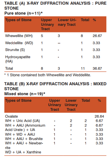
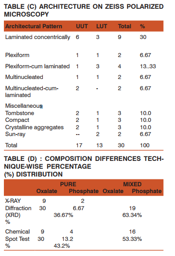
Result: X-ray diffraction patterns obtained was unique to the particular crystalline constituents with unique diffraction angles, corresponding \"d\" value (lattice spacing) and relative line intensities. X-ray diffraction showed a significant difference in detection
of pure stone 36.67% compared to 43.2% on chemical spot test. Polarized transmission microscopy revealed the crystal matrix
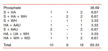
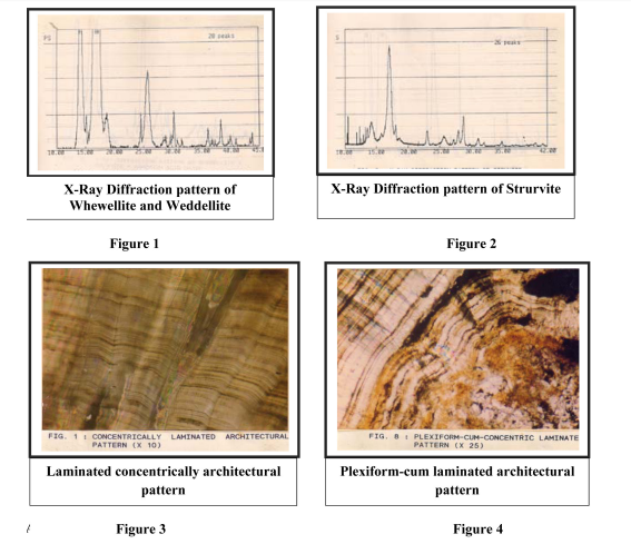
inter relationship and architectural design accounted for concentrically laminated 30% followed by plexiform-cum laminated in 13.33% tombstone coffin -lid, compact and crystalline aggregated observed in 10% each.
Conclusion: Crystallographic method of urinary analysis by X-ray Diffraction and Polarized Transmission Microscopy can identity
quickly and certainly different Urates, Oxalates and Phosphates with a certainty that cannot be equaled or attempted by any
other technique. They are considered a reference technique for identification and differentiation of crystals. Identification of the
constituents have bearing in prevention and recurrent of future stone formation.
Keywords: Crystallography, Urinary calculi, X-Ray Diffraction Optical Microscopy, Chemical Spot Test.
Full Text:
INTRODUCTION
Urinary calculi have been analyzed and studied by different techniques with diverse merits and demerits[1] [2]. The urinary stones are multi component systems containing crystalline and non-crystalline materials, crystallographic methods of analysis yield more information than other techniques. Crystallographic analysis yield more information than other techniques. Crystallographic analysis done by X-ray diffraction and optical microscopy provide specific X-Ray Diffraction patterns and optical properties of calculi that cannot be equaled or attempted by any other technique [3].
RESEARCH METHODOLGY
This study was carried out in the Department of Urology, I.M.S B.H.U in collaboration with Departments of Geology and Metallurgical Engineering, IIT B.H.U. 30 randomly selected urinary calculi were picked from Urology ‘Rockery’
(A) FOR X-RAY DIFFRACTION
The stone was crushed in a mortar and pestle to form a fine powdered specimen in order to produce smooth diffraction lines. The powder was produce smooth diffraction lines to cover an area approximately 5-12m.m. The specimen was placed over the holder in the Rigaku Xray Diffractometer and X-ray diffracted were recorded by linking a strip chart recorder to the Diffractometer . The diffraction pattern unique to the particular crystalline constituent present with diffraction angles and ‘d’ value (or lattice spacing) relative intensities matched with standard values. First 3 most intense lines with d-values were matched with reference of standards of Sutor [4].
(B) FOR POLARIZING MICROSCOPY
The urinary calculus was cut into two halves through the center using cut a fine hack-saw. The cut surface was polished on a glass plate using carborundum powder. The polished surface of the stone was mounted on a glass slide with the help of cooked Canada balsam as a cementing material. The other side of the stone was then grounded over the rotating disc using carborundum powder till the thickness of the stone became quite thin(0.03m.m) This thickness of section show great degree of transparency and interference of colors. The final preparation of the slide was done by washing excess of carborundum powder and Canada balsam removed with cotton soaked in methylated spirit or Xylol. The cover slip finally applied over the section with help of warm Canada Balsam. Care taken not to introduce air bubble. The slide prepared was ready for petrological assessment using Fuji Film 125 and analyzed for the architectural pattern of a given stone from nucleus, midzone and periphery of a given stone.
OBSERVATION
A retrospective stone analysis of 30 uroliths clinically showed preponderance in third and fourth decades of life. Male: Female ratio 2:1. Majority stones were oval grey colored, uneven with hardness of 2-3 (Moh’s scale). Chemical analysis revealed 53.33% stones were mixed as against 46.67% pure. Oxalate was the commonest stone in pure variety and oxalate-phosphate uric acid 20% in mixed stones. The X-ray diffraction patterns of the powdered specimen recorded on film as spectrum of peaks of varying intensity unique for the particular crystalline constituent present in the sample as the finger print of that substance. Simple measurement permit the calculation of the characteristic distance between lattice plane of atom compared with published reference standards of Sutor[4]. The commonest pure variety of stones we Whewellite 26.67%.Of 63.33% mixed stones, 13 possible combinations were encountered. Majority of the mixed urinary calculi were Phosphate 36.69% whereas oxalate was 26.64% .While pure uric acid calculous was non-entity in the present series. It was a solitary minor constituent in 6.67% more commonly with the oxalate type, more common was the acid urate admixed with oxalate 20%. Both uric acid and ammonium acid urate was uncommon associate (6.67%). Pure oxalates show a specific architectural anatomy of concentrically lamination 30% Phosphates do not typify for a fixed design due to diverse admixture constituents. Presence of micro channels, subsidiary channels and whorls formation showed presence of intercommunicating channels, festoon cross bending and contemporaneous deformation. Petrographic architectural pattern were assessed by hand lens magnification and Polarized Transmission Microscopy to revel architectural anatomy of the calculi. On hand lens magnification (x2) showed Rath and Nath design, a majority of urinary calculi were Type A 60% which contained ill defined central nucleus surrounded by a few laminations at periphery. 33.33% calculi were Type B with distinct central nucleus with concentric light and dark colored laminations. Pure oxalates show a specific architectural anatomy of concentrically lamination 30% Phosphates do not typify for a fixed design due to diverse admixture constituents. Presence of micro channels, subsidiary channels and whorls formation showed presence of intercommunicating channels, festoon cross bending and contemporaneous deformation. Phosphates do not typify for a fixed design due to diverse admixture constituents. Presence of micro channels, subsidiary channels and whorls formation showed presence of intercommunicating channels, festoon cross bending and contemporaneous deformation.
DISCUSSION
Chemical “ spot test ” showed 2% error in detecting calculi components[5].In pure stones 6.67 phosphates were detected on X-Ray Diffraction and 13.2% on spot test whereas in mixed stones 63.34% were oxalates and phosphates on X-Ray Diffraction and 53.33% by spot test. X-Ray Diffraction distinguished different urates oxalates and phosphates with certainty that cannot be equal led or attempted by any other techniques and case considered as the reference technique for identification and differentiation of crystals. X-Ray Diffraction analysis in my series is in confirmation with the survey of Sutor, Wooley, Illingworth and Rodgers 1974[6]. Under Zeiss polarizing microscope irrespective of the chemical nature of stone, formation of stone is an active process with centrifugal growth. Stones show presence of micro channel both central and dispersed micro units joined by subsidiary channels. Whorls formation looks like festoon cross bending on contemporaneous deformation. The presence of intercommunicating micro channels lamellar arrangement of the deposit, festoon cross bending and contemporaneous deform action indicate feeding micro channels Oxalates had concentric laminated pattern with striation whereas phosphates had diverse architectural patterns. Whewellite:Weddellite ratio in the current series was 4:1 similar to other workers and Rodgers 1974[6]. Under Zeiss polarizing microscope irrespective of the chemical nature of stone, formation of stone is an active process with centrifugal growth. Stones show presence of micro channel both central and dispersed micro units joined by subsidiary channels. Whorl formation looks like festoon cross bending on contemporaneous deformation. The presence of intercommunicating micro channels, la- mellar arrangement of the deposit, festoon cross bending and contemporaneous deformations indicate feeding micro channels Oxalates had concentric laminated pattern with striation whereas phosphates had diverse architectural patterns.
SUMMARY AND CONCLUSION
Crystallographic analysis done by X-ray diffraction and optical microscopy provide specific X-Ray Diffraction patterns and optical properties of calculi that cannot be equaled or attempted by any other technique. X-Ray Diffraction analysis showed a significant difference in detection of pure stone 36.67% in X-Ray Diffraction as compared to 43.2% on Chemical spot test. X ray diffraction is considered as a reference technique for identification and differentiation of crystals (Khan et al 1981) Polarized transmission microscopy revealed the crystal matrix interrelationship the predominant architectural design accounted was concentrically laminated (30%) followed by plexiform-cum-laminated in 13.33%.


Of 63.33% mixed stones, 13 possible combinations were encountered. Majority of the mixed urinary calculi were Phosphate 36.69% whereas oxalate were 26.64% .

ACKNOWLEDGEMENT
The author acknowledges the immense help received from the scholars whose articles are cited and included in references of this manuscript. The author is also grateful to authors/ editors/ publishers of all those articles, journals and books from where the literature of this article has been reviewed and discussed. The author is thankful to Prof. Dr. G.M.K. Sharma Dept. of Metallurgy IIT, BHU and Prof. M.S. Srinivasan Dept. of Geology BHU for their supervision and guidance. The author is thankful to Prof. Dr. V.N.P. Tripathi Ex. Director Institute of Medical Sciences BHU for the completion of his multi disciplinary work.

References:
1. Finlayson,B:Symposium on renal lithiasis. Renal lithiasis in review. Uro Clinic North Am.,1:181,1974
2. Carr , R.J: A new theory on the formation of renal calculi. Br.J. Uro, 26:105-117,1954
3. Beeler, M.F;Veith, DA:Analysis of urinary Calculus – Comparison of methods. Am.J.Clin.Patho 1964.
4. Sutor, D.J and WooleyS.E:Composition of urinary calculi by X-Ray Diffraction Br.J. Urology 46,229,1974.
5. AggrawalS.L : Chemical Composition of urinary calculi. Ind.Med.Asso.57,171,1971
6. Rodgers AL et al : A multiple technique approach to the analysis of urinary calculi. Urol. Res. 10:177-184, 1982.
7. Bailey, C.B., : A scanning electron microscope study of siliceous urinary calculi from cattle. Investigative Urology, 10(2) : 178-185, 1972
8. Beeler, M.F., Veith, D.A., Monis, R.H. and Biskind, G.R. : Analysis of Urinary Calculus – comparison of methods. Am. J.Clin. Patho., 41(5) : 553-560, 1964
9. Brien. G, Schubert, G and Bick, C. : 10,000 analysis of urinary calculai using X-ray diffraction and polarizing microscopy. Eur. Urol.., 8:251-258, 1982
10. Hazarika, E.Z., Balakrishna and Rao, B.N. : upper urinary tract calculi analyzed by X-ray diffraction and chemical methods. Indian J.Med. Res., 62:443, 1974
11. Kabra, S.G., Patni, M.K., Sharma, G.C., Banerji, P. and Gaur, S.V. : Urolithiasis : Histochemical study of microscopic section of urinary calculi prepared by petrographic method. Indian J. Surg., 34:309, 1972
12. Kirby, J.K., Pelphrey, C.F. and Roi-Ney, Jr : The analysis of urinary calculi. Am J.Clin. Path, 27:360-362, 1957
13. Malek, R.S. , and Boyce, W.H. : observation on the ultrastructure and genesis of urinary calculi. J. Urol. , 117:336, 1977
14. Winer J.H and Mattice, M.R. : Routine analysis of urinary calculi, rapid, simple method using spot test – J. Lab & Clin Med , 28:898-904, 1943 Quoted by Beeler et at 1964
15. Spector, M.Garden, N.M and Rous, S.N. : ultrastructure and pathogenesis of human urinary calculi. Br. J. Urol.., 50:12- 15, 1978
16. Prien, E.L., Prien, E.L. Jr. : Composition and structure of urinary stone. Am J. Med 45:655, 1988
|


 This work is licensed under a Creative Commons Attribution-NonCommercial 4.0 International License