IJCRR - 7(12), June, 2015
Pages: 48-51
Print Article
Download XML Download PDF
PHOTOGRAPHY AND PIXEL ISSUES IN UNDERGRADUATE SLIDES IN PATHOLOGY
Author: Jyothi B. Lingegowda, Prakash H. Muddegowda, Ramkumar Kurpad R., Prasanna G. Konapur, Sathiyamurthy K., Thamilselvi R.
Category: Healthcare
Abstract:Digital images have become an important tool in pathology. Telepathology towards consultation is rapidly developing. Teaching through digital slides is growing fast due to easily availability of digital cameras, computer hardware and the internet. Objective: Our purpose was to evaluate the resolution requirement for digital images. Size of data of these digital images and there upload speed was also evaluated. Methods: Digital images of 29 selected slides showing characteristic lesions were produced in five different resolutions each, ranging from 640x480, 2048x1536, 2592x1944, 3264x2448 and 4000x3000---. They were compared individually by a group of two experienced pathologists regarding the diagnoses and level of confidence. Results: Images at the resolution of 2048x1536 were perceived as equivalent to higher resolutions, Data upload and image loading was significantly better in 2048x1536 rather than 4000x3000 images Conclusion: For digital images in dermatology a resolution of 768 x 512 x 24 is suitable to recognize the relevant details of the source image.
Keywords: Adenocarcinoma stomach, Basal cell carcinoma, Fatty liver, Follicular adenoma
Full Text:
INTRODUCTION
Instances of imaging in pathology can date back to 1968 in Boston, where live images were captured or to 1974, where data was transferred from a ship in Brazil to Washington DC. Pathology is the driving field for microscopic imaging. In microscopy, pictures are data. The need to maintain and record morphological findings is necessary both at macroscopic and microscopic level for diagnosis, consultation, documentation and education. Images document the true appearances and eliminate inaccuracies in the reporting process.(1-5) Previously, elaborate photography sets, black and white images were not conducive and cost effective. Now, with the advent of digital photography, low cost of digital cameras and the ability to view on mobile, tablets or computers without the need to print along with expanding internet use has made major inroads in documenting pathological changes at both macroscopic and microscopic levels. Digital microimaging consists of digital camera, computer, imaging software and an optical connector to the microscope. Images can also be captured with a cell phone camera with or without adapter. Digital image format is appropriate for computer analysis, as the images are already in digital format.(4,6,7) In the present study, undergraduate slides digital images were prepared and evaluated to identify the appropriate megapixel for teaching, storage and transfer purposes.
MATERIALS AND METHODS
This study was conducted in department of Pathology, VMKV medical college, Salem. Selected undergraduate slides images were taken at various resolutions like 640x480, 2048x1536, 2592x1944, 3264x2448 and 4000x3000 (VGA mode, 2 MP, 5 MP, 8 MP, 13 MP respectively) using a Sony DSC W220 aim and shoot camera.
Standard binocular microscope was used for capturing images, and all were taken at 10X magnification. The camera was hand held against the microscopic eye piece to take photographs. No zoom function or adapters were used. The images were viewed using a laptop and was evaluated by two pathologists. The pathologists were completely blinded regarding the image resolution. The images were evaluated for the ability to make diagnosis, upload speed and also speed of image processing during viewing. They were asked to evaluate the image within 60 seconds per image and viewer recorded the diagnosis and level of confidence. Images were jumbled up and the resolutions of images were only known to the author and were blinded to the reviewers to prevent conscious and unconscious bias. Slides were taken from routine undergraduate student slides. The slides selected were from actinomycosis, acute appendicitis, adenocarcinoma stomach, basal cell carcinoma, capillary hemangioma, cavernous hemangioma, chronic pyelonephritis, chronic venous congestion of lung, cirrhosis liver, colloid goiter, fatty liver, follicular adenoma, granulation tissue, leiomyoma, lobar pneumonia, maduramycosis, malignant melanoma, osteoclastoma, osteosarcoma, papillary carcinoma thyroid, pleomorphic adenoma, renal cell carcinoma, rhinosporidiosis, squamous cell carcinoma, squamous cell papilloma, seminoma, teratoma, hydatidiform mole and Tuberculous lymphadenitis. All the slides were stained using Hematoxylin and Eosin stain.
RESULTS
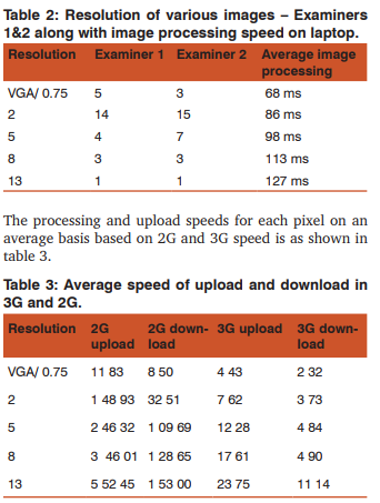
29 histopathology slides were selected and images were taken at various resolutions like 640x480, 2048x1536, 2592x1944, 3264x2448 and 4000x3000 (VGA mode, 2 MP, 5 MP, 8 MP, 13 MP respectively)
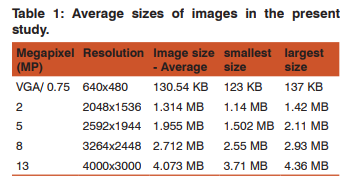
The average size of images in each megapixel is as shown in table 1
All the images were evaluated by two experienced pathologists and each one applied the image based on the appropriate for diagnosis. The collective decision regarding resolution is as shown in table 2. Images from conditions like actinomycosis, maduramycosis, osteoclastoma and colloid goiter were easily identified at the lowest resolution. Higher resolution was required for fatty liver and Tuberculous lymphadenitis. The processing of images varied with each resolution. The average duration is as shown in table 2. Both examiners agreed about larger resolutions taking upto 2 seconds at times and as the resolution increased, so did the image processing and loading time.
 .
.
DISCUSSION
Telepathology is defined as the acquisition of histological or microscopic images for transmission along telecommunication pathways for diagnosis, consultation or continuing medical education. It may be for primary opinion, frozen section consultation or second opinion. It is especially useful for supplying the remote areas with specialist medical advise. Images are also used for proficiency testing, web pages like blogs, facebook, image analysis, web based learning/ teaching, etc. (1,5,8,9,10) Various methods to take microphotography has been discussed previously. Digital zooms should be avoided, as it blurs the image, while optical zoom can help in a closer look. The images can be used for various purposes, in-cluding teaching and clinical meetings, conference presentations, diagnosis, image analysis, archiving, etc. In the present study, images no zoom was used and images were used for teaching purposes.(3,4) Telepathology can be simple store and forward or dynamic real-time telepathology. In few countries, immunohistology laboratories have employed the internet to post images of stains to avoid delay. Here, we used simple, store and forward method for evaluation purpose.(4) With the professionals using their education, experience and intuition, selection bias or sampling error, where an area is chosen for selecting for a closer look, leaving out other areas is of particular concern. To thwart this, image scanners are commonly used, which includes all images. Here pathologists with more than 5 years undergraduate teaching experience were involved in selecting areas. The evaluators agreed the chosen field was important and adequate for diagnosis for undergraduates.(2,4) There is a long list of manufacturers who have produced slide scanners for pathology. The setup includes preparation of microscopic images through computer controlled image capture, focus stacking, and image mosaic stitching. However, this has not been widely accepted due to high cost of specialized equipment, difficulty in capturing and stitching single images and also difficulty in manipulating and viewing smaller images. Here, we used hand held camera without any adapter. Images were found to be good enough for diagnosis.(2) With image storage, the size of physical storage and methods of transfer becomes important. Hard disks are large, bulky but can store plenty of data. While smaller data storages like pendrive, cannot carry multiple larger images and require much data storage. CD and DVD can also be used for permanent storage. Compressed images for storage are not advisable as poorly compressed images bring out poor quality photographs. Publishers always seek uncompressed files. For transfer of images, compression would be necessary to reduce use of bandwidth and faster transfer speeds. In our study, smaller uncompressed images were better appreciated. A resolution of 2048x1536 was found to be adequate mostly. However, some images, which were not classical, required larger resolutions for zoom and more depth evaluation. Transfer with 3G network was way better than 2G. The difference was astounding. Even though better, took a bit longer for larger images and in future could be use for transfer of uncompressed large data image files. With the advent of 4G in the country, larger data, if necessary especially stitched images, can be transferred easily. We also noticed, larger resolution images, occupied larger spaces and were cumbersome to transfer. (3,4,5) Traditional glass side teaching can never be replaced. However, interesting case slides, as it ages, is prone to fade, difficult to transport or share and is fragile, easily damaged and cannot be replaced. Digitalization of these slides can provide a solution to many of these drawbacks. (1,4) Problems faced include naming, storage of data and differences in identifying the magnification. Any model cameras can be used. Now a days, mobile phone with excellent cameras are available and immediate sharing through whatsapp and if necessary online is also being done. Problems of scientific fraud have been put forward by few critics; however, fraud existed before too due to falsification of data. Also problems like poor slide preparation can affect focus plane, which would make it difficult to work with relevant data. (4,5) Whole slide educational sets similar to what we have done can be made available on internet or intranet. Studies have shown, computer based teaching to be effective and could be an additional attractive way for medical students to self educate. Students need not own a microscope, but can examine virtual slides and learn the techniques. This helps in the improvement of efficiency and distribution of available resources.(5,10) In India, the potential is high for using digital images for teaching and consultation purposes. Given the growth of mobile use and its penetration, and reduced transmission costs, this could be very useful for both service providers and patients.
CONCLUSION
Even though digital imaging in microphotography is relatively developing, it has made rapid development because of low cost, rapidity and convenience of usage. Application of the same in teaching undergraduates could go a long way in better teaching. The usage of mobile cameras can be used for instant teaching along with immediate uploading and better management of this virtual scenario. The students will be benefitted with immediate help and interest in learning pathology at its microscopic level could go a long way in furthering the knowledge.
ACKNOWLEDGEMENT
Authors acknowledge the immense help received from the scholars, whose articles are cited and included in references of this manuscript. The authors are also grateful to authors/ editors/ publishers of all those articles, journals and books from where the literature for this article has been reviewed and discussed.
References:
1. Leong FJW-M, Graham AK, Gahm T, McGee JO’D. Telepathology: clinical utility and methodology. In: Lowe D. Underwood JCE, eds. Recent advances in histopathology 19, Edinburgh: Churchill Livingstone; 1999;217-39.
2. Longson J, Cooper G, Gibson R, Gibson M, Rawlins J, Sargent R (2010). Adapting Traditional Macro and Micro photography for scientific gigapixel imaging. Proceedings of the fine internatioanal conference on gigapixel imaging for Science, November 11-13, 2010. Available from: http:// repository.cmu.edu/cgi/viewcontent.cgi?article=1001andco ntext=gigapixel
3. Bagnell CR Jr. Photomicrography [Internet]. 2013.[cited 2014 April 13]. Available from : https://www.med.unc. edu/microscopy/files/courses/spring-2013-lm/path-464- class-notes/lm-ch-14-photomicrography
4. Leong FJW-M, Leong ASY. Digital photography in anatomic pathology. J Postgrad Med. 2004;50(1):62-9.
5. Schmitz J, Bollmann O, Vogel C, Bollmann R. Virtual microscopy (remote patchwork) as a new technique for teleconsultation and tele-education. Electronic J Pathol Histol 2003;9.2:32-0005.
6. Ying X, Monticello TM. Modern imaging technologies in toxicologic pathology: An overview. Toxicologic pathology 2006;34:815-26.
7. Bellina L, Missoni E. Mobile Cell-phones (M-phones) in telemicroscopy: increasing connectivity of isolated laboratories. Diagnostic Pathology 2009;4:19.
8. Khalbuss WE, Pantanowitz L, Parwani AV. Digital imaging in Cytopathology. Pathology Res Int. 2011;2011:264683.
9. Marcano F, De Armas N, Diaz-Cardama, Ferrer-Roca O. Collaborative systems for pathology applications. The Open Pathology Journal 2007;1:1-4.
10. Rozman P. The impact of telemedicine on the organization of blood transfusion services. Bilt Transfuziol. 2006;52(2- 3):22-6.
|
 IJCRR
IJCRR 


 This work is licensed under a Creative Commons Attribution-NonCommercial 4.0 International License


