IJCRR - 12(13), July, 2020
Pages: 01-05
Date of Publication: 06-Jul-2020
Print Article
Download XML Download PDF
Morphological and Morphometrical Analysis of First Rib: An Anthropological Study
Author: Nisha R Pillai, Jyothi K C, Shailaja Shetty
Category: Healthcare
Abstract:Introduction: The first rib is the most curved rib and very distinct from other ribs. The significant landmarks on the first rib include the head, tubercle, vascular groves on the superior surface and the scalene tubercle. Anomalous ribs are often discovered incidentally on chest radiographs. Such anomalies, maybe associated with the compression of the neurovascular bundle at the root of the neck. Further research on the first rib may also yield information that substantiates the growing relevance of first rib in sex identification and age estimation, particularly when the skull and pelvis are damaged to a significant extent.
Objectives: This study aims to analyze the morphological and morphometrical variations of first rib and understand the significance of such variations.
Materials and Methods: 35 right and 35 left first ribs were used for the purpose of this study. All the measurements were taken with digital Vernier calipers and flexible cloth tape. The findings were recorded and analyzed statistically. The study was conducted in Department of Anatomy, M. S. Ramaiah Medical College, Bengaluru.
Results: As far as the morphological parameters were concerned, scalene tubercles were either absent or rudimentary in nearly 50% of the ribs on both right and left sides. Similarly, vascular grooves were either absent or insignificant in approximately half of the ribs on both right and left sides. The variations of the head and the tubercle of the first rib were not encountered frequently.
Conclusion: Malformations of the first rib are common. When present, it may lead to compression of neurovascular bundle at the root of the neck causing thoracic outlet syndrome. Awareness of such anomalies are important for anatomists, radiologists and thoracic surgeons dealing with this region.
Keywords: First rib, Angle of rib, Scalene tubercle, Malformation, Thoracic outlet syndrome
Full Text:
INTRODUCTION
The thoracic wall is made up of sternum anteriorly, twelve thoracic vertebrae and their intervertebral disc posteriorly and twelve pairs of ribs and their costal margins laterally. The first rib is the most curved and frequently the shortest1.It forms the boundary for the thoracic inlet. The scalenus anterior is inserted into the inner border of the first rib, producing the scalene tubercle. Independent studies by Wattanasirichaigoon and Castriota suggested that rib anomalies are common, with approximately 2% in the general population. It is of paramount importance for radiologists to be able to identify these, so as to avoid misinterpretation of radiographs2. Most familiar rib anomalies include cervical rib, bifid rib, rib dysplasia.
The dimensions of the ribs are recognized to have a bearing on the development of thoracic outlet syndrome3,4.
The first rib is gradually gaining importance and being ratified for estimation of the age of young adult skeletons5.The use of first rib for sex identification is also being explored under the domains of Forensic Anthropology. Its importance is further amplified by the fact that it is less liable to be damaged as against other skeletal remains6.
Measurements of ribs have also been employed in biomechanical formulae for respiration, truncal structure and identifying lateral asymmetry in diagnosis of scoliosis7.
The present study aims to analyze the morphological and morphometrical variations of the first rib and understand its significance.
MATERIAL AND METHODS
70 first ribs (35 right and 35 left first ribs) were procured from the Department of Anatomy, M S Ramaiah Medical College.
Inclusion criteria- All intact right and left ribs were included.
Exclusion criteria- Damaged first ribs.
Morphological Study: Variations regarding the scalene tubercle, vascular grooves, head and tubercle of the rib were recorded carefully and expounded.
-
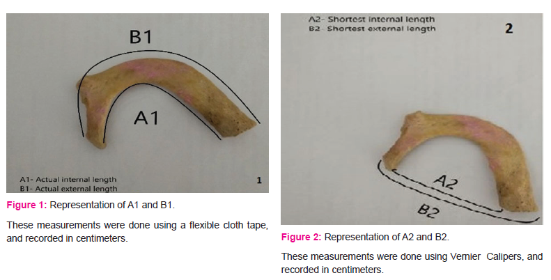
Actual Internal Length (A1): measured along the inner curvature of the first rib from the posterior sternal end to the medial side of the head of the rib.
-
Actual External Length (B1): measured along the outer and greater curvature of the first rib from the anterior sternal end to the lateral side of the head of the rib. ( Fig 1)
-
Both A1 and B1 were measured with the aid of a flexible cloth tape and recorded in centimeters.
-
Shortest Internal Length (A2): Measured from the posterior sternal end to the medial side of the head of the rib.
-
Shortest External Length (B2): Measured from the anterior sternal end to the lateral side of the head of the rib. ( Fig 2)
-
Both A2 and B2 were measured using a digital Vernier caliper and recorded in centimeters.
-
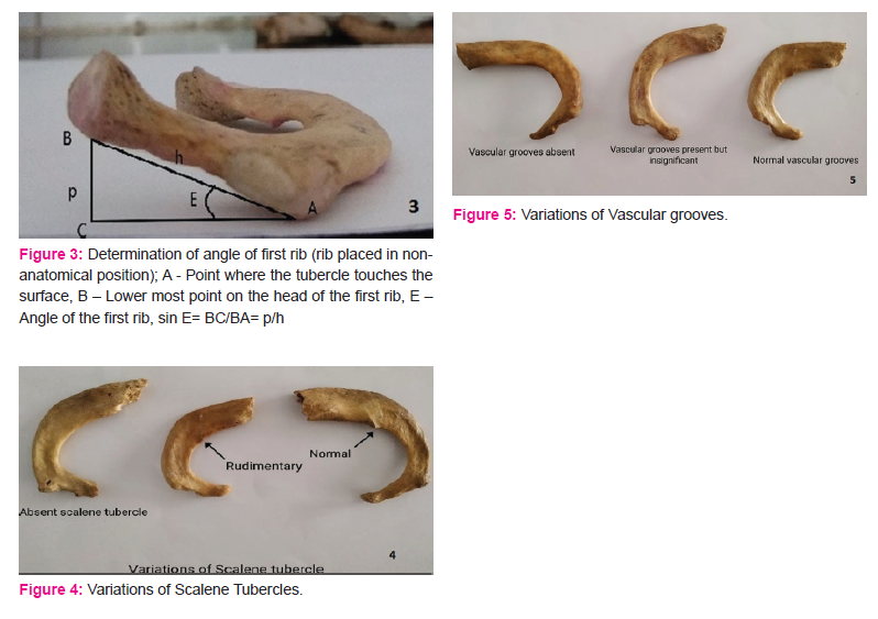
Angle of the first rib: The rib was placed on an even flat surface in the non-anatomical position as shown in figure 1. Inverse sine function in Microsoft Excel was used to determine the angle formed between the neck of the rib and the flat surface on which it was placed. ( Fig 3)
Statistical Analysis:
RESULTS
-
In this study, 35 right-sided and 35 left-sided first ribs were analyzed for their morphology and morphometry. All the morphometric parameters were found to be higher for the right ribs than those on the left, however it was not statistically significant.
-
The mean and the standard deviation of the angle on the right side was 15.82° ± 6.97, the maximum angle recorded being 28.42º and minimum being 0º. On the left side, the mean and standard deviation was 14.76° ± 4.89, the maximum being 26.74º and minimum being 8.53º.
-
Absent or insignificant vascular grooves were noted in nearly 50% of the ribs. Variations of the head and tubercle were not very common. Variations of the head and tubercle were not very common. 97.1% of the right ribs and 91.4% of the left ribs did not have a rudimentary head or tubercle. Only 2.9% ribs on either side showed rudimentary head and tubercle. Rudimentary or absent scalene tubercle were observed in about 50% of the ribs. (Fig 4,5)
-
The results are tabulated (Table 1- 5)
-
DISCUSSION
First rib studies have been conducted on radiological grounds as well as by inspection of dry bones. A study on rib anomalies by Etter encompasses a comprehensive radiological assessment of 40,000 cases, the main inference from it being that the most frequently encountered anomaly in the course of the study was forked rib8.
Another study evaluated the accuracy of CT derived first rib measurements for the determination of sex9.
In a study conducted by Sunita Bharati et al, with a sample size of 48 first ribs, 18.75% of the ribs did not have a Scalene tubercle and 18.75% of the ribs did not have vascular grooves. The mean values for the Actual External Length (B1) on right and left side were found to be 7.63cm and 7.86cm respectively10.
Rashia et al. studied 50 first ribs, and reported absent scalene tubercles in 46% of the ribs. As per the study, 28% of the ribs did not have vascular grooves, while rudimentary tubercle and head were found in 12% and 5.7% of the ribs respectively11.
D Souza et al. conducted a study to assess the adequacy of the first rib in identification of the sex. The mean of the Actual External Length (B1) on the right side was estimated to be 12.13cm, while the mean for the same on the left side was12.19cm. The mean of the angle of the rib on the right and left sides were 13.5
 and 15.1° respectively12.
In our study, we found that 22.5% of the ribs lacked a scalene tubercle and 50% of the ribs did not have vascular grooves. Rudimentary tubercle and head was reported in 24% and 2.85% of the ribs respectively. The mean values of B1 were found to be 12.97cm and 12.81cm on the right and left side respectively. The lack of coherence with the study conducted by Sunita et al. maybe attributed to racial differences.
A study conducted by Elrod suggests that sex of the individual maybe determined by using the angle of the first rib alone with a probability of 60.2%. The probability is enhanced to 70.5% if the angle of the first rib and its total length is taken into consideration13.
Similarly, another study which analyzed the utility of the first rib in sexing individuals stated that a combination of metric and geometric morphometric variables could yield a correct sex classification in European Americans and African Americans as high as 88.05% and 70.86% of the times respectively, thereby highlighting the role of ancestry and race in determining the characteristics of the rib14.
CONCLUSION
From the above comparisons, it can be inferred that the morphometric parameters are not significantly different; however the morphological parameters show wide variations.
The racial and the regional factors are likely to have a bearing on the morphological features of the ribs. Further research conducted in this light may help establish a more concrete association between race and rib characteristics.
The differences in the right and left values of the morphometric parameters is a chance occurrence as evidenced by the p-values obtained from the paired t-test.
Acknowledgements:
Authors acknowledge the immense help received from Mrs Radhika our statistician for the statistical analysis. Authors acknowledge the immense help received from the scholars whose articles are cited and included in references of this manuscript. The authors are also grateful to authors/ editors/ publishers of all those articles, journals and books from where the literature for this article has been reviewed and discussed.
SOURCE OF FUNDING: N/A
CONFLICT OF INTEREST: Nil


References:
1. S Standring, Ed., Gray’s Anatomy: The Anatomical Basis of Clinical Practice, Churchill Livingstone-Elsevier, Philadelphia, Pa, USA, 41st edition, 2016. 936p.
2. Aign?toaei AM, Moldoveanu CE, C?runtu ID, Giu?c? SE, Vicoleanu SP, Nedelcu AH. Incidental imaging findings of congenital rib abnormalities- a case series and review of developmental concepts. Folia Morphologica 2018;77(2)386-392.
3. Chang CS, Chuang DC, Chin SC, Chang CJ. An investigation of the relationship between thoracic outlet syndrome and the dimensions of the first rib and clavicle. J Plast Reconstr Aesthet Surg 2011;64(8):1000-1006.
4. Weber AE, Criado E. Relevance of bone anomalies in patients with thoracic outlet syndrome. Ann Vasc Surg 2014;28(4):924-932.
5. Rios L, Cardoso HF. Age estimation from stages of union of vertebral emphyses of the ribs. Am J Phys Anthropol 2009;140(2):265-274.
6. Kurki H. Use of First Rib for Adult Age Estimation: A Test of One Method. International Journal of Osteoarchaeology2005;15:342-50.
7. WhiteT.D, Black M.T, Folkens P.A. Human Osteology. 3rd Edition.London: Elsevier 2012. 157p.
8. Etter LE. Osseous abnormalities in thoracic cage seen in forty thousand consecutive chest photoroentgenograms. AMJ Roentgenol. Radium Ther1994;51:359-363.
9. Kubicka AM, Piontek J. Sex estimation from measurements of the first rib in a contemporary Polish population. International Journal of Legal Medicine 2016;130(1):265-272.
10. Bharati S, Jothi S S. Morphometric and Morphological Study of First Rib. International Journal of Biomedical Research 2017;8(1):49-50.
11. Rashia S, Zaidi SHH. A morphological study of First Rib Anomalies. International Journal of Advanced & Integrated Medical Sciences 2017;2(2):70-72.
12. D Souza A, Hosapatna M, Ankolekar VH, D Souza AS. The Angle of the First Rib and its Implication in Forensic Anthropology: A Morphometric Study. Journal of Medical and Health Sciences 2014;58(2):189-191.
13. Elrod PW. The potential of the angle of the first rib, head to tubercle, in sexing adult individuals in forensic contexts 2012. LSU Master’s Theses. 3714.
14. Lynch JJ, Cross P, Heaton V. Sexual Dimorphism of the First Rib: A Comparative Approach Using Metric and Geometric Morphometric Analyses. Journal of Forensic Sciences 2017;62(5):1251-1258.
|


 This work is licensed under a Creative Commons Attribution-NonCommercial 4.0 International License