IJCRR - 10(16), August, 2018
Pages: 05-07
Date of Publication: 27-Aug-2018
Print Article
Download XML Download PDF
Clinical and Anatomical Study of Retromolar Foramen on Adult Dry Mandible in Uttarakhand Region in India
Author: Shikha Malik, Sunita, Alok Choudhary
Category: Healthcare
Abstract:Aim and Objective: The aim of the present study is to observe and analyze the morphological variations found in retromolar foramen and to assess the average width of Retromolar foramen.
Material and Methods: In this study all available 72 dry, fully ossified adult mandibles from the department of Anatomy, Shri Guru Ram Rai institute of medical and health sciences, Dehradun, Uttarakhand north region of India were included without gender and age distinction. In 72 dry mandibles, the mandible which has retromolar foramen was studied for its diameter along with side preference.
Result: The diameter of the Retromolar foramen varies between 0.53 to 1.64 mm and the mean Diameter of foramen was 1.01mm. The mean width of Retromolar foramen in right side was 1.01mm which is same as of Left side (1.01mm).
Conclusion: The retromolar foramen and canal act as a common risk factor while performing conventional anesthetic techniques so the knowledge of the variation is relevant and after evaluating the width of the foramen which is not negligible surgeons must be careful about the anatomical structures that enter via Retromolar foramen, which can be injured during surgical approaches..
Keywords: Retromolar Triangle, Retromolar foramen, Retromolar canal
Full Text:
INTRODUCTION
A triangular area posterior to the third molar tooth on the alveolar surface of mandible known as retromolar triangle. Posterior to the lower third molar is a cribose triangle, the retromolar triangle, and laterally, the retromolar fossa.1 The boundary of retromolar fossa is formed from the medial side by temporal crest and from the lateral side by the anterior border of ramus.2 The retromolar foramen (RMF) of the mandible is a variant anatomical structure which is present on the retromolar fossa along with retromolar canal in Retromolar Triangle. It is sharp and narrow, and medially borders the extramolar sulcus (at the buccal edge of the third molar).3 This foramen runs straightly to the retromolar canal (RMC), which diverges from the mandibular canal and present as a morphologic and morphometrical variability.4 In the content of the Retromolar canal there is an artery which is a branch of the inferior alveolar artery, and a nerve derived from the inferior alveolar nerve and went towards the third molar region, the retromolar triangle mucosa, the buccal mucosa, the vestibular gingiva of the premolar region, and the inferior molars.5 Some researchers4,6,7 studied about the content of the canal and suggested that they emerges from the mandibular neurovascular bundlebefore enters into the mandibular canal and some other researchers5,8,9,10 studied that it arise from neurovascular bundle in mandibular canal and lie in buccal area.
According to the Japanese research studies retromolar canal is an extension through retromolar foramen on retromolar fossa of the body of the mandible through which the neurovascular bundle passed which has nerve and artery. This neurovascular bundle provided supply to the lower third molar, mucosa of retromolar triangle and the buccal gingival of premolar and molar area. The artery anastomosed with the branches of buccal artery and the facial artery after arising from the mandibular canal. Anthropological studies conducted that included 2500 number of mandibles of different human groups indicated that the Retromolar foramen can be found in diverse populations more common in native populations of North America than other populations of Africa, Europe, India and Northeast Asia.11 The information about the morphology of the mandible and its anatomical differences is very important in the preparation of various dental clinical procedures which rests on the presence of the neurovascular bundle, which passes through it.
MATERIAL AND METHOD
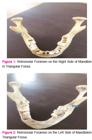
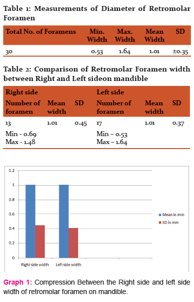
In this study all available 72 dry, fully ossified adult mandibles from the department of Anatomy, Shri Guru Ram Rai institute of medical and health sciences, Dehradun, Uttarakhand north region of India were included without gender and age distinction. In 72 dry mandibles each mandible was observed for morphological variations that is retromolar foramen and the mandible with foramen were evaluate for the diameter of retromolar foramen along with side preference (Fig no.1 and 2).
Mandibles showing signs of pathology or other processes which may have influenced result (osteopetrosis, advanced alveolar bone resorption in the retromolar area) etc. and mandibles with the signs of damage to the retromolar area whether due to the ageing or handling of mandibles were excluded.
Result: -
In our findings it shows that the diameter of the Retromolar foramen varies between 0.53 to 1.64 mm and the mean diameter of the Retromolar foramen is 1.0 mm and the mean width of the RMF on right side (1.01mm) is same as that of the left side (1.01mm) showing in Table no.1, 2 and Graph no.1.
Discussion: -
A triangular area behind the third molar tooth on the alveolar surface of mandible known as retromolar triangle. 72 human mandibles are examined in the present study for the diameter of the retromolar foramen along with the side preference. The diameter of the Retromolar foramen varies between 0.53 to 1.64 mm and the mean Diameter of foramen is 1.01mm. The mean width of Retromolar foramen in right side is 1.01mm which is same as of Left side (1.01mm). The above findings show that the right Retromolar Foramen is of same width as Left foramen.
In findings it shows that the mean diameter of the Retromolar foramen is 1.01 mm which was in part accordance with the study conducted by Md. A. Khan12 which showed that the mean diameter of the Retromolar foramen was 1.25 mm. The variation is possible because of the sample size. In our finding it is showed that the width of the Retromolar foramen on right side (1.01mm) is same as that of the left side (1.01mm), which was not in accordance with the study conducted by the Md. A. Ashraf Khan and Senthil Kumar.S13 both stated that the left Retromolar foramen was larger than right. The differences in these studies may be due to differential origin in Indian population and their sample size.
Conclusion: -
The diameter of Retromolar foramen through which the nerves and artery passes less than 1mm was not considered as foramen and the diameter of the Retromolar foramen on both right and left side were found same. The constituent of retromolar foramen and canal are the nerves that provides innervation to the pulp of third molar, retromolar area, fibers of the temporalis and buccinator muscle.14 Presence of retromolar foramen and canal and its contents have great clinical implication and can be damaged in various surgical activities like flap lifting, bone tissue for autologous bone grafts, osteotomy for the surgical removal of third molar, placement of Osseo integrated implants for orthodontic or in sagittal split osteotomy surgeries15 so retromolar foramen is considered as potential route for further innervation to the lower third molar area causing failure in anesthetic mandibular blocking.
References:
-
Suazo GI, Cantín LM, López FB, Valenzuela UV, Valenzuela RR. Morphometric study of the retromolar triangle. Int J Odontostomat. 2007. Vol.1 (2):129-32.
-
Susan Standring, “Infratemporal and pterygopalatine fossae and temporomandibular joint” in Grays Anatomy: The Anatomical Basis of Clinical Practices, S.Standring, H. Ellis, J.C. Healy, D. Jhonson and A. Williums, Eds., Churchill Livingtone, New York, NY, USA, 40 th ed, 2008. Pp. 532.
-
M. Lipski, I.M. Tomaszewska, W. Lipska, G.J. Lis, K.A. Tomaszewski. The mandible and its foramen: anatomy, anthropology, embryology and resulting clinical implication. Folia Morphol.2013.vol.72, no.4, pp.285-292. DOI: 10.5603/FM.2013.0048.
-
K Narayana, U A Nayak, W N Ahmad, J G Bhat, B A Devaiah. The retromolar foramen and canal in south Indian dry mandibles. European Journal of Anatomy. 2002; 6 (3): 141-146.
-
Kodera H, Hashimoto I. A case of mandibular retromolar canal; elements of nerves and arteries in this canal. Kailbogaku Zashi. 1995; 70 (1): 23-30
-
Kaufman E, Serman NJ, Wang PD: Bilateral mandibular accessory foramina and canals: A case report and review of the literature. Dentomaxillofac Radiol 29:170, 2000
-
Wang PD, Serman NJ, Kaufman E: Continious radiographic visualization of the mandibular nutrient canals. Dentomaxillofac Radiol 30:131, 2001
-
Ikeda K, Ho KC, Nowicki BH, et al: Multiplanar MR and anatomic study of the mandibular canal. Am J Neuroradiol 17:579, 1996
-
Ossenberg NS: Temporal crest canal: Case report and statistics on a rare mandibular variant. Oral Surg Oral Med Oral Pathol 62:10, 1986
-
Ossenberg NS. Retromolar foramen of the mandible. Am J Phys Anthropol. 1987; 73: 119-28.
-
Silva FM, Cortez AL, Moreira RW, Mazzonetto R. Complications of intraoral donor site for bone grafting prior to implant placement. Implant Dent. 2006. vol. 15; 420-26.
-
Md. A.Khan, S.Agarwal, R.S. Mandloi. Prevalence of Retromolar Foramen in Dried Mandible along with Morphometric and Analytical study in North India. Natl J Med Dent Res. 2013. Vol. 2(1): 11-14.
-
Senthil Kumar S, Kesavi D. A Study on the incidence of Retromolar Foramen and Canal in Indian Dried Human Mandibles and its Clinical Significance. International Journal of Anatomical Sciences. 2010. Vol.1: 14-16.
-
Reyneke J. P, T sakiris P and Becker P. Age as a factor in the complication rate after removal of unerupted/ impacted third molars at the time of mandibularsagittal split osteotomy. J. Oral Maxillofac. Surg. 2002; 60(6): 654-659.
-
Boronat Lopez A and Penarrocha Diago M. Failure of locoregional anesthesia in dental practice. Review of the literature. Med. Oral Patol. OralCir. Bucal. 2006; 11(6): 510-513.
|
 IJCRR
IJCRR 


 This work is licensed under a Creative Commons Attribution-NonCommercial 4.0 International License


