IJCRR - 5(16), August, 2013
Pages: 31-34
Date of Publication: 28-Aug-2013
Print Article
Download XML Download PDF
VASCULAR VARIATIONS IN AXILLARY ARTERY - A CADAVERIC STUDY
Author: M. Gurushanthaiah, Satheesh Naik K, S. Lokanadham
Category: Healthcare
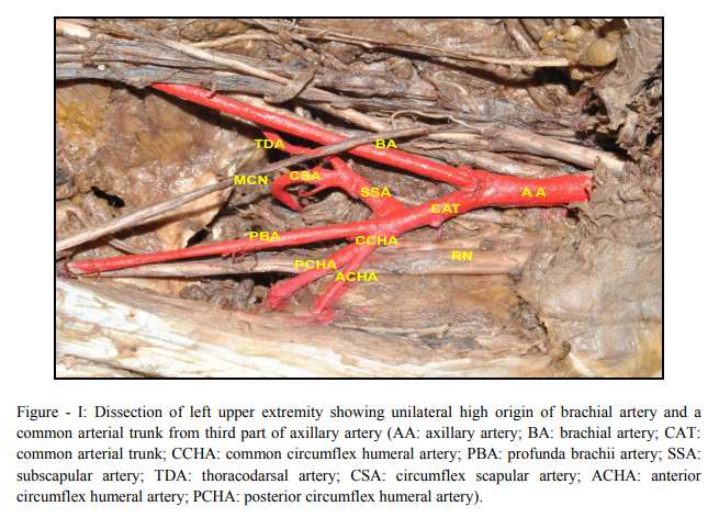
Abstract:An Unilateral variation in the branching pattern of third part of axillary artery was observed on the left upper extremity of a 52 years old embalmed cadaver, during routine dissection for medical undergraduate students. In the present study we observed high origin of brachial artery and a common arterial trunk from the third part of axillary artery. The common arterial trunk gave rise to subscapular, profunda brachii, and common circumflex humeral arteries. The subscapular artery divided into thoracodarsal and circumflex scapular arteries. The common circumflex humeral artery gave rise to anterior circumflex humeral and posterior circumflex humeral arteries. On the right upper extremity branching pattern of third part of axillary artery was normal. Arterial anomalies in upper limb are important for surgeons and anaesthesiologists for reparative surgery and angiography.
Full Text:
INTRODUCTION
The axillary artery is the direct continuation of the subclavian artery, begins at the outer border of the first rib, and ends normally at the inferior border of teres major muscle where onwards it continues as the brachial artery. Pectoralis minor muscle crosses it and so divides it into three parts which are proximal, posterior and distal to the muscle. Conventionally, the proximal part (first part) gives superior thoracic artery, the posterior part (second part) gives thoraco-acromial and lateral thoracic arteries and distal part (third part) gives subscapular artery, anterior and posterior circumflex humeral arteries (1).Many Authors have described the variations in the branching pattern of the axillary artery [2, 3, 4–10]. During routine dissection of the upper limb, we came across a unilateral variation in the branching pattern of third part of Axillary artery. The variation observed is not only rare but seems to be relevant and significant for understanding the formation of the arteries of the arm. There is an extensive collateral circulation associated with the branches of subclavian and Axillary arteries, particularly around the scapula. This clearly becomes of clinical significance during injury of the axillary artery.
MATERIALS AND METHODS
A 52 years male cadaver, bilateral upper extremity was dissected for medical under graduate students in the department of anatomy, Basaveshwara medical college, Chitradurga. We came across a unilateral variation in the branching pattern of third part of Axillary artery in the left upper extremity. Variations were studied and photographed. On the right side the branching pattern of third part of axillary artery was normal. Case Study A 52 years male cadaver was dissected to study the bilateral arterial pattern of upper extremity. We exposed the axillary artery from outer border of first rib to lower border of teres major muscle. From the third part we observed high origin of brachial artery and an unusual arterial trunk gives rise to subscapular artery, profunda brachii artery and common circumflex humeral artery (Figure – 1). The Subscapular artery divided into thoracodarsal, and circumflex scapular arteries, common circumflex humeral divided into anterior and posterior circumflex arteries. The course and distribution of the first, second part arteries and profunda brachii were normal. DISCUSSION Vascular anomalies in the upper limb are unilateral and occasionally bilateral (11, 12). Magden et a, has been reported that "abnormal" branching pattern of the axillary artery The lateral thoracic and thoracodorsal arteries arose together from the third part of the axillary artery as "a lateral thoracicthoracodorsal" common trunk. The superior thoracic artery was out of the position and the subscapular artery was not present. In our observation subscapular artery gives rise to thoracodarsal and circumflex scapular arteries. Johnson and Ellis studies show that in up to 30 - 40% of cases, the subscapular artery can arise from a common trunk with the posterior circumflex humeral artery. The posterior circumflex humeral artery may arise from the profunda brachii artery and pass back below the teres major instead of passing through the quadrangular space (13). In this study common arterial trunk gives rise to subscapular, profunda brachii and common circumflex humeral arteries. In another report by Samuel et al. (2006) the third part of the axillary artery gave a common arterial trunk, which further gives rise to anterior and posterior circumflex humeral, subscapular, radial collateral, and middle collateral and superior ulnar collateral arteries with absence of profunda brachii artery (14). Our cadaveric study reveals subscapular, profunda brachii and common circumflex humeral arteries was of axillary origin as a common arterial trunk. Anatomic variations in the major arteries of the upper limb have been reported. These include absence of the radial artery and the presence of a superficial brachial artery as well as a superficial ulnar artery (15). Our present report differs from this earlier report in branching pattern as well as course of these branches. Such anomalous branching pattern may represent persisting branches of the capillary plexus of the developing limb buds and their unusual course may be a cause for concern to the vascular radiologists and surgeons and may lead to complications in surgeries involving the axilla and the pectoral regions. The seventh cervical segmental artery gives rise to axillary artery and any abnormality during development results in the unusual branching pattern (16).
CONCLUSION
Anomalies in the branching pattern of axillary artery having practical importance for the radiologists and vascular surgeons. In our study high origin of brachial artery and a common arterial trunk arises from third part of axillary artery is uncommon and which gives rise to subscapular, common circumflex humeral and profunda brachii arteries, further supplies the axilla and fore arm. Variation in the principal artery branches are used for coronary bypass and important for vascular radiologist and surgeons of various clinical disciplines.
ACKNOWLEDGEMENT
Authors are thankful to Dr. G.M. Mahesh, Principal and Professor of Anatomy. Special thanks to Miss. Sudharani, Assistant professors and PG Students in Department of Anatomy, BMCH, Chitradurga, Karnataka.
References:
1. Standring, S.; Johnson, D.; Ellis, H. and Collins, P. Gray's Anatomy. 39th Ed. Churchill Livingstone, London, 2005. p.856.
2. Saeed M, Rufai AA, Elsayed SE, Sadiq MS. Variations in subclavian-axillary arterial system. Saudi Med J. 2002; 23: 206–212.
3. Yang HJ, Gil YC, Jung WS, Lee HY. Variations of the superficial brachial artery in Korean cadavers. J Korean Med Sci. 2008; 23: 884–887.
4. Susan Standring ed. Gray’s Anatomy. 40th Ed., London, Churchill Livingstone. 2008; 815–817.
5. Patnaik VVG, Kalsey G, Singla RK. Branching pattern of axillary artery – a morphological study. J Anat Soc India. 2000; 49: 127–132.
6. Rodriguez-Baeza A, Nebot J, Ferreira B, Reina F, Perez J, Sanudo JR, Roig M. An anatomical study and ontogenetic explanation of 23 cases with variations in the main pattern of the human brachio-antebrachial arteries. J. Anat. 1995; 187: 473–479.
7. Patnaik VVG, Kalse G, Singla RK. Bifurcation of axillary artery in its 3rd part – a case report. J Anat Soc India. 2001; 50: 166– 169.
8. Bhat KM, Gowda S, Potu BK, Rao MS. A unique branching pattern of the axillary artery in a South Indian male cadaver. Bratisl Lek Listy. 2008; 109: 587–589.
9. Tan CB, Tan CK. An unusual course and relations of the human axillary artery. Singapore Med J. 1994; 35: 263–264.
10. Saralaya V, Joy T, Madhyastha S, Vadgaonkar R, Saralaya S. Abnormal branching of the axillary artery: subscapular common trunk. A case report. Int J Morphol. 2008; 26: 963–966.
11. Yucel A.H. Unilateral variation of the arterial pattern of human upper extremity with a muscle variation of the hand. Acta Med Okayama.1999;53(2): 61 – 65.
12. Icten N, Sullu Y. Tuncer I.. Variant high origin radial artery: a bilateral case. Surg Radiol Anat 1996;18:63 – 66.
13. Johnson, D. and Ellis, H. Pectoral girdle and upper limb. In: Standring, S. Ed. Gray's Anatomy. 39th ed. Edinburgh, Elsevier, 2005. p. 845.
14. Samuel, V. P.; Vollala, V. R.; Nayak, S.; Rao, M.; Bolla, S. R. and Pammidi, N. A rare variation in the branching pattern of the axillary artery. Indian J. Plast. Surg., 39:222- 3, 2006.
15. Venieratos, D. and Lolis, E. D. Abnormal ramification of the axillary artery: subscapular common trunk. Morphologie., 85(270):23-4, 2001.
16. Wollard, H. H. The development of the principal arterial stems in the forelimb of the pig. Contrib. Embryo.
|
 IJCRR
IJCRR 


 This work is licensed under a Creative Commons Attribution-NonCommercial 4.0 International License


