IJCRR - 5(24), December, 2013
Pages: 41-45
Date of Publication: 31-Dec-2013
Print Article
Download XML Download PDF
VARIATIONS IN THE MORPHOLOGICAL APPEARANCE OF LINGULA IN DRY ADULT HUMAN MANDIBLES
Author: Smita Tapas
Category: Healthcare
Abstract:Objective: This study aims to analyse the different forms of presentation of lingula in dry adult human mandibles. Materials and Methods: Fifty dry adult human mandibles (100 sides) were studied to analyse the different forms of presentation of lingula. Results: Four different shapes of lingula were identified: triangular, truncated, assimilated and nodular type. The triangular shape of the lingula was noticed in 42 sides (42 %). The truncated shape lingula was noticed in 36 sides (36 %). The assimilated lingula was noticed in 12 sides (12 %). The nodular lingula was noticed in 10 sides (10 %). Conclusion: A prior knowledge of such variations in the morphological appearance of lingula will minimise the damage to the important nerves and vessels related to it during anaesthetic block or during operative procedures on the lower jaw. Morphological types of lingula can also be useful as anthropological marker.
Keywords: mandible, lingula, variations
Full Text:
INTRODUCTION
Lingula also known as Spix's ossicle or Spine after Johannes-Baptist Spix.1 Ireland called it as ligula mandibulae or spixs spine.2 Lingula is a tongue shaped bony projection located on the medial surface of mandibular ramus. It lies in close proximity to the mandibular foramen through which inferior alveolar nerves and vessels passes into the mandibular canal.3 Due to the close proximity of lingula to the mandibular foramen and in turn to the neurovascular bundle, it is used as an important landmark by the oro - maxillofacial surgeons for injection of local anaesthetics during dental surgical procedures.3,4 Lingula is also an important clinical landmark for bilateral sagittal split osteotomy, the most common surgical method to correct mandibular deformities.5 Structural variability in lingula can account for failure in inferior alveolar nerve block,6 as well as inaccurate localization may lead to the intraoperative complication like haemorrhage, fracture and nerve injury.5
Keeping the above factors in view, the present study is undertaken to analyse the different forms of presentation of lingula in dry adult human mandibles.
MATERIALS AND METHODS
The study conducted on fifty dry adult human mandibles (100 sides) to analyse the different forms of presentation of lingula. The morphological appearances of lingula were compared on either side.
OBSERVATION AND RESULTS
Four different shapes of lingula have been identified: triangular, truncated, assimilated and nodular type.
A. Triangular – The lingula with wide base and narrow rounded or pointed apex and apex being directed postero superiorly i.e., towards condyle or towards posterior border of ramus of mandible was classified as triangular. (Fig 1)
B. Truncated: The lingula with quadrangular top with superior, inferior and posterior borders was classified as truncated. (Fig 2)
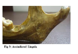
C. Assimilated: Lingula completely incorporated into ramus of mandible was classified as assimilated. (Fig 3)
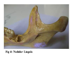
D. Nodular: Entire lingula except for its apex merged into the ramus of mandible was classified as nodular. (Fig 4)
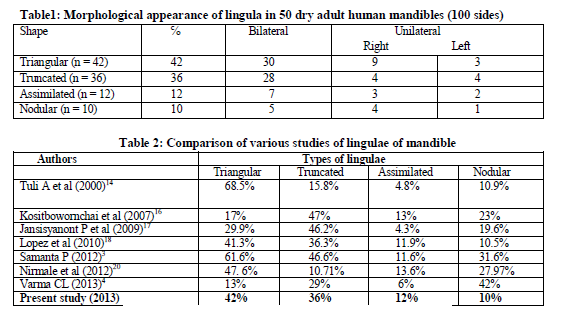
The triangular shape of the lingula was most prevalent (42 %). It was noticed bilaterally in 30 mandibles and unilaterally in 9 right and 3 left mandibles. The truncated shape lingula was noticed in (36 %). It was noticed bilaterally in 28 mandibles and unilaterally in 4 right and 4 left mandibles. The assimilated lingula was noticed in (12 %). It was noticed bilaterally in 7 mandibles and unilaterally in 3 right and 2 left mandibles. The nodular lingula was noticed in (10 %). It was noticed bilaterally in 5 mandibles and unilaterally in 4 right and 1 left mandibles. (Table 1)
DISCUSSION
The medial surface of the ramus of mandible is characterized by the lingula, a small tongue of bone at the anterior margin of mandibular foramen7 to which the sphenomandibular ligament is attached. Another end of sphenomandibular ligament is attached to the spine of sphenoid.8 The spine of sphenoid, the sphenomandibular ligament and the part of the mandible bearing the lingula have a common origin from the Meckels cartilage of first branchial arch.9
Earlier studies have reported the presence of various shapes of lingula but did not provide details about the various types and incidence.6, 10 Standard books describe the shape of this lingula to be triangular.7, 8 Truncated type was described by Hollinshead (1962)11, nodular by Berkovitz et al. (1978)12, and assimilated type by Morgan et al. (1982)13. Tuli et al (2000), have carried out a study on 165 dry mandibles of Indian origin, to determine the shape, direction and borders of lingula. They found triangular lingula in 68.5% mandibles, truncated, nodular and assimilated shape in 15.8%, 10.9% and 4.8% respectively.14 According to Devi, Arna et al (2003), the truncated and nodular types of lingula are most frequent.15 Study on 144 dry mandibles of Thai population by Kositbowornchai et al (2007) showed truncated (47%) to be most common followed by nodular, triangular and assimilated in 23%, 17% and 13% respectively.16 Jansisyanont et al (2009) studied on 92 Thai cadavers and found truncated lingula in 46.2% cases, triangular, nodular and assimilated shape in 29.9%, 19.9% and 4.3% respectively.17 Lopes et al (2010) did a study on 80 dry mandibles in south of Brazil. In their study the triangular shape of lingula was found in 41.3%, truncated in 36.3%, nodular in 10.5% and assimilated in 11.9%.18According to the Khan et al (2011), Triangular shape lingula is more prevalent in males (59.25%). The least prevalent in males is nodular (4.5%) and in females is assimilated (0%). The truncated type is almost twice as common in males (6%) than females (3.5%).19 Samanta et al (2012), reported the most prevalent shape of lingula was triangular and the least prevalent shape of lingula was assimilated type.3 Nirmale et al (2012) reported most prevalent shape of lingula was triangular and the least prevalent shape of lingula was truncated type.20 Varma et al (2013) study shows nodular lingula in 42%, truncated in 29 %, triangular in 13 %, assimilated in 6 % and M shaped in 4%.4 (Table 2). Gite et al studied location and the distance of lingula from sigmoid notch on panaromic radiograph.21
In present study, triangular shape of lingula was most prevalent and the least prevalent shape of lingula was nodular (Table 1) which is in accordance with the study of Lopes et al (2010) in southern brazil6, but contradictory to the findings of varma et al.1 where nodular shape of lingula is most prevalent.
As to why the shape of the lingula varies is not understood. According to Tuli et al14, the sphenomandibular ligament which is attached to the tip of the lingula is an accessory ligament to the temporomandibular joint and has minimal influence on altering the shape of the lingula.14
CONCLUSION
In present study, four different shapes of lingula have been identified: triangular, truncated, assimilated and nodular type. Triangular shape of lingula was most prevalent and nodular shape of lingula was least prevalent. It becomes a necessity to know the morphology of lingula so as to preserve the important structures during surgical interference of mandible around the lingula region.


References:
- Dobson J., Anatomical Eponyms, 2nd edition. Edinburgh, London: E. and S. Livingstone. 1962; Pp: 194
- Ireland R, Oxford Dictionary of Dentistry. Oxford University Press, New York. 2010; 1: Pp: 410
- Samanta PP, Kharab P. Morphological Analysis of Lingula in Dry Adult Human Mandibles of North Indian Population. J Cranio Max Dis. 2012; 1:7-11.
- Varma CL, Sameer PA. Morphological Variations of Lingula in South Indian Mandibles. Res and rev J Med Health Sci. 2013; 2(1): 31-34.
- Behrman SJ. Complications of sagittal osteotomy of the mandibular ramus. J oral surg. 1972; 30: 554-561.
- Nicholson ML. A study of position of the mandibular foramen in the adult mandible. Anat Record. 1985;212:110-112.
- Sinnatamby CS. Mandible, Osteology of skull and hyoid bone. Last’s Anatomy, Regional and Applied. Eleventh edition. Churchill Livingstone, Elsevier. 2006. Pp. 532-533.
- Standring S, Collins P, Healy JC, Wigley C, Beale TJ. Mandible: Infratemporal and pterygopalatine fossae and temporomandibular joint. Gray’s Anatomy - The Anatomical Basis of Clinical Practice, Fortieth edition. Churchill Livingstone, Elsevier. 2008. Pp. 530-532.
- Moore KL, Persaud TVN. The Developing Human-Clinically Oriented Embryology, Seventh edition, Saunders, Philadelphia. 2003. Pp: 204.
- Dubrul E L, Sicher. DuBrul's Oral Anatomy, Eighth edition Tokyo and New York: Ishiyaku Euro America.1988; Pp: 32-35.
- Hollinshead W H. Textbook of Anatomy. First edition. Calcutta, India: Harper and Row.1962; Pp: 855- 856.
- Berkovitz BKB, Holland GR, Moxham BJ. Colour atlas and textbook of oral anatomy. Second edition. London: Wolfe Medical Publication. 1978; Pp:15.
- Morgan DH, House LR, Hall WP, Vamuas S J. Diseases of temporomandibular apparatus. Second edition. Saint Louis: CV Mosby. 1982 ; Pp: 19.
- Tuli A, Choudhry R, Choudhry S, Raheja S, Agarwal S. Variation in shape of the lingula in the adult human mandible. J Anat. 2000; 197(2):313-317.
- Devi R, Arna N, Manjunath KY, Balasubramanyam M. Incidence of morphological variants of mandibular lingula. Indian J Dent Res. 2003; 14(4):210-213.
- Kositbowornchai S, Siritapetawee M, Damrongrungruang T, Khongkankong W et al. Shape of the lingula and its localization by panoramic radiograph versus dry mandibular measurement. Surg Radiol Anat. 2007; 29(8):689-694.
- Jansisyanont P, Apihasmit S, Chompoopong. Clin Ana. 2009; 22:787-793.
- Lopes PTC, Periera GAM, Santos AMPV. J Morphol Sci. 2010; 27(3-4):136-138.
- Khan TA, Sharieff JH. Observation on Morphological Features of Human Mandibles in 200 South Indian Subjects. Anatomica Karnataka. 2011; 5(1): 44-49.
- Nirmale VK, Mane UW, Sukre SB, Diwan CV. Morphological Features of Human Mandible. Int J of Recent Trends in Sci Technol. 2012; 3 (2): 38-43
- Gite M, Padhye M. Location of lingula from sigmoid notch in an Indian population - A Radiographic study. Scientific J. 2007; 1.
|


 This work is licensed under a Creative Commons Attribution-NonCommercial 4.0 International License