IJCRR - 6(1), January, 2014
Pages: 01-05
Print Article
Download XML Download PDF
USE OF CHROMOGENIC MEDIUM FOR SPECIATION OF CANDIDA ISOLATED FROM CLINICAL SPECIMENS
Author: Dharmeswari T., Sheela Devi Chandrakesan, Nagaraja Mudhigeti, Anitha Patricia, Reba Kanungo
Category: General Sciences
Abstract:Aim: To explore the usefulness of chromogenic medium in speciating clinical isolates of Candida and to determine their antifungal susceptibility. Methodology: A total of 50 Candida isolated from various clinical specimens were included in the study. Speciation of Candida was done based on the growth on chromogenic medium and other conventional methods. Antifungal susceptibility testing was performed against fluconozole and amphotericin B. Results: Among the 50 clinical Candida isolates, 45 could be speciated with the help of chromogenic medium. Only 30% of the Candida isolates were identified as Candida albicans and the rest were non albicans Candida species. Among the non albicans species Candida tropicalis was the commonest isolate followed by C. glabrata and C. parapsilosis. None of the strains were resistant to amphotericin B or fluconazole. Conclusion: Use of chromogenic medium with the morphology on corn meal agar provides rapid identification of commonly isolated Candida species from clinical specimens. This will be useful to initiate appropriate antifungal therapy thereby reducing morbidity and mortality.
Keywords: Candida albicans, Chromogenic medium, Antifungal susceptibility, speciation, Non albicans Candida
Full Text:
INTRODUCTION
The incidence of various fungal pathogens has increased dramatically over the past few decades. Candida species are the most common of these pathogens and have emerged as important opportunistic pathogens in the last decade. Infections by this group are often severe, rapidly progressive and refractory to therapy. Suppressed host defense and exposure to multiple risk factors are responsible for their emergence. Use of newer and broad spectrum antibacterial agents, increasing patient population living with AIDS, haematological malignancies or solid organ transplantation have been the contributory factors for the emergence of Candida species as important pathogens (1,2) Though Candida albicans is considered to be the commonest species causing human diseases, there are increasing reports of non-albicans Candida species emerging as important pathogens. (1,3) A rise in less common Candida sp. has been associated with significant morbidity and mortality. (4) The emergence of drug resistant Candida sp. which is largely attributed to use of prolonged and irrational empirical therapy has further complicated patient management. Some of the non-albicans Candida sp. are known to have varied susceptibility pattern to the routinely used antifungal agents especially the azoles. Conventional methods available to speciate the non albicans Candida sp. are time consuming and cumbersome. (5) To overcome this problem CHROM agar, a chromogenic differential culture medium was introduced. The manufacturers claimed that this chromogenic agar can facilitate the isolation and presumptive identification of certain clinically important Candida species. With the current scenario where non albicans Candida species are frequently isolated from clinical specimens, an early identification and speciation of positive Candida cultures is important . The present study was undertaken to explore the usefulness of chromogenic medium for speciation of Candida isolated from clinical specimens and to determine their drug susceptibility patterns.
MATERIALS AND METHODS
Isolation and identification of yeast
A total of 50 Candida isolated from various specimens (blood, clean-voided mid stream urine, pus, broncho-alveolar lavage and tracheal aspirates) submitted to the clinical microbiology laboratory from different clinical units of Pondicherry Institute of Medical Sciences (PIMS), a tertiary care hospital, were included in the study. All suspected yeast colonies on sheep blood agar were confirmed by Gram staining. All such yeast isolates that grew on sheep blood agar (HiMedia, Mumbai, India) were sub cultured on chromogenic medium (HiChrom agar Candida; HiMedia, Mumbai, India) and incubated at 37o C. The isolates grew well and developed distinctive colored colonies after overnight incubation. The plates were further incubated for 24 h to observe well distinguished colonies. Presumptive identification was made by n o t i n g t h e color of the colonies as per the manufacturer's Instructions. All isolates were further inoculated on corn meal agar (CMA) by slide culture method to determine microscopic morphological features of various Candida species. (6) Antifungal susceptibility testing by disc diffusion method was performed as per CLSI M44A (7) document against fluconazole discs (Hi Media, Mumbai, India) for all the 50 isolates. Briefly the yeast suspension was prepared in 5ml of sterile normal saline and turbidity was adjusted to 0.5McFarland standard. Modified Muller Hinton agar (Muller-Hinton agar + 2% glucose + 0.5 µg/ml of Methylene blue, pH 7.2) plates were inoculated and fluconazole (25 µg) discs (Hi Media, Mumbai, India) was placed on each plate. Results were obtained after 24 hours of incubated at 37 o C. MIC of Amphotericin B was determined by agar dilution method for 37 isolates. Amphotericin B (Hi Media, India) stock solution and working concentration were prepared as per CLSI guidelines. (8) As the study did not involve human subjects, obtaining informed consent was waived by the Institute Ethics Committee. Throughout the study C.albicans (ATCC90028), C.parapsilosis (ATCC90018), and C.krusei (ATCC6258), ATCC standard strains were used as Quality controls.
RESULTS
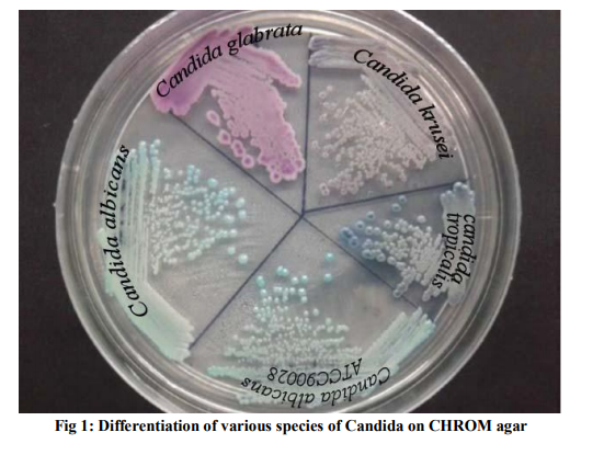
Only 15/50 (30%) of the strains were identified as C. albicans. Nearly half of the remaining 35 strains were C. tropicalis (43%), followed by C.glabrata (25%) and C.parapsilosis ( 17%). Candida species identified by the color on the chromogenic medium is shown in fig 1. Out of 50 isolates, 45 isolates showed characteristic morphology for that particular Candida species identified by CHROM agar. Three Candida isolates did not correlate with morphology on corn meal agar and hence did not fit with any Candida species. However on CHROM agar t h e s e were identified as Candida glabrata, Candida albicans and Candida krusei. One Candida albicans which showed green colored colonies on CHROM agar and was positive for Germ tube test (GTT) did not show any chlamydospores on Corn meal agar, but only elongated yeast cells. The other two isolates, one identified as Candida glabrata on CHROM agar resembled Candida parapsilosis in morphology on corn meal agar (short pencil shaped yeast cells) and the one identified as Candida krusei on CHROM agar gave a typical morphology of Candida tropicalis (oval blastoconidia and long pseudohyphae) on corn meal agar. Two other isolates did not produce any color on Chrom agar and showed round to oval yeast cells on corn meal agar. Antifungal susceptibility of all the 50 Candida isolates for fluconazole was performed by disc diffusion method. None of the isolates in the study were resistant to fluconazole. Only 3 Candida isolates were intermediately susceptible showing that they were dose dependent susceptible (two Candida glabrata and one Candida tropicalis). The two Candida glabrata strains were isolated from high vaginal swab and one Candida tropicalis was isolated from blood. All the other Candida isolates were sensitive to fluconazole. Minimum Inhibitory concentration (MIC) was performed by agar dilution method for amphotericin B for thirty seven Candida isolates. It ranged from <0.062μg/ml to 1μg/ml for the 37 Candida isolates tested, which were sensitive.
DISCUSSION
The incidence of fungal infections particularly those caused by Candida species, has increased markedly over the past decade, especially in immuno-compromised patients. Although the recent use of combination anti-retro viral therapy in AIDS patients may help to reverse the trend, Candida infection remain by far the commonest opportunistic infection in AIDS patients. There is a striking increase in the frequency with which non albicans species are isolated primarily Candida tropicalis, Candida glabrata, Candida krusei, and Candida parapsilosis. (9) . In the present study 70% were non albicans compared to 30% of the Candida isolates were Candida albicans. Among the non albicans, Candida tropicalis was the most commonly isolated (30%). This indicates that it is no longer sufficient to confirm or exclude only Candida albicans from clinical specimens. The conventional test used for speciation are time consuming and cumbersome. Moreover the germ tube test, which is the commonest test employed and gives rapid results, is not very accurate as more than 5% Candida albicans can be negative. Some non albicans species like Candida tropicalis and Candida parapsilosis are occasionally germ tube positive.(10) . It is necessary to evaluate simple, cost effective and rapid method like chromogenic medium to identify Candida to species level. Chromogenic agar is a newer and more rapid method to speciate Candida, contains enzymatic substrates that are linked to chromogenic compounds. When specific enzyme cleaves the substrate, the chromogenic substances produce colour. The action of different enzymes produced by yeast species results in color variation which is useful for the presumptive identification of some yeast (11) In our study 45 isolates could be speciated based on the color on the chromogenic medium and the morphology on Corn meal agar blocks. In the present study CHROM agar could identify, Candida albicans, Candida tropicalis, Candida glabrata and Candida paraopsilosis. Similar findings have been reported by different workers in the literature. (5, 12, 13) . Some of these studies have shown that Candida krusei could be identified by CHROM agar, but in our study 2 isolates identified as Candida krusei on CHROM agar did not show the typical morphology on corn meal agar. The Chromogenic medium facilitates presumptive identification of yeast isolates upto the species level within 24hrs of incubation. Primary inoculation of the clinical specimen on chromogenic medium which are positive for yeast cells on direct gram stain can hasten the presumptive species identification of yeast in clinical specimens. This will allow early initiation of appropriate therapy. The antifungal susceptibility of the Candida isolates to Amphotericin B revealed that all the isolates were sensitive to it. But some studies have stated that amphotericin B resistance is on the rise. A study from South India has shown that 100% of Candida albicans isolates and 88.9% of Candida parapsilosis were sensitive to Amphotericin B (14) The fluconazole susceptibility was tested by disc diffusion method and showed that 47(94%) Candida isolates were sensitive. The three isolates (2 identified as Candida glabrata and 1 Candida tropicalis) were dose dependant susceptible. Studies from India have shown that fluconazole resistance varies from zero to 30.6%. (14-16) The limitation in this study was that no other method was used to confirm the identity of the Candida isolates like the conventional carbohydrate fermentation and assimilation tests or the Vitek system.
CONCLUSION
Use of chromogenic medium with the morphology on corn meal agar provides rapid identification of commonly isolated Candida species from clinical specimens. This will be useful to initiate appropriate antifungal therapy thereby reducing morbidity and mortality. The findings of this study have helped our clinical laboratory to use chromogenic agar for presumptive identification of Candida species from clinical specimens.
ACKNOWLEDGEMENT
This study was supported by the Indian Council of Medical Research (ICMR) as a short term research project awarded to Ms.Dharmeswari T, under the guidance of Dr Sheela Devi C (ICMT STS project No.2011-01728).
References:
REFERENCES
1. David Trofa, Attila Gácser and Joshua D. Nosanchuk. Candida parapsilosis, an Emerging Fungal Pathogen. Clin. Microbiol. Rev. 2008; 21(4):606.
2. Jacqueline M. Achkar and Bettina C. Fries. Candida Infections of the Genitourinary Tract. Clin. Microbiol. Rev. 2010; 23(2):253.
3. Wingard JR. Importance of Candida species other than Candida albicans as pathogens in oncology patients. Clin. Infect. Dis.1995;20:115-125.
4. Chakrabarti. A, Singh. S and Das. S. Changing pace of nosocomial candidemia. Indian. J.Med. Microbiol.1999;17:160-166.
5. Li-Ung Huang,Chi-Hsiang Chen,Chu –Fang Chou, Jang- Jih Lu,Wei-Mingchi, Wei-Hwa Lee. A comparison of methods for yeast identification including CHROM agar Candida , Vitek system YBC and a traditional biochemical tests. Chinese Medical Journal (Taipei.).2001;64:568-574.
6. Agarwal S, Manchanda V, Verma N, Bhalla P. Yeast identification in routine clinical microbiology laboratory and its clinical relevance. Ind J of Med Microbiol 2011; 29 : 2:172-177.
7. Wayne Pa. National committee for antifungal disk diffusion susceptibility testing yeast: approved guidelines; CLSI;M-44A;2004.
8. Wayne Pa. Clinical and laboratory Standards Institute Reference method for broth dilution antifungal susceptibility testing of yeasts; approved standard-third edition; CLSI document M27-A3;2008(a)
9. Malini R Capoor, Deepthi Nair, Manorama Deb, Pradeep Kumar Verma, Lakshmi Srivastva and Pushpa Aggarwal. Emergence of Non albicans Candida species and antifungal resistance in a tertiary care hospital. Jpn J Infect Dis.2005; 58:344- 348.
10. J.E. Hoppe, P.Frey: Evaluation of six commercial tests and the germ tube test for the presumptive identification of Candida albicans. Eur. J. Clin. Microbiol. Infect. Dis.1999; 18:188-191.
11. Odds, F. C., and R. Bernaerts. 1994. CHROM agar Candida, a new differential isolation medium for presumptive identification of clinically important Candida species. J. Clin. Microbiol. 32:1923-1929.
12. Ann P Koehler, Kai Cheong Chu, Elizabeth T S Houang and Augustine F B Cheng. Simple, reliable and cost effective yeast identification scheme for the clinical laboratory. J. Clin. Microbiol.1999;37(2):422- 426.
13. Pfaller M A, Houston A and Coffmann S. Application of CHROM agar Candida for rapid screening of clinical specimens for Candida albicans, Candida tropicalis, Candida krusei and Candida glabrata. J Clin Microbiol.1996;34:58-61.
14. L Srinivasan and J Kenneth. Antibiotic susceptibility of Candida isolates in a tertiary care hospital in Southern India .Indian J Med Microbiol.2006;24(1):80-81
15. Adikary.R. Joshi.S. Species distribution and anti-fungal susceptibility of candidemia at a multi- super specialty center in southern India. Indian J Med Microbiol.2011; 29(3):309- 311.
16. Rizvi MW, Malik A, Shahid M, Singhal S. Candida albicans infections in a North Indian tertiary care hospital: Antifungal resistance pattern and role of SDS-PAGE for characterization. Biology and Medicine 2011; 3(2):176-181.
|
 IJCRR
IJCRR 


 This work is licensed under a Creative Commons Attribution-NonCommercial 4.0 International License


