IJCRR - 6(6), March, 2014
Pages: 71-74
Print Article
Download XML Download PDF
A STUDY OF FIBULAR PLANTAR MARGINAL ARTERY WITH ITS CLINICAL PERSPECTIVE
Author: Anupama K., Saraswathi G. , Jyothi K. C. , Shanmuganathan K.
Category: Healthcare
Abstract:Introduction: The plantar arterial arch is formed by anastomosis of the deep plantar branch of the dorsalis pedis artery and the deep transverse branch of the lateral plantar artery. The plantar arch gives off three perforating and four plantar metatarsal branches. An additional lateral digital branch to the fifth toe arises directly from the lateral plantar artery near the base of the fifth metatarsal bone and this artery is named as Fibular Plantar Marginal artery or Lateral Marginal Artery. The study was undertaken because of its varied origin and clinical importance in reconstructive surgery, vascular surgery and auto graft transplantation. Objectives: To study the incidence, origin and variations of Fibular Plantar Marginal Artery. Materials and methods: 50 feet of the formalin fixed adult human cadavers were dissected and studied, in the Department of anatomy, JSS Medical College, Mysore. Results: The presence of fibular plantar marginal artery was observed in 14 specimens (28%) and absent in 36 specimens. In 12 specimens it was originating from Lateral plantar artery, in 2 specimens the origin was from Dorsalis pedis artery. Conclusion: Knowledge of the variations in the anatomy of plantar arteries and the presence of fibular plantar marginal artery are important for vascular surgeons to carry out reconstructive surgeries and to obtain neurovascular flap for auto graft transplantation.
Keywords: Plantar Arch, Fibular Plantar Marginal Artery, Reconstructive Surgeries, Neurovascular flaps.
Full Text:
INTRODUCTION
The human foot is a complex structure, adapted to allow orthograde, bipedal stance and locomotion. It is the only part to support the entire body weight during locomotion. The blood supply of the foot is rich and is derived from branches of the major arteries, namely the Dorsalis pedis, Posterior tibial and Peroneal.1 The plantar arch is formed by anastomosis of the deep plantar branch of the dorsalis pedis artery and the deep transverse branch of the lateral plantar artery. The plantar arch gives off three perforating and four plantar metatarsal branches and numerous cutaneous and muscular branches in the sole.2 An additional lateral digital branch to the fifth toe arises directly from the lateral plantar artery near the fifth metatarsal base. This artery is given the name Fibular Plantar Marginal artery or lateral marginal artery .3 In the present study the incidence, origin and variations of fibular plantar marginal artery were observed. This study was undertaken because of its clinical importance in reconstructive surgery, vascular surgery, and autograft transplantation.
MATERIALS AND METHOD
Dissection method
50 feet (25 right and 25 left) from 25 embalmed adult cadavers (21 male and 4 female, 35-70 years of age) were dissected and studied in the Department of anatomy, JSS Medical College, Mysore. The superficial and deep plantar regions of the foot were dissected carefully. The oblique head of adductor hallucis was dissected to visualize the full course of lateral plantar artery and plantar arterial arch. The plantar metatarsal branches were traced and cleaned. The presence of fibular plantar marginal artery and its origin was noted. The arterial arch and its branches were coated with acetone and quickfix solution, painted using synthetic enamel asian paints and photographed. In the present study, the incidence and origin of fibular plantar marginal artery was studied.
RESULTS
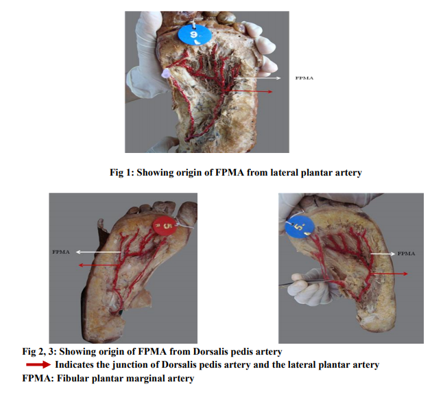
The fibular plantar marginal artery was observed in 14 specimens (28%) and absent in 36 specimens. In 12 specimens it was taking origin from lateral plantar artery (Fig 1); in 2 specimens the origin was from dorsalis pedis artery. (Fig 2, 3)
DISCUSSION
The lateral marginal artery may arise independently from lateral plantar artery or as a common trunk with a fifth superficial plantar metatarsal artery as described by Murakami. It pierces the lateral intermuscular septum and courses distally between the flexor digiti minimi brevis and adductor minimi muscles and its incidence being 22.5% 3 . In the present study it was found to be present in 28% of the specimens. The extrinsic circulation to the fifth metatarsal area is provided by the dorsal metatarsal artery, the plantar metatarsal arteries, and the fibular plantar marginal artery. These three source arteries supply branches to the metatarsal and adjacent joints. In the absence of fibular plantar marginal artery the dorsal and the plantar metatarsal arteries take over the vascularity in this region4 . The architecture of the skeletal and fibromuscular components is well designed to support the body weight in erect posture. The integrity of the structure invariably depends upon the blood supply5 . In the past few decades’ change in the lifestyle and increase in the use of vehicles have drastically increased the incidence of accidents and trauma all over the world. A thorough knowledge of topography and relations of the plantar arteries help the vascular surgeons in successfully performing microvascular surgeries of the foot. It helps in reconstructive surgeries of the foot, to avoid amputation in cases of trauma in industrial and automobile accidents. The knowledge of vascular variations are indispensable to the surgeons undertaking surgeries of the foot and ankle6, 7 . In diabetics and some non diabetics with atherosclerotic ischemic symptoms in the foot, the main occlusive process affects the pedal arteries. With the efforts of vascular surgeons restoring vascularisation by bypass surgery in patients with critical leg ischemia, the number of amputations as primary treatment has been greatly reduced8 . The neurovascular flaps are being utilized frequently in reconstructions in conditions such as burns, amputation following malignancy or due to accidents and in cosmetic correction of congenital defects. The dorsalis pedis artery was first used as the vascular basis for such neurovascular flap by O’Brien et al. Almost simultaneously it was found that the plantar vascular system involving first and the second intermetatarsal arteries can also be used for such reconstructions. The fibular plantar marginal artery when present can be an additional source of such neurovascular flap, without compromising the function of the foot 9, 10 . Angiographic investigations specially intraarterial digital arteriography and contrast enhanced magnetic resonance angiogram, not only indicates the pattern of the plantar arterial arch but also the presence of fibular plantar marginal artery, and helps to identify the site of occlusion of the arteries if any. This helps to determine the nature of treatment11 . In the year 1900, a pedicle graft of a toe to replace a missing digit in the hand was first described, but owing to the complicated nature of the operation this method for reconstruction of a digit had never been popular. The advent of microvascular surgery enabled one to use toes for free vascularised complex tissue grafts12, 13 . Hence the present work was undertaken which has relevance and significance for surgeons in the field of reconstructive surgeries.
CONCLUSIONS
The foot plays an important role in recreational, sporting and occupational activities, with the integrity of its component parts depending on its blood supply. Consequently knowledge of arteries of the foot is necessary for advances in arterial reconstruction. The study was undertaken because of its varied and important clinical application in reconstructive surgery, vascular surgery and autograft transplantation. Knowledge of the variations in the anatomy of plantar arteries and the presence of fibular plantar marginal artery are of immense help to the plastic surgeons while harvesting the neurovascular flaps and for vascular surgeons to carry out plastic and reconstructive surgeries. The purpose of this study is to draw the attention to this most exciting field and its ongoing developments. If accessory arteries are present in the foot they can be used as vascular autograft for surgeries in other parts of the body
ACKNOWLEDGEMENT
I sincerely thank all the editors /authors of all the articles, journals and books whose references I have quoted in this article. I also thank my Head of Department Dr Shailaja Shetty for her constant support and encouragement.

References:
REFERENCES
1. Pomposelli FB, Kansal N, Hamdan AD, Belfield A, Sheahan M, Campbell DR et al., A decade of experience with dorsalis pedis artery bypass. Analysis of outcome in more than 1000 cases. Journal of vascular surgery. 2003; 37: 307-315.
2. Mahadevan V. Pelvis and lower limb. Chapter 84 in Gray’s Anatomy 40th ed. Standring S. Churchill Livingston. Elsevier; 2008: 1455-1457.
3. Sarrafian SK. Anatomy of the foot and ankleDescriptive, topographic, functional. JB. Lippincott Company. Philadelphia; 1983: 281- 291.
4. Kitaoka HB, Holiday Jr AD, Metatarsal head resection for bunionette: Long term followup. Foot and ankle international, an official journal of AOFAS. 2004; 25: 521-525.
5. Gabrielli C, Olave E, Mandiola E, Rodrigues CFS, Prates JC. The deep plantar arch in humans. Constitution and topography. Surg radiol anat. 2001; 23(4): 253-258.
6. Strauch B, Sharzer LA, Brauman D. Innervated free flap for sensitivity and coverage, chapter 14 in Microsurgery for major limb reconstruction. Urbaniak JR. Mosby publication; 1990: 112- 116.
7. Ozer MA, Govsa F, Bilge O. Anatomic study of the deep plantar arch. Clinical anatomy. 2005; 18(6): 434-442.
8. Hughes K, Domenig CM, Hamdan AD, Schermerhorn M, Aulivola B, Blattman S et al., Bypass to plantar and tarsal arteries: An acceptable approach to limb salvage. Journal of vascular surgery. 2004; 40: 1149-1157.
9. Cobbett JB. Free digital transfer. Report of a case of transfer of a great toe to replace an amputated thumb. The journal of bone and joint surgery. 1969; 51: 677-679.
10. Papon X, Brillu C, Fournier HD, Hentati N, Mercier P. Anatomic study of the deep plantar artery: Potential bypass receptor site. Surg Radiol Anat. 1998; 20(4): 263-6.
11. Sebastien C, Douek P, Moulin P, Vaudoux M, Marchand B. Contrast-Enhanced MR Angiography of the Foot: Anatomy and Clinical Application in Patients with Diabetes. American Journal of Radiology. 2004; 182: 1435-1442.
12. Biemer E. Toe transfer for thumb and finger replacement in Textbook of Microsurgery. Lie TS. 3rdedition. International Congress Series 465 Excerpta Medica. Amsterdam; 1979: 17-19.
13. Foucher G, Merle M, Maneaud M, Michon J. Microsurgical free partial toe transfer in hand reconstruction: A report of 12 cases. Plastic and Reconstructive Surgery. May 1980; 65 (5): 616-626.
|


 This work is licensed under a Creative Commons Attribution-NonCommercial 4.0 International License