IJCRR - 6(7), April, 2014
Pages: 52-57
Print Article
Download XML Download PDF
A STUDY ON EVALUATION OF SURFACE ROUGHNESS AND ANTI-STANING PROPENSITY OF NANO - COMPOSITE DENTURE TEETH
Author: Jyoti Kundu , Ravinder Kumar , Shivam Seshan
Category: Healthcare
Abstract:Background and Objective: The introduction of nano-filled resin systems has resulted in considerable controversy. Lack of evidence-based scientific information and unavoidable time lag in establishing the precise relationship between their physicomechanical properties and clinical performance sought us to substantiate & qualify relative surface roughness & anti-staining characteristics of three commercially available type of artificial teeth. Materials and Methods: Three brands of three types of artificial teeth were examined .The staining behavior of the artificial teeth after immersion in tea solution for one hour was evaluated by spectrophotometeric analysis. Qualitative SEM analysis was used to assess the surface appearance after treatment with 2% citric acid for four hours. Results: The difference in mean optical density values for unstained and stained specimen suggested least staining with nanocomposite among the combinations used. Examined teeth when subjected to citric acid retreatment showed no qualitative surface changes in nano and micro filled composite but significant surface alterations were observed in dual cross-linked acrylic teeth. Conclusion: Within the limitations of this study, Nano composite showed significantly improved surface smoothness and stain resistance when compared to microfilled composite and dual cross-linked teeth tested.
Keywords: Nanocomposite denture teeth, surface finish, stain resistance, scanning electron microscope, spectrophotometry.
Full Text:
INTRODUCTION
Artificial teeth are often necessary for prosthodontic rehabilitation when natural teeth are lost. Acrylic resins and porcelains have been used for the fabrication of artificial teeth; however, neither type completely accomplishes the requirements for an ideal prosthetic tooth.1 It is well known that some dietary factors, such as tea lead to extrinsic tooth discoloration. 19Also citric acid , an organic acid found in high percentages in many dietary supplements, cause dental erosion and produces surface roughness of denture teeth. 20 Its been observed that during wear of resin composite teeth, inorganic fillers debond from the resin matrix and leave a void, increasing the surface roughness and forming a surface susceptible to exterior stain3 . The amount of filler content, the geometry and size of the filler particles,and the properties of the polymer matrix have been reported to influence the properties of polymer materials.2,4-12 A new type of denture tooth, fabricated of nano-composite resin, has recently been developed as a highly polishable, stain and impact resistant material.14 Few laboratory tests have been able to substantiate and quantify the surface roughness and anti staining property of polymeric denture teeth. Also, evidence-based scientific information regarding these new types of artificial teeth with respect to composition and physicomechanical properties is lacking. Therefore, studies critically discussing latest peer- reviewed reports and evaluating properties of commercial artificial teeth become necessary.
MATERIALS AND METHODS
Three groups of teeth (dual cross- linked acrylic resin, microfilled composite resin & nanofilled composite resin) were analysed for study.
SURFACE ROUGHNESS ANALY
SIS Preparation of samples and methodology for surface roughness evaluation
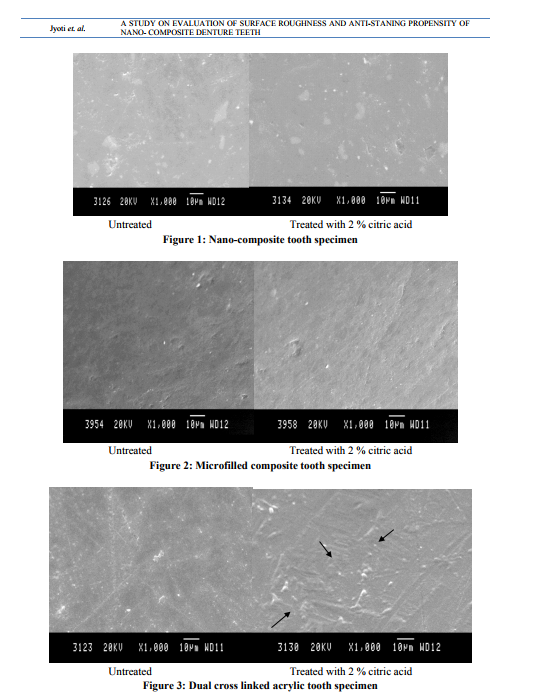
Fourteen specimens of maxillary central incisors from each type were used for SEM analysis using the sophisticated Scanning Electron Microscope (SEM). (JEOL, JFC - 1100E, Hitachi, HighTechnologies Corp, Tokyo, Japan). After vaporcoating with gold by ion sputtering device, the untreated incisal surfaces were examined in the SEM with the back-scattered electron images under high magnification of 1000x operating at 20Kv . Subsequently, these specimens were soaked in l0ml of 2% citric acid solution for 4 hours (assuming that average exposure is 40 sec. per day, thus simulating 1 year of exposure)20 . This was followed by qualitative SEM analysis to assess the surface appearance of the resultant acid treated specimens
STAIN RESISTANCE EVALUATION
Specimen preparation Fourteen specimens of maxillary 2nd molar from each type were used for stain resistance evaluation. Perspex strips of dimension 5x1 cm were prepared and maxillary 2nd molar was mounted at a height of 2.5 cm at an angulation of 45 degrees such that occlusal surface facing outward direction. This was the standardized guideline followed for specimen preparation such that focus of UV- Light of spectrophotometer is identical in position and location for all the specimens to be evaluated. Tea Solution Preparation 100ml of double distilled water was taken in a beaker & allowed to boil . After that 1gm of green tea leaves (Elixir, rohini estate, Darjeeling), measured in electronic balance, were brewed for 5 min. As temperature affects staining reaction ( Addy et al, 1985) so, experiment was planned to be conducted at room temperature 27 . The tea solution was cooled to room temperature and filtered with Whitman filter paper no.6. Wavelength Selection Absorbance decreases gradually from 360nm to 600nm wavelength. At 360nm absorbance of unstained specimen at constant stable position, optical density was 0.513 and at 600 nm, it was 0.153. Since 360-370 nm wavelength is the transition zone, so we opted for 395nm wavelength as the dominant wavelength showing peak absorbance of 0.418. Method of Data Collection Optical density of each of the unstained forty two specimen at selected wavelength of 395nm was noted. Then, all the forty two specimens were immersed in freshly prepared tea solution for 60 minutes & later on washed with distilled water for 30sec & bench dried27. Stained dried specimens were then subjected to spectrophotometer UVlight at same constant stable position and wavelength. Optical density of dried, stained specimens was noted and difference in the optical density of the specimens, before and after staining, was taken as a criteria to measure stain resistance. Lesser the difference, more the stain resistance. Statistical technique used ANOVA One way analyses of variance were used to test the difference between groups. To find out which of the two groups means is significantly difference scheffe’F’ test is used.\
RESULTS
Surface Roughness Analysis Qualitative Assessment as shown in figure 1, 2, 3 ? The SEM image of untreated nanocomposite tooth surface shows the small angular splintered nano-filler complexes of various sizes distributed in the matrix. While on other hand, SEM image of 2% citric acid treated nanosurface looks like the mirror image of untreated one, depicting the excellent surface smoothness even after one hour of citric acid treatment. ? In case of microfilled composite, untreated tooth surface analysis shows angular and spherical prepolymerised microfiller complexes incorporated in organic matrix.Whereas for the treated surface, SEM image shows no topographic changes suggestive of no significant alterations in the surface smoothness. ? Untreated Dual cross-linked acrylic SEM images shows macrofillers of various sizes of identical or different composition admixed in organic matrix of Urethane Dimethacryl (UDMA). But here in this case, noticeable difference was seen on surface treatment with citric acid and prominent surface irregularities were seen, indicative of certain qualitative changes in the surface topography debarring the surface smoothness.
Stain Resistance Evaluation
Table 1 shows the calculated mean and standard deviation of optical density of unstained and stained specimens among three groups. With tea, least staining was seen with nanocomposite while minimum stain resistance was shown by Dual Cross linked Acrylic (DCL) specimen teeth, although data may vary depending upon evaluation designs. The difference in mean absorbance value (optical density) of three groups exist on account of material composition & homogenity.
DISCUSSION
New materials, even if they are proved excellent, often have one or the other limitation, because they may be associated with a re - evaluation of the established systems of use and may not readily be amenable for use. Furthermore, there is an unavoidable time lag in establishing the precise relationship between their properties and clinical performance. Thus, the introduction of nanofilled resin systems has led to considerable controversy, both from the standpoint of the dentist and within the scientific community. However, it is possible to evaluate newer composite resins systems on the basis of their microstructure. Earlier researchers and manufacturers have reported that nano-composites were made up of homogenous urethane organic matrix reinforced by heterogeneous, pre-polymerized silica fillers13,14,22. While the micro-filled composite denture teeth (Endura) are heterogeneous, microfilled composite resins with agglomerated microfillers are similar to traditional macro-filled ones in size and chemistry, but not in structure. Further, they allow a substantial increase in the micro-filler content when admixed to an organic matrix. Such a resin composite has been known to demonstrate excellent finishing and perfect surface qualities. However, not much is known about the in vivo performance of composites with nano fillers. Consequently, available property data on these composite materials is rather limited. The absence of such vital data was the basis for taking up the study reported here. Results of this study clearly indicate that the hybrid (especially the nano-filled) resin composites are markedly superior to the traditional composites and acrylic resins in terms of surface smoothness and anti-staining tendency. Further, as the filler particle size is reduced, the polishability, permanence of surface smoothness, and esthetics of the nano-filled composites improve.
CONCLUSION
Judging by these results, it can be authentically concluded that nano-composite denture teeth may be one of the most promising and appropriate materials for denture teeth in near future. However, further investigation of other characteristics such as wear, impact resistance, and bonding to reparative autopolymerizing resins should be performed.
ACKNOWLEDGEMENT
We acknowledge to Geetanjali Medical College and Hospital, Udaipur and Indian Institute of Sciences, Bangalore for their immense support.
Conflict of Interest
None declared
References:
REFERENCES
1. Zarb GA, Bolender CL. Eckert SE, Jacob RF,Fenton AH, Merickske-stern RM. Prosthodontic treatment for edentulous patients:Complete denture and Implantsupported prosthesis. 12th ed. St. Louis: Mosby; 2004. p. 195-8.
2. Zeng J, Sato Y, Ohkubo C, Hosoi T. In vitro wear resistance of three types of composite resin denture teeth. J Prosthet Dent 2005; 94: 453–457.
3. Huan lu,:leslie b. roeder,: Effect of Surface Roughness on Stain Resistance of Dental Resin Composites J Esthet Restor Dent 17:102–109, 2005.
4. Kim KH, Ong Okuno O. The effect of filler loading and morphology on the mechanical properties of composites.J Prosthet Dent 2002;87:642-9.
5. Condon JR, Ferracane JL. In vitro wear of composite with varied cure, filler level, andfiller treatment. J Dent Res 1997;76:1405- 11.
6. Li Y, Swartz ML, Philips RW, Moore BK,Roberts TA. Effect of filler content and size on properties of composites. J Dent Res1985;64:1396-401.
7. Jaarda MJ, Wang RF, Lang BR. A regression analysis of filler particle content to predict composite wear. J Prosthet Dent 1997;77:57- 67.
8. Hashinger DT, Fairhurst CW. Thermal expansion and filler content of composite resins. J Prosthet Dent 1984;52:506-10.
9. Soderholm KJ. Influence of silane treatment and filler fraction on thermal expansion of composite resins. J Dent Res 1984;63:1321-6.
10. Schwartz JI, Soderholm KJ. Effect of filler size, water, and alcohol on hardness and wear of dental composites. Acta Odontol Scand 2004;62:102-6.
11. Turssi CP, Ferracane JL, Vogel K. Filler features and their effects on wear and degree of conversion of particulate dental composites.Biomaterials 2005;26:4932-7.
12. Miyasaka T. Effect of shape and size of silanated fillers on mechanical properties of experimental photo cure composite resins.Dent Mater J 1996;15:98-110.
13. Lutz F, Philips RW. A classification and evaluation of composite resin sytems. J Prosthet Dent 1983;50:480-8.
14. Suzuki S. In vitro wear of nano-composite denture teeth.J Prosthodont 2004; 13: 238– 243.
15. Fumiaki Kawano, Takafumi Ohguri, Tetsuo IchikawaIwate Mizuno, Akira Hasegawa. Shock absorbability and hardness of commercially available denture teeth. Int J Prosthodont 2002;15: 243-247.
16. Mandikos MN, McGivney, Davis E,Bush PJ, Carter JM. A comparison of the wear resistance and hardness of indirect composite resins. J Prosthet Dent 2001;85:386-95.
17. Kawano F, Ohguri T, Ichikawa T, Mizuno I, Hasegawa A. Shock absorbability and hardness of commercially available dentureteeth. Int J Prosthodont 2002;15:243-7.
18. Okada K, Tosaki S, Hirota K, Hume WR.Surface hardness change of restorative filling materials stored in saliva. Dent Mater 2001;17:34-9.
19. Leard A. Addy:The propensity of different brands of tea and coffee to cause staining associated with chlorhexidine.JClin Periodontol 1997:24:115-118.
20. Asher C, Read MJF: Early enamel erosion in children associated with the excessive consumption of citric acid.Br. HP.HI J 19X7; 162:384-387.
21. Mui S. Soh, Adrian U. J. Yap , Alan Sellinger (2007): Physicomechanical evaluation of lowshrinkage dental nanocomposites based on silsesquioxane cores . European Journal of Oral SciencesVolume 115, Issue 3, Pages 230- 238
22. Paola G. Loyaga-Rendon, Hidekazu Takahashi,Iwao Hayakawa, c and Naohiko Iwasaki (2007) : Compositional characteristics and hardness of acrylic and composite resin artificial teeth. (J Prosthet Dent 2007; 98: 141-149.)
23. Muhamad Ghazal and Matthias Kern (2009): The influence of antagonistic surface roughness on the wear of human enamel and nanofilled composite resin artificial teeth .J Prosthet Dent 2009;101:342-349.
24. M. Addy and W. R. Roberts ( 1981): Comparison of the bisbiguanide antiseptics alexidine and chlorhexidine. II. Clinical and in vitro staining properties. . Journal of Clinical Periodontology 1981: 8: 220-230.
25. T. Stober , H. Gilde, P. Lenz (2001): Color stability of highly filled composite resin materials for facings. Dental Materials 17 (2001) 87±94.
26. JM Brady and RD Wood (1977) : Scanning microscopy of cervical erosion .J Am Dent Assoc, Vol 94, No 4, 726-729.1977.
27. A.Leard and M.Addy (1997) : The propensity of different bunds of tea and coffee to cause staining associated with chlorhexidine. J Clin Periodontol 1997; 24: 115-118
|
 IJCRR
IJCRR 


 This work is licensed under a Creative Commons Attribution-NonCommercial 4.0 International License


