IJCRR - 6(9), May, 2014
Pages: 124-130
Print Article
Download XML Download PDF
A STUDY OF VARIATION OF CIRCLE OF WILLIS IN ADULT HUMAN BRAINS IN NAGPUR REGION OF MAHARASHTRA, INDIA
Author: Saniya H. Lade, Sonal Talokar, Ashish Radke
Category: Healthcare
Abstract:Objective: The purpose of the study was to examine the variations in arteries contributing in the formation of Circle of Willis in adult human brains from Nagpur region of Maharashtra. Material and methods: The external diameters of posterior cerebral artery (PCA), posterior communicating artery (PCOM), internal carotid artery (ICA), anterior cerebral artery (ACA) and anterior communicating artery (ACOM) in 50 male and 50 female brains were measured with verniercaliper. Photographs were taken and results studied by applying t.test and z test. Results: In this study 99% circles were complete.One circle with absent right sided PCA was found.Hypoplasia was noted in about 37 to 39% circles for PCA, about 39% circles for PCOM, 20% circles for ICA, 44% circles for ACA and 44% circles for ACOM.6% ACOM showed duplication. Regarding gender differences, hypoplasia of vessels overall was found to be more common in females than males. Duplication of ACOM was another finding found in female circles only. Conclusion: In present study it appears that there do exist variation in arteries forming circle of willis. Some preponderance to some arteries is also found in this study. Again male and female comparison is also helpful as it has revealed some significant findings. Such knowledge of circle of willis is of utmost importance to surgeons before performing any surgery related to ICA and also important for physicians to lead to conclusions in stroke and infarct patients where MRI can be used to find out status of arteries.
Keywords: Circle of Willis, Anterior Cerebral Artery, Anterior Communicating Artery, Posterior Cerebral Artery, Internal Carotid Artery, Posterior Communicating Artery.
Full Text:
INTRODUCTION
Thomas Willis is considered as one of the greatest anatomist of all times. His name is associated with the Circle of Willis, an anastomotic circle at the base of the brain. His work also formed the foundation of basic neuroanatomical description and nomenclature and comparative neuroanatomy. Willis provided a complete description of this vascular pattern and indicated that he understood the probable function of the circle. (Cagatay Ûstun ) 1 . It is a circle that supplies blood to the brain and is also known as Willis Polygon. It comprises ofanterior cerebral artery, anterior communicating artery, posterior cerebral artery, internal carotid artery, posterior communicating artery.It is an anastomosis of basilar system and internal carotid system lying in the interpeduncular cistern. (Satheesha Nayak)2 The Circle of Willis has an important role in maintaining a stable and constant blood flow to the cerebral hemisphere especially old people who may have reduced brain blood supply. The most common reason for this is senile arterioscelosis. Researchers have found a close correlation between a low capacity circle and an increased risk of stroke.Collateral ability of circle of willis be best used when there is emergency which again depends on the size of lumen and caliber of its component vessels. (K Ranil D De Silva) 3 Abnormalities in diameter of vessels forming circle of willis is found by many workers .Two reports appear in literature that of Alpers, Berry andPaddison(1959) and that of Fetterman and Moran(1941) stating that an external diameter of 1mm and 0.5mm or less, respectively would be considered abnormal. (Sylvia Kamath) 4 Against this background present study is carried out with following aims and objectives: 1. To study the variations of vessels forming the circle of willis 2. To find out any preponderance of variation in any particular vessel in this region. 3. To find out differences in males and females if any. 4. To compare the frequencies of different variants with previous autopsy studies
MATERIAL AND METHODS
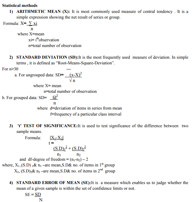
The present study was carried out in the department of anatomy from July 2009 to August 2011.It includes100 human brains (50 males and 50 females) irrespective of the cause of death. Brains were obtained from Forensic department of IGGMC, Nagpur and from the cadavers in the dissection hall of our college. Steps in studying the circle of willis: 1. Skull bones were carefully cut with hammer and chisel. Vault was removed and brain was taken out carefully after cutting the dura folds. 2. The removed brains were dipped in water and then washed under running water for 15 mins and now placed inverted over a clean surface to expose the base of brain; the water was soaked with tissue paper. 3. The circle of willis was observed at the base of brain. Measurements of arteries forming the cicle of willis were taken with vernier calipers graduated to measure upto 0.5mm, at two ifferent points and values were noted down on a preformed data sheet. 4. Finally, photographs were taken with digital cameras in order to avoid errors due to different angles of view, images were taken almost perpendicular to the plane of the circles. Arteries of 1mm and less diameter were considered abnormal, barring the communicating arteries, where 0.5 mm and less was considered abnormal. The measurements were then subject to stastisticalanalysis
Morphology of vessels forming Circle of Willis: Segments from the following corresponding regions were included in study by K Ranil D De Silva5 as right and left internal carotid arteries (ICA) close to their distal ends, precommunicating and postcommunicating part of the anterior cerebral arteries (a1), (a2) and the posterior cerebral arteries (p1), (p2) close to their origin, right and left posterior communicating arteries (PCOA) at their middle point and anterior communicating artery (ACOA) (with its variations if present) at its middle point. (K Ranil D De Silva)5 In this study following points were taken into consideration for measurement of the external diameter of arteries with the vernier calipers. A 1-PCA just before bifurcation on right side B 1- PCA after bifurcation on right side A 2- PCA just before bifurcation on left side B 2- PCA after bifurcation on left side C 1-PCOM near PCA right side D 1- PCOM near ICA right side C 2 - PCOM near PCA left side D 2- PCOM near ICA left side E 1-ICA before forming circle of willis on right side F 1- ICA after forming circle of willis on right side E 2- ICA before forming circle of willis on left side F 2- ICA after forming circle of willis on left side G 1-ACA at its origin on right sideH 1-ACA near ACOM on right side G 2- ACA at its origin on left side H 2-ACA near ACOM on left side Diameter of ACOM at its center point After the collection of data mean value of the measurements from the above two different points was taken out. Single value of right sided vessels was now ready for comparison with single value of left sided vessel.
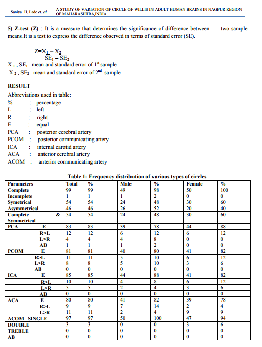
Symmetrical circle: Circle which has the external diameters of vessels on right side exactly equal to that on left side is the symmetrical circle. (Prof.E.Fawcett 1905)6 . Circle with variation in external diameters of corresponding vessels are considered as asymmetrical circles. The arteries were measured and mean was taken.Normal measuring arteries, hypoplastic arteries and absent arteries were noted. Posterior cerebral artery: 63% of right sided arteries and 61% of left sided arteries were of normal measurement while 37% of right side and 39% of left sided arteries were hypoplastic. One artery on right side in a male brain was found to be absent. Posterior communicating artery: 64% arteries on right side and 61% arteries on left side were normal. Hypoplasia was observed in 36% on right side and 39% on left side. Internal carotid artery: 80% of arteries were normal on both side while only 20% showed hypoplasia. Anterior cerebral artery: 56% arteries on right side and 58% arteries on left were normal .44% and 42% arteries on right and left side respectively showed hypoplasia. One circle of a male brain showed stenosis. Anterior communicating artery: 56% arteries were normal while 44% were hypoplastic. Gender differences In posterior cerebral artery more percentage ofhypoplasia was seen in males (48%) than in females(42%). 2% of male brains show absent right sided artery.No aplasia was observed in female brains. There was a significant finding regarding hypoplasia of posterior communicating artery, internal carotid artery, anterior cerebral artery and anterior communicating artery. Frequency of hypoplasia was more in females than males. Posterior communicating artery: Frequency of vessels measuring >0.5mm is more in females both for right (74%) and left(62%) as compared to males where the values are 54% for right and 60% for left. Internal carotid artery: In females hypoplasia is more (32%) as compared to males (8%) on both right and left side. Anterior cerebral artery: In males right sided artery is larger than left side in 14% circles which is more than females where the value is 4%.viceversa is true where 9% of females show left sided artery larger than right .In males this value is only 2%. Frequency of hypoplasia is more in females with preponderance to right side (54%).Only 46% circles show hypoplasia on left side.Male circles show only 32% and 30% hypoplastic arteries on right and left side respectively. Anterior communicating artery:Frequency of hypoplasia is more common in females which is 62% than males which is 26%.Duplication is seen in 3% of female circle .In present study duplication is 0% in male circles.
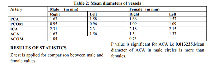
P value is also significant for ACOM i.e 0.001209. Mean diameter of ACOM in males is larger than females.
DISCUSSION
The study reveals some unknown facts regarding vessels contributing in formation of CirleOf Willis in adult human brains in Nagpur region with preponderance to some arteries.Complete circles are more common with aplasia of any vessel being a rare finding. (Prof. E Fawcett)6 Aplasia of PCA on right side is noted in one male brain which is a significant finding with no literature coinciding with this finding. Frequency of hypoplasia in ICA is more as compared to previous findings. In present study it is 20%. It means that people in this region have more hypoplastic ICA as compared to other places when the previous literatures are reviewed. ACA again show higher percentage of hypoplasia (44%) which is too high to coincide with the previous studies. Abubakhr7 and Bertram8 revealed only 0.7% and 10% hypoplasticACA.Stenosis is noted on left side which is very significant finding in present study. Hypoplasia is a common finding in communicating vessels which is a finding in this study also.Range of hypoplasia is very wide from 0.2% to 86% as literature states.Bertram8 noted duplication as most common abnormality.In present study 6% of ACOM showed duplication which can be compared to the finding of Prof. Fawcett6 which is 7.2%. Symmetrical circles are more common in females than males. Mean diameter of anterior cerebral artery is significantly larger in males than in females. (p<0.05) Mean diameter of anterior communicating artery in males is significantly larger than females. (p<0.05).Posterior cerebral artery is absent in one male case. No absence of any artery is noted in females. Frequency of hypoplasia is significantly higher in females than in males for both sided anterior cerebral arteryduplication of anterior communicating artery is higher in females than males.
CONCLUSION
We can concludethat, the findings of study are very important from surgeons point of view.There is a close correlation between a low capacity circle of willis and an increased risk of stroke. Degree of stenosis decides incidence of a particular stroke pattern. Hypoplastic posterior communicating artery is one of the common finding in this study. A small or absent ipsilateral posterior communicating artery is a risk factor for ischaemic cerebral infarction in patients with internal carotid artery occlusion. After the occlusion of an internal carotid artery the principal source of collateral flow is through the arteries of the circle of willis, but the size and patency of these arteries are quite variable. Study of the anatomy of the collateral pathways in patients with internal carotid artery occlusion with or without infarction in the watershed area of the deep white matter may identify patterns that afford protection from ischaemic infarction.Hypoplasia of internal carotid artery is more common in females as compared to males. Stenosis of anterior cerebral artery which is one of our finding is a risk factor for ischaemia in anterior part of cerebral hemisphere. Hypoplasia is a comparatively more in females than in males. Hypoplasia of anterior communicating artery is seen in this study. Duplication is more common in females than in males. Studies show that duplication of any artery is a site of structural wall weakness and may lead to aneurysm which is more common in anterior communicating artery Thus from present study it appears that there do exists variation in arteries forming Circle of Willis. Some preponderance to some arteries is also found in this study. Again male and female comparison is also helpful as it has revealed some significant findings. Such knowledge of Circle of Willis is of utmost importance to surgeons before performing any surgery related to internal carotidartery and also important for physicians to lead to conclusions in stroke and infarct patients where MRI can be used to find out status of arteries.
ACKNOWLEDGEMENT
These words are not borne of customs, they carry a far greater feeling in many ways. I sincerely wish to place on records my profound sense of gratitude to my revered guide Dr. D. D. Kheersagar (M.S.), Professor and Head of Department, Department of Anatomy, N K P Salve Institute Of Medical Sciences and Research Center, Nagpur, for his fatherly help,invaluable guidance, healthy criticism through evaluation of this paper and encouragement from time to time.He kindled a spirit of research and guided me to pursue the subject in depth with keen interest. I also thank Dr.A.P.Dongre, Dean, I.G.G.M.C., Nagpur for permitting me to carry on my paper work and utilize the facilities of postmortem in Department Of Forensic Medicine And Toxicology. I owe special obligation to Dr. Y. K.Kulkarni, Associate Professor, Department Of Anatomy, I.G.G.M.C,Nagpur for helping me to carry on my work in his department. I acknowledge the immense help received from the scholars whose articles are cited and included in references of this manuscript. I am also grateful to authors/editors/publishers of all those articles, journals and books from where the literature for this article has been reviewed and discussed. My special thanks to all my colleagues for their constant motivation throughout the study. I will be failing in my duty if I do not extend my thanks to my husband Mr. Amit Malani for his unparallel contribution in my study.
References:
REFERENCES
1. CagatayÛstunDr. Thomas WillisÕ Famous Eponym: The Circle of Willis;Turk J Med Sci 34 (2004) 271-274.
2. SatheeshaNayak,Somayaji S N,Saumya K V ;Variant arteries at the base of the brain; International Journal of Anatomical Variation[2008]60-61.
3. K Ranil D De Silva, RukmalSilva, W.S.LGunasekera , R W Jayesekera; Prevalence of typical circle of willis and variation in anterior communicating artery :A study of a Sri Lankan population;Annals of Indian Academy of Neurology; July-Sept 2009;vol.12;issue 3;157- 161.
4. Sylvia Kamath;Observations on length and diameter of vessels forming the circle of willis;Journal of Anatomy(1981);133;3;419-423. 5
. K Ranil D De Silva ,Rukmal Silva , DhammikaAmaratunga , WSL Gunasekera and Rohan W Jayesekera.Types of the cerebral arterial circle (circle of Willis) in a Sri Lankan Population .BMC Neurology 2011, 11:5.
6. ProfessorE.Fawcett,Dr.J.V.Blachford;The circle of willis:An examination of 700 specimens; journal of anatomy and physiology; 1905;vol.XL ,pg.83.
7. Abubakr HM Alawad, Mustafa A Hussein , Mohamed A Hassan. Morphology and normal variations of the Cerebral Arterial Circle ?of Willis? in Khartoum Diagnostic Centre. Khartoum Medical Journal (2009) Vol. 02, No. 02, pp. 215 – 219.
8. BertramC.A.Windle-On arteries forming the circle of willis;journal of anatomy and physiology;1888 Jan;22[pt.2];289-293.
9. Paul S And Mishra S;Variation of the anterior cerebral artery in human cadavers:A dissection study;Journal of Anatomical society India 53[1]15-16[2004]
10. Sylvia Kamath;Observations on length and diameter of vessels forming the circle of willis;Journal of Anatomy(1981);133;3;419-423.
|
 IJCRR
IJCRR 


 This work is licensed under a Creative Commons Attribution-NonCommercial 4.0 International License


