IJCRR - 6(9), May, 2014
Pages: 115-117
Print Article
Download XML Download PDF
CYTOLOGICAL EVALUATION OF THYROID LESIONS BY FINE NEEDLE ASPIRATION VERSUS NON-ASPIRATION CYTOLOGY TECHNIQUES- A COMPARATIVE STUDY
Author: Purushotham Krishnappa, Sowmya Ramakrishnappa
Category: Healthcare
Abstract:Aim: To compare the fine-needle non-aspiration cytology (FNNAC) with fine-needle aspiration cytology (FNAC) in providing the adequate samples for the evaluation of thyroid lesions. Materials and Methods: A total of 48 patients presenting with thyroid lesions at Standard Diagnostics Laboratory, Bangalore during period of Jan 2009 to October 2010, underwent both FNAC and FNNAC techniques. All the needle-sampling procedures were done by a single pathologist. The obtained samples were assessed for cytological evaluation based on sample adequacy, aspiration of blood, cellular features and possible diagnosis. Results: FNNAC produced less inadequate samples (20.9 %) than FNA (31.25%). Conclusion: The FNNAC is a better technique of sampling thyroid nodules and appears to produce more adequate specimen.
Keywords: Thyroid lesions, Fine Needle Aspiration Cytology, Non-aspiration
Full Text:
INTRODUCTION
Femoral shaft fracture is an incapacitating pediatric injury3 .There are a wide variety of non surgical treatment and surgical options available such as spica casting, traction followed by casting, plate fixation and flexible intramedullary nails with no clear consensus as to the preferred treatment4 . Internal fixation of paediatric shaft femur fracture by elastically stable intramedullary nail (ESIN) is gradually replacing conservative treatment5 . The material properties of titanium confer advantages for an implant used to stabilize pediatric femur fractures6 .Although good results have been reported with elastic intramedullary nails, plate fixation continues to be a viable alternative in surgical treatment of femoral shaft fractures7 . It is also considered that, compression plate fixation is a safe and effective treatment in children with both isolated femoral shaft fracture and those associated multiple injuries8 . The aim of this study was to identify the implant of choice for femoral diaphyseal fractures in paediatric age group with regard to immediate and long term functional outcomes
MATERIAL AND METHODS
The ethical clearance for the study was obtained from the Institutional Ethical Committee of our hospital on 21-07-2008 and Informed consent was taken from each of the parents/guardians of the patients in their own vernacular language. We conducted a prospective comparative study. Patients who presented to the out-patient department and casualty of the hospital with femoral diaphyseal fractures during August 2008 to August 2010 were considered for the study and each case was followed up for a period of three years from the date of enrollment. Closed femur diaphyseal fractures in the age group of 6-14 years were included in the study. Children aged < 6 years and > 14 years, children with open fractures and in whom metaphyseal extension was present were excluded from the study. DCP Procedure: Lateral approach for femur was used in all patients. After achieving the temporary anatomical stable reduction with k-wires, it was fixed with Dynamic Compression Plate. TENS Procedure: Nail diameter was determined by using the formula, Nail Diameter=Diameter of the medullary cavity at its narrowest part/2 - 0.59 . The nails were prebent to three times the diameter of the narrowest part of the medullary cavity to generate optimal resistance to malaligning forces10.The insertion points on femur were marked 2 to 3 cms proximal to the distal epiphyseal plate, determined under image intensifier. Nails of predetermined diameter were prebent and inserted to the medullary cavity with the help of a nail inserter. Postoperatively 2 days of intravenous third generation cephalosporin was given. Isometric quadriceps strengthening exercise, hip and knee joint mobilization exercises were advised on first post operative day. Toe touch walking was delayed till the appearance of callus radiologically. All the patients had regular follow ups at an interval of 4 weeks till six months and then, they were followed up once in every three months for one year and once in six months for next two years. There were no drop outs. The TENS outcome score suggested by Flynn et.al1 was applied to all the cases in the study after fracture union, irrespective of the mode of treatment. The outcome was graded as Excellent / Satisfactory / Poor. Then Harris hip score2 was applied the end of second and third year of follow up.
RESULTS
Femur shaft fractures were found to have high incidence in the age group of 12-14 years with Mean ± SD 10.85±2.26. Out of the 120 cases there were 93 (77.5%) males and 27 (22.5%) females. The most common mode of injury in our study was road traffic accident (RTA). In the study 96 cases (80%) had femur shaft fracture in the middle 3rd. There were 54 cases (45%), 51 cases (42.5%) and 15 cases (12.5%) of transverse, oblique and spiral fractures respectively. The time taken for the completion of procedure in group I was 95.60±8.47 minutes and in group II was 93.±9.04 minutes with a p value of 0.496. The amount of blood loss in group I was 96.5± 13.02 ml and in group II was 36.75± 8.77ml with a p value of <0.001. Three case (5%) in group I was immobilized with hip spica. Twelve (20%) cases in group II were immobilized with hip spica. Group I had a mean hospital stay of 15.30 ± 6.14 days, mean duration for toe touch walking was (started after the appearance of callus radiologically) 7.85 ± 2.23 weeks and mean duration of union was 17.90 ± 5.09 weeks. In group II mean hospital stay was 11.80 ±. 5.87 days, mean duration for toe touch walking was 3.97 ± 1.68 weeks and the mean duration of union was 13.00±1.37 weeks.
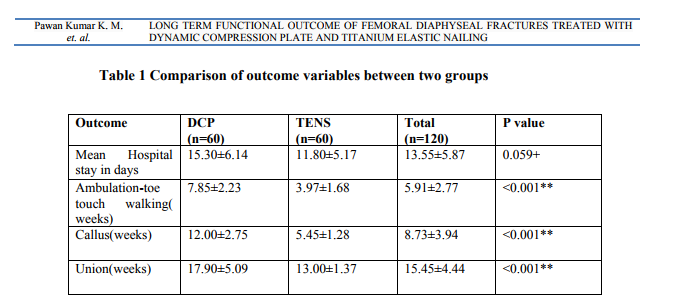
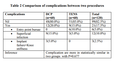
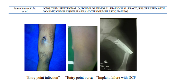
Limb length inequality was found in 9 cases (15%) and 6 cases (10%) in group I and group II respectively at the end of first year. In all these cases it was <2cms. At the end of third year only 2 cases of group I had limb length inequality in the form of lengthening. Malalignment was not observed in group I and was noted in 6 cases (10%) of group II. In group II malalignment was in coronal plane and was < 50 in all the cases. At the end of three years none of the cases had malalignment in any plane .There were 12(20%) cases with complications in group I, of which 9 cases had superficial infection and 3 cases had implant failure. Nine cases (15%) developed complications in group II 6 in the form of entry point bursitis and 3 cases had superficial infection. Functional outcome was assessed with Flynn’s TENS outcome score2 , applied to both the groups at the end of first year of follow up. Functional outcome at the end of first year-Group I had poor result in 3 cases (5%), satisfactory in 15 cases (25%) and excellent in 42 cases (70%). In group II satisfactory results was observed in 12 cases (20%) and excellent results in 48 cases (80%). Harris hip score at the end of second year of follow up had 6 cases (10%) in group I and 4 cases (6.6%) in group II with good functional outcome rest had excellent outcome. At the end of third year all cases in each group showed excellent outcome according to Harris hip score2 .
DISCUSSION
As surgeons consider different methods to treat pediatric femur fractures and mobilize the injured child, the ideal mode of treatment remains controversial11 . Titanium elastic nails are popular for the management of lengthstable diaphyseal femoral fractures in school-age children. Recently, sub muscular plating has been found to be a successful alternative option for management of length-unstable femoral fractures in school-age children11 . In the present study the average time taken for union in patients treated with DCP (Group-I) was 17.90±5.09 weeks and that in patients treated with TENS (Group-II) was 13.00±1.37 weeks which was statistically significant. In Group-I the union time is slightly higher when compared to reported results8,13, 14, 15 and in Group-II it is comparable to reported time for union6, 1, 16, 17. In our study we advised the patients, pain tolerated toe touch weight bearing with assistive devices as soon as the callus was visible radiologically. Group-I patients started toe touch walking at around 7.85±2.23 weeks where as Group-II patients started toe touch walking early at 3.97±1.68 weeks which is statistically significant with p value <0.001. The results are similar to Fyodorov I et al13 (6 weeks) and Agus H14 (8.5 weeks) in those treated with DCP for femur shaft fractures. And the study involving TENS as treatment modality reports of about 4 weeks (Flynn JM et al1 ). Three cases in group I (5%) were immobilized with hip spica. Twelve cases in group II (20%) were immobilized with hip spica. We found that incidence of hip spica was higher in Group-II (TENS) but was not statistically significant. Eren OT et al1 reported about 25% incidence of immobilization in patients treated with DCP and Flynn JM et al1 reported around 29.3% incidence of immobilization in femur shaft fractures treated with TENS, similarly Moroz LA et al18 report it to be around 22.2%.As the decision to immobilize was based on fracture anatomy, the strength of the fixation and confidence of the operating surgeon on the fixation, it is difficult to draw a statistically valid conclusion. In our study, after the procedure, except the patients who were immobilized the rest were advised to move the hip and the knee while lying on the bed from the second day. None of the patients moved their limbs on second post operative day in Group- I where as in Group-II 16(80%) patients started hip and knee mobilization from second day onwards, which was statistically significant with p value <0.001. The presence of the large surgical wound and the associated pain in patients treated with DCP may have caused the delay in mobilization. Carey TP et al16 report an average time for mobilization of 5.5days with TENS, similarly Flynn JM et al1 report it as 9 days. Timothy W et al19 report average time for mobilization as six weeks in patients treated with DCP. Our study noted a mean duration of hospital stay in Group-I to be 15.30±6.14 days and in Group–II to be 11.80±5.17, which was not statistically significant. Many reports suggest decreased hospital stay in patients treated with TENS6, 9, 17 compared to those treated with DCP, but this could not be observed in our study, probably because the time of discharge was seldom decided by the surgeon. It was more commonly influenced by the financial constrains of the patient. In our study we noted the limb length inequality was around 15 %( 9) in Group-I and around 10% (6) in Group-II but the distribution was statistically similar in both the groups and it was in all cases 50 of malalignment. Carey TP et al16, Ligier JN et al9 , Saikia KC et al17 and Roop Singh et al6 report 8%, 11%,9.09% and 8.57% incidence of malalignment in cases treated with TENS respectively. In our study 12 cases out of 60 in group I developed complications in the form superficial infection in 9 and implant failure in 3 cases. The cases which developed superficial infection resolved with regular dressings and extended oral antibiotics. The patients (3) who had implant failure were dealt individually. Group-II had 9 cases with minor complications. Six cases (10%) had burse at the site of entry point in distal femur and 3 (5%) case developed superficial infection. Superficial infection was resolved with regular dressings and extended oral antibiotics, which could have been avoided by leaving shorter length of nail outside femur and proper trimming of the nail ends. Functional outcome was assessed in both Group-I and Group-II by applying the TENS outcome scoring1 system at the end of one year. Group I had poor result in 3 cases (5%), satisfactory in 15 cases (25%) and excellent in 42 cases (70%). In group II satisfactory results was observed in 12 cases (20%) and excellent results in 48 cases (80%). Harris hip score at the end of second year of follow up had 6 cases (10%) in group I and 4 cases (6.6%) in group II with good functional outcome rest had excellent outcome. At the end of third year all cases in each group showed excellent outcome according to Harris hip score2 .
CONCLUSION
In TENS blood loss is minimal, minimally invasive, has got good early union rate. Implant failure rate is less and easy to remove. Only disadvantage is radiation and some cases required short term immobilization. Even though functional outcome at the end of one year and three years are statistically similar, TENS has a number of statistically significant advantages over DCP in terms patient morbidity. Hence TENS is the implant of choice at present for femoral diaphyseal fractures in children aged 6-14 years.
ACKNOWLEDGMENTS
We are extremely thankful to the immense help provided from the scholars whose articles are cited and included in references of this manuscript. We are also grateful to authors / editors / publishers of all those articles, journals and books from where the literature for this article has been reviewed and discussed.
References:
REFERENCES
1. Flynn JM, Hresko T, Reynolds RA. Titanium elastic nails for pediatric femur fractures: A multicentric study of early results with analysis of complications. J Pediatr Orthop 2001; 21:4-8.
2. Harris WH. Traumatic arthritis of the hip after dislocation and acetabular fractures: treatment by mold arthroplasty. An end-result study using a new method of result evaluation. J Bone Joint Surg Am. 1969 Jun;51(4):737-55.
3. Flynn JM, Skaggs DL , Sponseller PD, Ganley Tj, Kay Rm, Leitch Kk. The operative management of pediatric fractures of the lower extremity. J Bone Joint Surg Am 2002; 84:2288-2300.
4. Clinscales CM, Peterson HA. Isolated closed diaphyseal fractures of the femur in children: Comparison of effectiveness and cost of several treatment methods. Orthopedics 1997; 20 (12) :1131-6.
5. Saikat Sarkar Ranadeb Bandyopadhyay Arindam Mukherjee Titanium elastic nail - complications in the treatment of paediatric diaphyseal fracture of femur .Open Orthop J. 2013; 7: 12–17
6. Roop Singh, SC Sharma , Magu NK , Amit Singla. Titanium elastic nailing in pediatric femoral diaphyseal fractures. Ind J Orthop 2006; 40(1) : 29-34.
7. Eren OT, Kucukkaya M, Kockesen C, Kabukcuoglu Y, Kuzgun U. Open reduction and plate fixation of femoral shaft fractures in children aged 4 to 10. J Pediatr Orthop 2003; 23(2):190-193.
8. Caird MS, Mueller KA, Puryear A, Farley FA. Compression plating of pediatric femoral shaft fractures. J Pediatr Orthop 2003; 23(4):448- 452.
9. Ligier JN, Metaizeau JP, Prevot J, Lascombes P. Elastic stable intramedullary nailing of femoral shaft fractures in children. J Bone Joint Surg Br 1988; 70: 74-77.
10. Titanium Elastic Nail- Surgical Techinique. Synthes (Original instruments and implants of the assosiation for the study of internal fixation-ASIF): 2- 24.
11. David AS, Theodore JG, John MF. Titanium elastic nailing of pediatric femur fractures. Oper Tech Orthop 2005; 15: 326-330.
12. Li Y, Hedequist DJ. Submuscular plating of pediatric femur fracture. J Am Acad Orthop Surg. 2012 Sep;20(9):596-603.
13. Fyodorov I, Sturm PF, Robertson WW. Compression-plate fixation of femoral shaft fractures in children aged 8 to 12 years. J Pediatr Orthop 1999; 19(5): 578-584.
14. Agus H, Kalenderer O, Erynilmaz G, Omeroglu H. Biological internal fixation of comminuted femur shaft fractures by bridge plating in children. J Pediatr Orthop 2003; 23(2): 184-189. 15. Sink EL, Hedequist D, Morgan SJ, Hresko T. Results and technique of unstable pediatric femoral fractures treated with submuscular bridge plating. J Pediatr Orthop 2006; 26(2): 177-181.
16. Carey TP, Galpin RD. Flexible intramedullary nail fixation of pediatric femoral fractures. Clin Orthop Relat Res 1996; 332: 110-118.
17. Saikia KC, Bhuyan SK, Bhattacharya TD, Saikia SP. Titanium elastic nailing in femoral diaphyseal fractures of children in 6-16 years of age. Ind J Orthop 2007; 41(4): 381-385.
18. Moroz LA, Launay F, Kocher MS, Newton PO, Frick SL, Sponseller PD, Flynn JM. Titanium elastic nailing of fractures of the femur in children. J Bone Joint Surg Br 2006; 88: 1361-1366.
19. Timothy W, Jon L, Andrew K. Compression plating for child and adolescent femur fractures. J Pediatr Orthop 1992; 12: 626-632.
|
 IJCRR
IJCRR 


 This work is licensed under a Creative Commons Attribution-NonCommercial 4.0 International License


