IJCRR - 9(2), January, 2017
Pages: 29-31
Date of Publication: 20-Jan-2017
Print Article
Download XML Download PDF
Formation of the Median Nerve by an Additional Lateral Root: Embryological Basis and its Clinical Implications
Author: Archana Srivastava1, Anita Rani2, Archana Rani3, Pradeep Kumar Sharma4
Category: Healthcare
Abstract:Aim: A complicated network of arrangement of brachial plexus lead to frequent variations in this region. Therefore, a careful dissection of cadavers was performed during routine dissection classes of first year MBBS students.
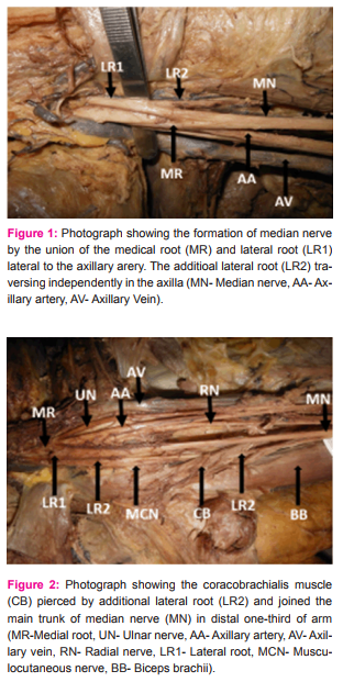
Case Report: During dissection of right arm of a 50 year old male cadaver the median nerve was found to be formed by the union of three roots instead of normally described two. An additional lateral root originated from the lateral cord of the brachial plexus. The medial root united with one lateral root in the axilla, lateral to the axillary artery to form the main trunk of the median nerve. The additional lateral root travelled independently in arm and pierced the coracobrachialis muscle without supplying it and then joined the main trunk of median nerve in the lower one-third of the arm. This type of variation in the median nerve is uncommon.
Discussion: Variations in median nerve are documented in the literature but the present case report is rare.
Conclusion: A detailed knowledge of normal and variant anatomy of median nerve should be kept in mind for correct diagnosis and appropriate treatment.
Keywords: Median nerve, Lateral root, Additional lateral root, Medial root
Full Text:
INTRODUCTION
The median nerve is formed in the axilla by fusion of two roots, the lateral and medial roots. Medial root of median nerve arises from the medial cord (C8,T1) of brachial plexus. The lateral cord (C5, C6, and C7) gives rise to lateral root of the median nerve. The two roots unite either anterior or lateral to the third part of the axillary artery.1 Median nerve then enters the arm lateral to the brachial artery. At the level of insertion of coracobrachialis muscle, it crosses the artery in front of it and then descends medial to it up to the cubital fossa. It enters the forearm between the two heads of pronator teres muscle. A high percentage of variations in the formation of median nerve have been reported. It includes the presence of additional roots, formation of the nerve in the arm and formation medial to axillary artery.2 The present case, reports an unusual formation of median nerve by three roots. The knowledge of these anatomical variations is of great importance to the surgeons in order to avoid injury to the median nerve during neck and axilla surgery.
CASE REPORT
During dissection of right arm of a 50- year old male cadaver, an additional third root of the median nerve was found. The main trunk of median nerve was formed by the union of the medial root and one lateral root in the axilla, lateral to the axillary artery. The additional root originated from the lateral cord, it travelled independently in arm and pierced the coracobrachialis muscle without supplying it and then joined the main trunk of median nerve in the distal one-third of the arm (Figure 1 & 2). The further course of median nerve in the arm was normal. The course and pattern of supply of the median nerve in the forearm and palm was normal. The musculocutaneous nerve showed no anatomical variation in the formation, course and supply. Vascular supply of the limb did not show any variation. No communication between the median and musculocutaneous nerves was observed. The formation, course and branching pattern of median nerve in the other limb was normal.

DISCUSSION
Formation of median nerve by two roots is seen in 48% to 88.5% of cases.2 Whereas formation by three or more roots has been well documented in the literature. These additional roots arise usually from the lateral cord of the brachial plexus.3-5 The reported incidence of this variation ranges from 14.2% to 20%.6,7 Rarely the additional root may arise from the medial cord or from the anterior division of the middle trunk of the brachial plexus.8,9 The median nerve may sometimes receive a communicating branch from the musculocutaneous nerve which is regarded by some authors as an additional lateral root.10 Sontakee et al. (2011) described a case where the median nerve was formed by three roots, two roots from the lateral cord and the third root from the medial cord. The first lateral root joined the medial root in the axilla and the second lateral root joined the medial root in the arm to form the median nerve.11 Pais et al. (2010) reported the formation of median nerve by three roots in their case study.12 The two lateral roots joined the medial root in the axilla to form the median nerve. A similar variation was reported by Satyanarayana et al. (2009).5
Das and Paul (2005) observed in their case that the median nerve was formed by two lateral and one medial roots. The upper lateral root crossed the third part of axillary artery anteriorly to unite with medial root to from the median nerve in the axilla. The median nerve thus formed was related medial to the axillary artery.13 A high incidence of variations in the site of union has also been documented.2,7
In our case, the median nerve was formed by union of two lateral roots and one medial root. The main trunk of the median nerve was formed in the axilla and the second lateral root joined the main median nerve trunk in the lower third of arm. This type of variation in the formation of median nerve is rare.
The variations in the formation of median nerve had been explained on the basis of embryological development. In human, the forelimb muscles develop from the mesenchyme of the para-axial mesoderm during fifth week of embryonic life. The upper limb buds lies at the level of lower five cervical and upper thoracic vertebrae. The axon of the spinal nerves then enters the mesenchyme of the limb buds and grows within it making an intimate contact with differentiating mesoderm. This directional growth of nerve fibres is controlled by cell surface receptors such as neural cell adhesion molecule (N-CAM) and L1 and transcriptional factors like cadherins.14,15
As the signalling of the developing axon is regulated by expression of these factors in a highly coordinated site specific fashion, any alteration in signalling between mesenchymal cells and neuronal growth cones can lead to significant variations.16
Knowledge about the variations in the formation of median nerve has clinical importance as these roots may be injured during surgery or trauma giving rise to unusual clinical symptoms. During brachial plexus block given by the anaesthetists, these variations may be responsible for failed or incomplete nerve block.
CONCLUSION
Embryological basis of different variations should be kept in mind to arrive at correct diagnosis. The knowledge of such anomalies are very important for the surgeons performing procedures in the axilla as well as for anatomists for academic purpose.
ACKNOWLEDGEMENTS
I express my gratitude to the staff of the Department of Anatomy for assistance in providing infrastructure facilities and necessary help. Authors acknowledge the immense help received from the scholars whose articles are cited and included in references of this manuscript. The authors are also grateful to authors / editors / publishers of all those articles, journals and books from where the literature for this article has been reviewed and discussed.
Source of Funding: Nil
Conflict of interest: All authors have none to declare.
ABBREVIATIONS USED: MN-Median Nerve, LR1- Lateral root, LR2- Additional lateral root, MR- Medial root, AA- Axillary artery, AV- Axillary vein, MCN- Musculocutaneous nerve, UN- Ulnar nerve, RN-Radial nerve, CB- Coracobrachialis, BB- Biceps brachii
References:
- Standring S. Gray’s Anatomy 39th ed. Churchill Livingstone. Elsevier, New York, 2005.
- Pandey SK and Shukla VE. Anatomical variations of the cords of brachial plexus and the median nerve. Clin Anat 2006; 20: 150-156.
- Saeed M and Rufai AA. Median and musculocutaneous nerves: variant formation and distribution. ClinAnat 2003; 16 (5): 453-457.
- Sargon MF, Uslu SS, Celik HH, Aksit DA. Variation of the median nerve at the level of brachial plexus. Bull Assoc Anat 1995;79 (246): 25-6.
- Satyanarayana N, Vishwakarma N, Kumar GP, Guha R, Dutta AK, Sunitha P. Rare variations in the formation of median nerve-embryological basis and clinical application. Nepal Med Coll J 2009; 11 (4):287-290.
- Budhiraja V, Rastogi R and Asthana AK. Anatomical variations of median nerve formation: embryological and clinical correlation. J MorpholSci 2011; 28 (4): 283-286.
- Channabasangouda Patil S, Shinde V, Jevor PS and Nidoni M. A study of anatomical variations of median nerve in human cadavers. Int J Biomed Research 2013; 4 (12):682-690.
- Paranjape V, Swamy PV, Mudey A. Variant median and absent musculocutaneous nerve. J Life Sci 2012; 4 (2): 141-144.
- Uzun A, Bilgi S. Some variations in the formation of the brachial plexus in infants. Tr J Med Sci 1999; 29: 573-577.
- Natsis K, Paraskevas G, Tzika M. Five roots pattern of median nerve formation. Acta Medica 2016; 59 (1):26-28.
- Sontakee BR, Tarnekar AM, Waghmare JE and Ingole IV. An unusual case of asymmetrical formation and distribution of median nerve. Int J Anat Variations 2011; 4: 57-60.
- Pais D, Casal D, Santos A, O’Neill JG. A variation in the origin of median nerve associated with an unusual origin of the deep brachial artery. J of Morph Sci 2010; 27 (1): 35-38.
- Das S and Paul S. Anomalous branching pattern of lateral cord of brachial plexus. Int J Morphol Sci 2005; 23 (4): 289-292.
- Brown MC, Hopkins WG, Keynes RJ. Essentials of neural development. Cambridge. Cambridge University Press, 1991; p. 46-66.
- Larsen WJ. Human embryology. 2nd ed. Edinberg. Churchill Livingstone. 1997; p. 311-339.
- Samnes DH, Reh TA, Harris WA. Development of nervous system. New York: Academic Press, 2000; p. 189-197.
|


 This work is licensed under a Creative Commons Attribution-NonCommercial 4.0 International License