IJCRR - 6(20), October, 2014
Pages: 35-38
Date of Publication: 20-Oct-2014
Print Article
Download XML Download PDF
ADENOMATOID ODONTOGENIC TUMOR OF MANDIBLE MIMICKING AMELOBLASTOMA: A DIAGNOSTIC CHALLENGE
Author: K. Vinay Kumar Reddy, R. Mounica, Kotya Naik Maloth, K. Sunitha, Govindraj S.J.
Category: Healthcare
Abstract:Adenomatoid odontogenic tumor is an uncommon benign Hamartomatous lesion of odontogenic origin, which affects young individuals, with female predilection and mainly occurring in second decade of life. Maxillary anterior region is most often involved, and associated with unerupted or impacted canine. We report a rare case of treated multilocular adenomatoid odontogenic tumor of posterior mandible in a 31 year old female patient.
Keywords: Adenomatoid Odontogenic tumor, Hamartoma, True neoplasm
Full Text:
INTRODUCTION
Adenomatoid odontogenic tumor (AOT) is a benign odontogenic lesion hypothesized develops from the enamel organ, dental lamina, reduced enamel epithelium or their remnants. It was first described by Dreibaldtin in 1907 as a Psuedo-adenoameloblastoma.1,2 Harbitz in 1915 reported AOT as cystic adamantoma.3 In 1948 Stafne considered it a distinct entity, but it was classified by others as a variant of ameloblastoma. The lesion is known by many names, including adenoameloblastoma, adenoameloblastic odontoma, and epithelial tumour associated with developmental cysts, ameloblastic adenomatoid tumour, adenomatoid or pseudo-adenomatous ameloblastoma, and teratomatous odontoma.4 The term AOT was proposed by Philipsen and Birn in 1969, was suggested that it is not to be regarded as a variant of ameloblastoma because of its different behaviour. In 1971 the term AOT was adopted in the initial edition of WHO histological typing of odontogenic tumors jaw cysts and allied lesions.4 WHO described the histological features of the tumor as follows: “A tumor of odontogenic epithelium with duct like structures and with varying degree of inductive changes in the connective tissue . The tumor may be partly cystic and in some cases the solid lesion may be present only as masses in the wall of a large cyst.1,2 It is generally believed that the lesion is not a neoplasm”. AOT accounts for 2.2 to 7.1 % of all odontogenic tumors and ranks for 4th or 5th among the odontogenic tumors. It is generally believed to be a hamartoma rather than a neoplasm.1,2
CASE REPORT
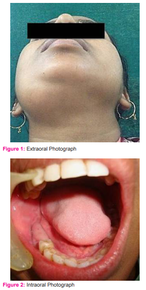
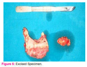
A 31 year old female patient presented with a chief complaint of swelling on her right lower one third of face since 1 year. Patient was asymptomatic 1 year back then she noticed a swelling on her right lower one third of face, which was initially small in size and gradually increased to present day size, with no history of pain, discharge and trauma. Patient gave a history of extraction of her lower right back tooth 3 years back. On extraoral examination a solitary diffuse swelling was seen on her right lower one third of face, which is roughly oval to dome shape, measuring about 5×4cm in size, extending anterio-posteriorly 2cm below the chin to angle of mandible, superiorly from the lower border of the mandible to inferiorly 5 cm below the lower border of mandible, surface over the swelling was smooth and skin over the swelling was stretched. On palpation it was mild tender, firm in consistency, compressible, non reducible with lo cal rise of temperature [Figure 1]. On intraoral examination buccal and lingual vestibular tenderness in relation to 45- 48 was noted with buccal vestibular obliteration in relation to 46, 47, 48 region [Figure 2]. Orthopantomograph was taken which revealed single well-defined multilocular radiolucency measuring about 6.5x6cm in size, extending anterio-posteriorly from mesial aspect of 45 to 1cm below sigmoid notch, superior-inferiorly 1.5cm above right alveolar ridge to 2cm below the lower border of the mandible with thinning of inferior border of mandible [Figure 3] CT-scan revealed a well-defined multiple radiolucent area with well-defined radiopaque border on right side of mandible with buccal and lingual cortical plate expansion [Figure 4]. Based on history, clinical and radiographic examination a provisional diagnosis of ameloblastoma on right body and ramus of mandible was given. Complete Surgical excision of the lesion was done under general anaesthesia [Figure 5]. Excised specimen was sent to histopathological examination which revealed epithelial and connective tissue components, solid nodules of epithelium arranged in the form of whorles which are cuboidal to columnar in nature. Some duct like spaces are noted with eosinophilic material and cords of the epithelium extending into the stroma. Cribriform pattern like tumor cell strands are also noted which are filled by dysplastic dentine/ amorphous eosinophilic like material. In addition connective tissue stroma showed odontogenic cell rests, proliferating blood vessels, large areas of haemorrhagic and few inflammatory cells. Bony trabeculae and surface epithelium are also noted [Figure 6: A, B]. Based on clinical, radiographic, and histopathological examination a final diagnosis of Adenomatoid Odontogenic tumour of Right body and ramus of mandible was given. Patient is under follow-up since 1 year without any recurrence [Figure 7].
DISCUSSION
Adenomatoid odontogenic tumour is a benign, noninvasive odontogenic lesion, with a predilection for the anterior maxilla (ratio of cases 2:1 relative to mandible) of young females.5 63% of AOT’s are diagnosed in the second decade of life, but in our case it was found to be in third decade of life. The maxilla is the predominant site of occurrence than mandible, and the anterior part of the jaw is more frequently involved than the posterior part.6 An unerupted tooth is most commonly associated with AOT but in our case it occurred in right posterior region of mandible and is not associated with an impacted tooth. Generally the tumors do not exceed 1–3 cm in greatest diameter, but they can be larger, as in the case reported here.7 The lesions are typically asymptomatic and growth results in cortical expansion, with displacement of adjacent teeth as in the case reported here.2 There are 3 variants of AOTs, it can occur both intraosseously and extraosseously. Intraosseous AOT may be radiographically divided into 2-types follicular / Pericoronal (73%) characterized as a well-defined unilocular radiolucent lesion surrounding the crown, and is a part of unerupted tooth, extrafollicular / (extra-coronal (24%) characterised as a well defined radiolucent lesion, but located between above, or superimposed upon the root of an erupted tooth. Minute radiopacities are frequently found within the lesion, but there are cases where the lesion has no radiopaque component as in our case.6 The extraosseous / peripheral, or gingival types of AOT (3%) are rarely detected radiographically, but there may be slight erosion of alveolar bone cortex, this lesion usually surrounds the crown of an impacted tooth.8 AOT is usually surrounded by a well-defined connective tissue capsule. It may present as a solid mass, a single large cystic space, or as a numerous small cystic spaces.3 AOT’s, accounting for approximately 3% of all odontogenic tumours, are less frequent than odontoma, cementoma, myxoma and ameloblastoma. It has been suggested that this tumor may be a hamartoma rather than a true neoplasm,9 but there is currently no evidence to resolve this dispute. For cases in which the lesion appears to surround an unerupted tooth and has no radiopaque component, dentigerous cyst may also be considered in the differential diagnosis. AOT often appears to envelop the crown as well as the root, whereas dentigerous cysts do not envelop the roots.10 The origin of AOTs is controversial. Because of its exclusive occurrence within the tooth bearing areas of jaws (most closely associates with an unerupted or impacted tooth) and its resemblance to the dental lamina, reduced enamel epithelium, enamel organ and or their remnants there is an agreement that the AOT is of odontogenic origin.1,2 The histological appearance of all variants is identical and exhibits a remarkable consistency. At low magnification most striking pattern is that of various sizes of solid nodules of columnar or cuboidal epithelial cells forming nests or rosette-like structures with minimal stromal connective tissue. Between the epithelial cells of the nodules and in the centre of the rosette-like configuration is found eosinophilic amorphous material, often described as tumour deposits. Conspicuous within the cellular areas are structures of tubular or duct-like appearance.11 A third characteristic cellular pattern consists of nodules of polyhedral, eosinophilic epithelial cells with squamous appearance and exhibiting well-defined cell boundaries and prominent intracellular bridges, islands may contain pools of amorphous amyloid-like material and globular masses of calcified material (thus the suggestion of a combination of calcifying epithelial odontogenic tumour and AOT). Another epithelial pattern has a trabecular or cribriform configuration. Ultra structurally, tumour epithelial cell types have been recognized, corresponding to the types that are evident on light microscopy.11 Immuno-histochemical studies of the lesion suggest expression of keratin and vimentin in the tumour cells at the periphery of the ductal, tubular or whorled structures. As all variants of AOT reveal an entirely benign biologic behaviour and are well encapsulated, conservative surgical enucleation or curettage has proven to be the treatment of choice.12 Recurrence has been reported in very few cases. In present case complete surgical excision was done under general anaesthesia and patient is under follow up since one year without any recurrence.
CONCLUSION
Our case report supports the general description of adenomatoid odontogenic tumor as in the previous studies except the multilocular variant mimicking ameloblastoma. So we conclude that the rarity of adenomatoid odontogenic tumor may be associated with its slowly growing pattern and symptomless behavior. Therefore, it should be distinguished from more common lesions of odontogenic origin.
ACKNOWLEDGEMENT
Authors acknowledge the immense help received from the scholars whose articles are cited and included in references of this manuscript. The authors are also grateful to authors / editors / publishers of all those articles, journals and books from where the literature for this article has been reviewed and discussed. Authors are grateful to IJCRR editorial board members and IJCRR team of reviewers who have helped to bring quality to this manuscript.
References:
1. Bhullar RPK, Brar RS, Sandhu SV, Bansal H, Bhandari R. Mandibular adenomatoid odontogenic tumor: A Report of an unusual case. Contemporary Clinical Dentistry 2011; 2(3):230-33.
2. Batra P, Prasad S, Parkash H. Adenomatoid Odontogenic Tumor: Review and Case Report J Can Dent Assoc 2005;71(4):250-53.
3. Garg D, Palaskar S, Shetty VP and Bhushan A. Adenomatoid Odontogenic Tumor – Harmatoma or True neoplasm: A case report. Journal of Oral Science 2009; 51(1):155-59.
4. Guruprasad Y and Prabhu PR. Adenomatoid Odontogenic Tumor of the Mandible. Indian J Stomatol 2011; 2 (3):190- 92.
5. Philipsen HP, Srisuwan T, Reichart PA. Adenomatoid odontogenic tumor mimicking a periapical (radicular) cyst: a case report. Oral SurgOral Med Oral Pathol Oral Radiol Endod2002; 94(2):246–48.
6. Philipsen HP, Reichart PA. Adenomatoid Odontogenic Tumor: facts and figures. Oral Onclogy 1998; 35:125-131.
7. Philipsen HP, Reichart PA, Zhang KH, Nikai H, Yu QX. Adenomatoid odontogenic tumor: biologic profile based on 499 cases. J Oral PatholMed1991; 20(4):149–58.
8. Rick GM. Adenomatoid Odontogenic Tumor. Oral Maxillofac Surg Clin North Am 2004;16: 333-54.
9. Regezi JA, Kerr DA, Courtney RM. Odontogenic tumors: analysis of 706 cases. J Oral Surg1978; 36(10):771–78.
10. Lee JK, Lee KB, Hwang BN. Adenomatoid odontogenic tumor: a case report. J Oral Maxillofac Surg2000; 58(10):1161–64.
11. Kramer IRH, Pindborg JJ, Shear M. WHO International histological classification of tumours. Histological typing of odontogenic tumors. 2nd ed. Berlin: Springer Vering; 1992.
12. Saku T, Okabe H, Shimokawa H. Immunohistochemical demonstration of enamel proteins in odontogenic tumors. J Oral Pathol Med 1992; 21(3):113–19.


|


 This work is licensed under a Creative Commons Attribution-NonCommercial 4.0 International License