IJCRR - 6(20), October, 2014
Pages: 11-15
Date of Publication: 20-Oct-2014
Print Article
Download XML Download PDF
MORPHOMETRIC ANALYSIS OF FORAMEN MAGNUM IN ADULT HUMAN SKULLS AND CT IMAGES
Author: Arthi Ganapathy, Sadeesh T., Sudha Rao
Category: Healthcare
Abstract:Aim: To provide basic osteometric data of the following diameters anteroposterior, transverse, right and left oblique, and shape of human foramen magnum in Indian skulls and CT images. A total of 100 adult human skulls from the Department of Anatomy and 100 CT Brain images taken in the Department of Radiology, Mahatma Gandhi Medical College and RI, Pondicherry were evaluated. Methodology: Maximum transverse, anteroposterior, right and left oblique diameters of foramen magnum were calculated using sliding vernier calipers to an accuracy of 0.1mm and visually assessed for foramen magnum shape classification into- oval, round, tetragonal, hexagonal and irregular. The same parameters were also evaluated in adult CT Brain images after 3D reconstruction. Results: The mean anteroposterior, transverse, right oblique and left oblique diameters in dry skulls and CT images were 3.39cm, 2.87cm, 2.90cm, 2.92cm and 3.49cm, 2.98cm, 3.04cm, 3.04cm respectively. The dimensions in CT images were significantly higher than dry skull and significantly higher in CT images of males compared to females. Commonest shape noted was oval followed by irregular and the least was round in both dry skull and CT images. Conclusion: The foramen magnum plays an important role as a landmark because of its close relationship to key structures such as the brain stem and the spinal cord. It is of particular interest in field of forensic medicine to identify fire victims and also used for intracranial surgical approaches. Size of foramen magnum has an etiological significance in herniaton of cerebellar tonsil. With such clinical significance there is paucity of literature regarding its variations in size and shape in context to different races. Hence the present study.
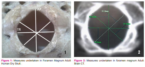
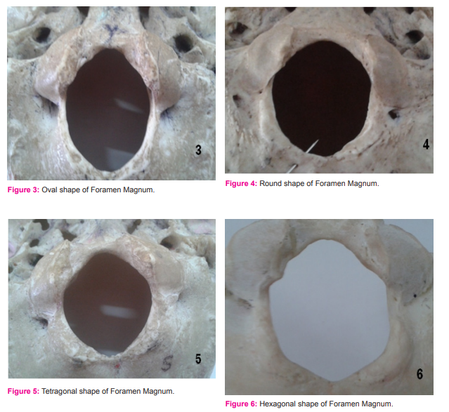
Keywords: Foramen magnum, AP- anteroposterior, TR- transverse, RO- right oblique and LO- left oblique
Full Text:
INTRODUCTION
Cranial morphometry is used for human studies like age estimation, stature, and ethnicity. These parameters are important for forensic investigations and anthropological examinations of unknown individuals 1, 2, 3. Foramen magnum plays an important role as a landmark in the region of skull and spine because it transmits key structures like the lower end of the medulla oblongata, meninges, vertebral arteries and the spinal accessory nerve. It is situated in the occipital bone2 . The foramen magnum in apes and in humans is formed by the fusion of the four individual parts of the occipital bone (pars squama, left and right pars lateralis, and pars basilaris)4 . Studies comparing the shape of human foramen magnum with other primates have been done earlier5 .The position of the foramen magnum in humans is unique compared to other mammals. In humans it has migrated well forward in the occipital bone from the back of the skull, to a position beneath the center mass of the skull and brain 6 . It is of particular interest for anthropology, anatomy, forensic medicine and other medical fields. Recent studies report that morphometry is a fast and efficient method for the evaluation of morphological characteristics, such as ethnicity, gender, age, genetic factors, dietary habits, and regional variations which can alter the shape and size of bone structures 1, 3. These aspects are significantly important in determining the anthropometric changes between different populations. Anatomical variations of morphology of foramen magnum are of clinical significance 7 . Dimensions of foramen magnum are of significance in field of forensic medicine to identify fire victims. This is because skull base is covered by a large mass of tissues that preserves the region of foramen magnum especially in standing position 1, 8. Hence morphometric study of foramen magnum has been done to assist in determining the ethinicity and gender when there is loss of other parts of the skeleton due to trauma, fire or severe destruction. It is also noted that the foramen magnum dimensions are specific for a particular population and becomes low when applied to populations with a large ethnic mix 9 . Size of foramen magnum has an etiological significance in cerebellar tonsil herniaton10. It has also been noted that longer antero-posterior dimension of foramen magnum permitted greater contralateral surgical exposure for condylar resection thus enhancing the feasibility of various intracranial surgical approaches11. Determining the size of foramen magnum in conditions like achondroplasia is of utmost importance to detect the risk of foramen magnum stenosis. Some authors have used the absolute dimensions of the foramen magnum as a guideline, and have found this to be helpful. In another study of patients with achondroplasia, it was found that anteroposterior and transverse measurements of the foramen magnum on computerized tomography scans aided to determine the risk factor for the need of a cervicomedullary decompression in case of foramen magnum stenosis 12. Despite such anatomical and clinical significance, there is still a lack of basic osteometric data of foramen magnum pertaining to a particular ethnic group. With the present study an attempt has been made to throw some light on the morphometry of foramen magnum in south indian population.
MATERIALS AND METHODS
100 adult human skulls of both sex (including occipital bones with intact foramen magnum) from the Department of Anatomy Mahatma Gandhi Medical College and Research Institute, Pondicherry and medical colleges in and around Pondicherry were evaluated. 100 CT Brain images taken from the Department of Radiology, Mahatma Gandhi Medical College and RI, Pondicherry were also evaluated.The fetal & children skulls, incomplete/ broken skulls were excluded. Anteroposterior, transverse, right and left oblique diameters of foramen magnum were measured using a Vernier Calliper to an accuracy of 0.1mm (figure 1). Anteroposterior diameter of foramen magnum is the distance between the opisthion(posterior border) and the basion (anterior border) in the mid sagittal plane. Transverse diameter is the maximum distance along the transverse plane. The right and left oblique diameters were measured from the midpoint of the corresponding occipital condyle to the point midway between posterior ends of opposite condyle to the opisthion. The same parameters were also evaluated in adult CT Brain images after 3D reconstruction (figure 2). All the dry bones were visually assessed to determine the shape of foramen magnum. Each foramen magnum was classified into one of the following five shapes- oval (figure 3), round (figure 4), tetragonal (figure 5), hexagonal (figure 6) and irregular.
STATISTICAL ANALYSIS
The mean and standard deviation were measured. The differences were analyzed using student’s t- test and a p value of <0.05 was considered significant.
RESULTS
The various parameters of foramen magnum recorded in dry skulls and CT images are represented in table 1. The shapes of foramen magnum were visually assessed and their frequency of occurrence in dry bones and CT images is represented in table 2. Difference in the CT images of male and female Foramen magnum were calculated and was found that the dimensions were significantly higher in males compared to females(p<0.05) as shown in table 3.
DISCUSSION
The findings of the present study were compared with previous studies based on different ethnic groups. Manoel C et al 1 in their study of 215 Brazilian skulls have shown that the mean Antero-posterior diameter was 3.52±0.33cm and the mean Transverse diameter was 3.03±0.20cm. A study on 110 Central European skulls by Gruber et al 13 in 2009 showed the mean anteroposterior diameter of 3.66±0.28cm and the mean transverse diameter of 3.11±0.27cm. Morphometric study of foramen magnum in 100 Nigerian skulls by Ukoha U et al8 in 2011 showed a mean Antero- posterior diameter of 3.62±0.23cm and a transverse diameter of 3.00±0.25cm. A study on 54 skulls of Indian origin done by Chethan et al 11 in 2011 showed results similar to the present study. The mean Antero- posterior diameter shown by them was 3.1±0.24cm and transverse diameter 2.52±0.24 cm. The various shapes of foramen magnum were also assessed by them and classified into round, egg, tetragonal, hexagonal, pentagonal and irregular shape. According to their study the most common shape observed by them was round unlike the present study where the most common shape observed was oval.
A review of literature showed two studies on CT images of healthy adults. Khalil et al 14 in 2003 have done a study on 110 healthy subjects in Turkey and showed the mean Antero- posterior diameter to be 3.46±0.31cm and transverse diameter 2.93±0.21cm. Another study was done by Fatma et al 15 in 54 subjects in Turkey showed the Antero- posterior diameter to be 3.41±0.38 and transverse diameter to be 2.98±0.27 cm. In both the studies the parameters measured were higher in males compared to females. These findings were similar to the present study.
CONCLUSION
The present study gives a morphometric reference to various types of foramen magnum in Indian population and its clinical significance. Evaluation of CT images has shown significant differences in the quantified parameters between males and females. Prospective studies will help surgeons with a proper reference value for determining feasibility of transcondylar surgical approaches for a particular ethnic group which are being done in an increasing trend in recent times for brain stem lesions16.
ACKNOWLEDGEMENT
We acknowledge the immense help received from the scholars whose articles are cited and included in the references of this manuscript. We are also grateful to authors/ editors/ publishers of all those articles, journals and books from where the literature for this article has been reviewed and discussed.
References:
1. Manoel C, Prado FB, Caria PHF, Grappo FC: Morphometric analysis of foramen mangnum in Human skulls of Brazillian individuals in relation to gender.Braz.J.Morphol. Sci.,2009;26(2):104.
2. Uthman A T, Al- Rawi N H, Al- Timmimi J F: Evaluation of Foramen Magnum in gender determination using helical CT scanning. Dentomaxillofac Radio.,2012; 41(3): 197- 202. Avcl E, Kim A H, Ozturk H, Kara E: Anatomical Variations of the Foramen Magnum,Occipital Condyle and Jugular Tubercle. Turkish Neurosurgery, 2011; 21(2): 181- 90.
3. Radhakrishnan SK, Shivaraman CH, Ramakrishna A, Bhagya B: Morphometric analysis of foramen magnum for sex determination in South Indian population. NJUHS, 2012; 2:20-2.
4. Avci E, Kara E, Ozturk N C, Uluc K: Anatomical variation of the foramen magnum, occipital condyle and jugular tubercle. Turkish Neurosurgery, 2011;2(2): 181-90.
5. Luboga SA, Wood BA: Position and orientation of foramen magnum in higher primates. American Journal of Physical Antvropology, 1990;81: 67-76.
6. Kimbel WH, Rak Y: The cranial base of Australopithecus afarensis: new insights from the female skull. Phil. Trans. R. Soc. B, 2010; 365: 3365-76.
7. Tubbs RS, Greissenever CJ, Loukar M, Shoja MM, Cohen Gadol AA: Morphological analysis of the foramen magnum: an anatomical study. Neurosurgery, 2010; 66(2):385-8.
8. Ukoha U, Egwu OA, Okafor IJ, Angabolu AE, Ndukwe GU: Sexual dimorphism in the foramen magnum of Nigerian adult.Int J Biol Med Res.,2011;2(4):878-81.
9. Galdames ICS, Russo PP, Matamala DAZ, Smith RL: Sexual Dimorphism in the Foramen Magnum Dimensions. Int. J. Morphol, 2009; 27(1):21-23.
10. Milhorat TH, Nishikawa M, Kula RW, D lugaz YD: Mechanism of cerebellar tonsillar herniation in patients with Arnold Chiari Malformations as a guide to clinical management. Acta Neurochir, 2010; 152: 1117-27.
11. Chethan P, Prakash JA, Murlimanju BV, Prashanth KV: Morphological analysis and morphometry of the foramen magnum: an anatomical investigation. Turkish neurosurg, 2012; 22(4): 416-9.
12. Bagley CA, Pindrik JA, Bookland MJ,Joaquin Q: Cervicomedullary Decompression for foramen magnum stenosis in Achondroplasia.J Neurosurg, 2006; 104:166-72.
13. Gruber P, Henneberg M, Boni T, Ruhli FJ: Variability of Human foramen magnum size. The Anatomical Record, 2009; 292:1713-9.
14. Mushed KA, Emine A, Tuncer I: Morphometric evaluation of the foramen magnum and variations in its shape: a study of CT images of normal adults. Turk J Sci., 2003; 33:301- 6.
15. Edril FH, Saban V, Cimen M, Isik O: Morphometric Evaluation of the foramen magnum by CT. Ericyes Medical Journal, 2010; 32(3): 167-70.
16. Muthukumar N, Swaminathan R, Venkatesh G, Bhanumathy SP: A Morphological Analysis of Foramen Magnum region as it relates to transcondylar approach. Acta Neurochir(Wien) 2005; 147(8): 889-95.

|
 IJCRR
IJCRR 


 This work is licensed under a Creative Commons Attribution-NonCommercial 4.0 International License


