IJCRR - 6(22), November, 2014
Pages: 25-28
Date of Publication: 21-Nov-2014
Print Article
Download XML Download PDF
METHOD OF COLOURING WET SPECIMEN IN ANATOMY
Author: Lalit Kumar Jain, Hitesh Babel
Category: Healthcare
Abstract:Aim: To develop a technique to colour the various important structures of a specimen which can be kept in 10 percent formalin filled jar in museum. Methodology: Previously several workers used different regins to colour the specimen but we used camlin oil paints mixed with white enamel paint to colour arteries, Veins, Nerves, Ligaments and Muscles of the specimens. These coloured specimens were kept in 10 percent formalin filled jar. Results: The painted colours remained stable in the formalin solution for several years without any visible changes. Conclusion: Colouring the important structures by using the anatomically correct colours make the monochrome specimens more attractive and lively and thus help students in identification and understanding of relations of various structures in a specimen.
Keywords: Museum, Artery, Vein, Nerve, Oil paint, White enamel paint, Formalin solution
Full Text:
INTRODUCTION
Museum in anatomy is a place where students get much information about the normal anatomical parts along with variation in the distribution of Nerves, vessels etc. Mounting the specimen as such in formalin solution filled jar may not look more informative as well as attractive. The text books which include coloured diagrams give full knowledge to the students. Many workers used different techniques to colour specimens. Congdon, E. D. (1932) used albuminous paints to colour the specimen (1). Intravascular injection of silicone, gelatin, latex or epoxy was used to highlight the vessels (Tiedemann,1982; von Hagens, 1985; Oostrom, 1987; Oostrom and von Hagens, 1988; Riepertinger and Heuckendorf, 1993; Grondin and Olry, 1996). Robert W. Henry, Larry Janick, and Carol Henry (1997) used silicon to colour plastinated specimens(2). Although everything of the specimen are essential but the important parts dissected may be highlighted in wet specimen with the permanent colours. We used camlin oil paints mixed with white enamel paint to colour the specimens. These coloured specimens can be kept in 10 percent formalin filled jar. By this technique arteries, Nerves, Veins, Ligaments, Tendons and even Muscles can be coloured which remain permanently in 10 percent formalin filled glass jar. Hence a technique has been developed which can be used to colour desired structures in wet specimen. Same technique was also used by the author to colour various structures of dry specimen (3).
MATERIAL AND METHOD
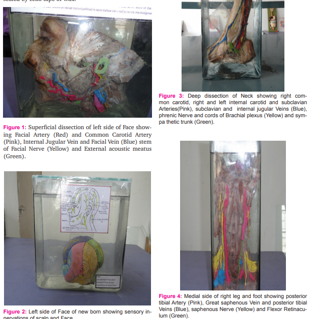
The material required for the technique are camlin oil paints, white synthetic enamel paint, painting brushes of different sizes and turpentine oil for cleaning the brushes. The specimen is prepared by fine dissection of the human body parts either fresh or left after dissection by students. When the specimen is prepared it is kept in a room for one or two days to make the surface of the structures to be coloured like vessels, Nerves, Ligaments Muscles, ducts, glands dry enough to apply colour. Now the vessels, Nerves, etc are raised from the underlying Muscles by putting swab of cotton under them which results in early drying of structures without drying of Muscles. The vessels and nerves dry earlier than other structures so colour is applied first on them and then on others. On a glass plate desired shade of colour is prepared by mixing the oil tube colour (camlin) with white enamel paint, when desired shade is prepared it is painted on the structures. We used Pink / Red colour for Artery, Blue for Vein, and Yellow for Nerve etc.
The oil paints are water proof and when mixed with white enamel colour they become adherent on the structures. After painting all the structures which are to be painted, specimen is kept overnight to make the paint dry. Now the specimen is put in the glass jar supported by cylindrical pieces of pet bottle(4) and jar is filled with 10 percent solution of formalin then the jar is covered with lid and sealed by cello tape or wax.


RESULT
By this technique various important structures of specimens were painted with anatomically correct colours. Colours evenly spread over the structures. The coloured specimens were kept in 10 percent formalin filled jars and have not shown any visible changes in colour for the last 5 years. With this technique it became easier for the students to understand relations of various structures in the specimen.
DISCUSSION
Many workers have worked on different techniques to colour to specimen with or without plastination. We used this technique to colour wet specimens which can be kept in jars filled with 10 percent formalin solution without any visible changes in colours. Cares to be taken: 1. Allow the Specimen to dry slightly before applying the paint 2. Structures which became dry should be painted first while others which are still wet may be coloured when they become dry.
ADVANTAGES OF USING COLOURED LABELLED SPECIMENS:
1. Colours aids memory. 2. Makes the specimen attractive. 3. Better distinguish anatomical Structures. 4. Creates interest of students. 5. Helps in better understanding of relations of different structures.
ADVANTAGES OF THIS TECHNIQUE:
1. Can be used for wet specimens.
2. Colours do not fade in formalin with time.
3. Convenient to perform.
4. Colours are well adherent to tissues.
5. Evenly colour the structures.
6. Colours can be stored at room temperature.
7. Great range of Colours are easily available.
CONCLUSION
From the results of above technique it can be concluded that coloured anatomical specimens are very valuable in teaching students of medical science. Colouring the important structures by using the anatomically correct colours make the monochrome specimens more beautiful and lively. As we human beings are very sensitive to colours it aids memory. So in brief it’s a better way of presentation of the specimens.
ACKNOWLEDGEMENT
Authors acknowledge the immense help received from the scholars whose articles are cited and included in references of this manuscript. The authors are also grateful to authors / editors / publishers of all those articles, journals and books from where the literature for this article has been reviewed and discussed. Authors are grateful to IJCRR editorial board members and IJCRR team of reviewers who have helped to bring quality to this manuscript.
References:
1. Congdon, E. D. The use of albuminous paints in anatomical preparations. The Anatomical Records, volume 51, issue 3, pages 327–331, January 1932.
2. Robert W. Henry, Larry Janick, and Carol Henry. Specimen Preparation for Silicone Plastination. Journal of the International Society for Plastination, Volume 12, Number 1:13- 17, 1997.
3. Lalit kumar Jain, Pooja Rajendra Gangrade, Neha Vijay. New technique for preparation of dry specimen using discarded cadaveric parts. International journal of Current Research and Review, September 2012/ Volume 04, issue18, pages 71-77.
4. Lalit Kumar Jain et al. New Technique to Mount Specimen in the Formalin Filled Jar for Anatomy Museum with Almost Invisible Support. International Journal of Current Research and Review, June 2013/ Volume 05, issue 12, Pages 45-50.
|


 This work is licensed under a Creative Commons Attribution-NonCommercial 4.0 International License