IJCRR - 6(23), December, 2014
Pages: 64-67
Date of Publication: 10-Dec-2014
Print Article
Download XML Download PDF
COMPUTED TOMOGRAPHIC ASSESMENT OF DIAMETERS OF ASCENDING AND DESCENDING AORTA IN INDIAN SUBJECTS
Author: Syed Naziya P., Althaf Ali S., Syed Imran S., Anand Abkari, Murthy G. S. N.
Category: Healthcare
Abstract:Background: An enlargement of the aortic diameter exceeding at least 50% of the normal range results in aneurysm formation. Endovascular stent grafting is a newer form of treatment for thoracic aortic aneurysms that is less invasive than open surgery and has the success rate around 95%. The ratio of stent graft size to aortic diameter should be of the order of 1.1\?1.15; higher diameters have to be avoided in order to limit the vessel wall stretch which may result in antegrade or retrograde dissection or perforation. After reviewing the literature thoroughly, it was found that even though the diameter of ascending and descending aorta has a wide range of clinical implications and applications, there is dearth of literature pertaining to the quantitative measurements of the diameters of ascending and descending aorta in Indian subjects. Aim: • To measure the diameter of ascending aorta (DOAA) and diameter of descending aorta (DODA) at the level of pulmonary artery bifurcation in Indian subjects using computed tomography. • To establish the upper normal limits of DOAA and DODA; to rule out aneurysm of thoracic aorta. Materials and Method: The CT chest images of 76 patients (44 males and 32 females) were studied. The DOAA and DODA were measured at the level of pulmonary artery bifurcation; across a line joining the ascending and the descending aorta. Results: 1. The average DOAA was 33.44 mm \? 1.12 mm in males and 31.43 mm \? 1.52 mm in females; the upper normal limits of DOAA were 35.68 mm for males and 34.47 mm for females. 2. The average DODA was 25.11 mm \? 1.34 mm in males and 23.04 mm \? 1.58 mm in females; the upper normal limits of DODA were 27.79 for males and 26.20 for females. Conclusion: A quantitative assessment of the diameters of ascending and descending aorta has been presented which will help the clinicians for diagnostic and therapeutic purpose especially in Indian subjects.
Keywords: Ascending aortic diameter (DOAA), Descending aortic diameter (DODA), Pulmonary artery bifurcation, Indian subjects
Full Text:
INTRODUCTION
The aorta represents a complex organ system which begins in the aortic ring adjacent to the aortic root with the origin of the two major coronary arteries, and ends at the iliac bifurcation.1 The aorta as an organ can be regarded as a biological ‘‘windkessel’’, storing kinetic energy during systole which is delivered during diastole in order to maintain a relative constant mean aortic pressure. The size of the aorta decreases with distance from the aortic valve in a tapering fashion.1 The ageing of the aorta is accompanied by a loss of compliance, and an increase of wall stiffness caused by structural changes including an increase in the collagen content and formation of intimal atherosclerosis with calcium deposits.2, 3 An enlargement of the aortic diameter exceeding at least 50% of the normal range results in aneurysm formation.1
Thoracic aortic aneurysm is a common, potentially lethal, but treatable disease, particularly if detected before dissection or rupture. Multiple imaging modalities are available for assessing the thoracic aorta, including X-ray angiography, transesophageal echocardiography (TEE), computed tomography (CT), and magnetic resonance imaging (MRI). Although all of these modalities have diagnostic value, CT has evolved to be the mainstay of evaluation owing to its accuracy and reproducibility, as well as its speed, simplicity, and true 3-dimensional capabilities. To distinguish the normal from the enlarged aorta, it is necessary to standardize the values of “normal” aortic dimensions.4 Accurate assessment of aortic size is a key component in this detection and in guiding therapeutic decisions.5 The importance of aortic diameter in determining risk for complications have been well established; 6 cm for the ascending aorta and 7 cm for the descending aorta.1, 5 Surgery is recommended well before these levels are reached. Endovascular stent grafting is a newer form of treatment for thoracic aortic aneurysms that is less invasive than open surgery and has the success rate around 95%.6, 7 The endovascular stent is placed inside the thoracic aorta to help reinforce the blood vessel and prevent the aneurysm from rupturing. Once placed in the correct location, the stent graft expands to fit within the diameter of the thoracic aorta and provides a new path for the blood flow.8, 9 The ratio of stent graft size to aortic diameter should be of the order of 1.1–1.15 with healthy areas before and after the aneurysm neck. Higher diameters should be avoided to prevent the vessel wall stretch which may result in antegrade or retrograde dissection or perforation.1 After reviewing the literature thoroughly, it was found that even though the diameter of ascending aorta and descending aorta has a wide range of clinical implications and applications, there is dearth of literature pertaining to the quantitative measurements of the diameters of ascending and descending aorta. Therefore, our aim was to determine the normal limits for ascending and descending thoracic aorta diameters in Indian subjects.
MATERIALS AND METHOD
Materials: The CT chest images using Somatom, four row CT scanner (Siemens) of 76 patients (44 males and 32 females) investigated in a private hospital were studied. Criteria of Inclusion / Exclusion: Only the patients of age group 18-60 years with normal CT report were included in the study. The patients with any space occupying lesion of mediastinum or any lung disease, patients with known coronary heart disease, hypertension, chronic pulmonary and renal disease, diabetes and severe aortic calcification were excluded. Method: It was a prospective study. The patient’s informed consent was taken to participate in the study. Patient was put in a supine position with both arms abducted and was instructed to hold the breath in full inspiration. 5mm thin sections were taken and the data obtained was analyzed at the work station of reporting room.
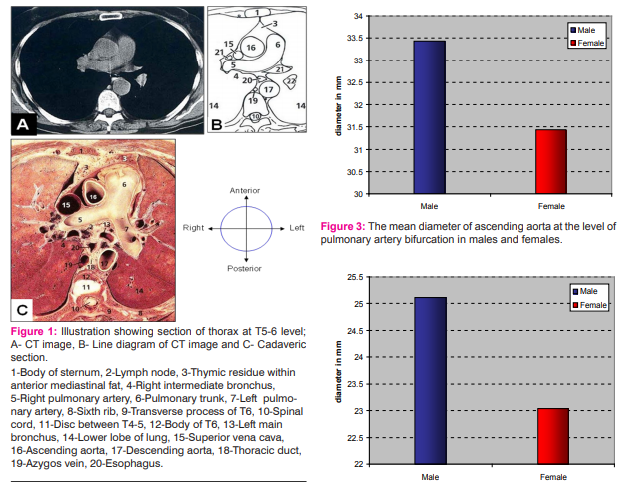
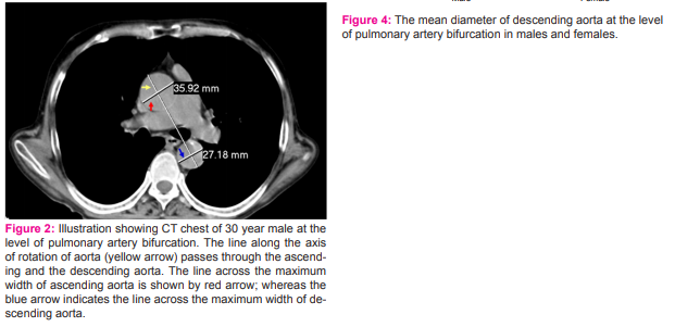
The diameter of ascending aorta and descending aorta were measured at the level of pulmonary artery bifurcation. The diameter were measured perpendicular to a line joining the ascending and the descending aorta; i.e. along the axis of rotation of aorta (Figure 1). The dimensions of arch of aorta were not studied because of lack of any specific level for its measurement. All these data were recorded as per the Performa in a master chart. Statistical Analysis: All these measurements were statistically analyzed by calculating the Mean and Standard Deviation (SD) using software SPSS version 22.0.
RESULTS
The following observations were made: The Diameter of Ascending Aorta The DOAA at the level of pulmonary artery bifurcation was measured and the findings were as shown in Table 1. The DOAA at the level of pulmonary artery bifurcation ranged between 30.94 mm and 35.92 mm in males and between 29.64 mm and 34.82 mm in females. The average DOAA was found to be 33.44 mm ± 1.12 mm in males and 31.43 mm ± 1.52 mm in females. The upper normal limits (mean + 2 standard deviations) of DOAA were 35.68 mm for males and 34.47 mm for females. The Diameter of Descending Aorta The DODA at the level of pulmonary artery bifurcation was measured and the findings were as shown in Table 2. The DODA at the level of pulmonary artery bifurcation ranged between 22.91 mm and 27.89 mm in males and between 20.12 mm and 26.48 mm in females. The average diameter was 25.11 mm ± 1.34 mm in males and 23.04 mm ± 1.58 mm in females. The upper normal limits of descending aorta diameter were 27.79 mm for males and 26.20 mm for females.
DISCUSSION
Kahraman et al, 10 in their study on 25 subjects from Turkey found the average DOAA to be 32.58 mm ± 3.2 mm and DODA to be 28.88 mm ± 3.45 mm. Hager et al, 3 in their study on 70 German subjects found the average DOAA to be 30.19 mm ± 4.1 mm and DODA to be 24.7 mm ± 4.0 mm. Mao et al11 studied 1422 cases and found out the average diameter of ascending aorta in males to be 33.6 mm ± 4.1 mm and 31.1 mm ± 3.9 mm in females. In present study the average diameter of ascending aorta was found to be 32.69 mm ±1.61 mm and that of descending aorta as 24.33 mm ± 1.74 mm. The average DOAA and DODA found in present study closely correspond to that found in the study by Kahraman et al 10 and Hager et al.3 However some of the differences from prior CT studies are related to differences in imaging methods including image acquisition modes, measuring site, imaging temporal resolution. Typically, invasive angiography and echocardiography use the intra-luminal diameter, while conventional cross sectional imaging includes the wall thickness. CONCLUSION Further studies with larger sample size are needed to rationalize the present findings. With more application of cardiac CT and thoracic CT, it is essential to define the normal thoracic aortic diameter changes with aging in both genders; especially in Indian subjects. Gender specific and age adjusted normals for aortic diameters are necessary to evaluate pathologic atherosclerotic changes and aneurysms of aorta.
References:
1. Raimund Erbel, Holger Eggebrecht. Aortic dimensions and the risk of dissection. Heart 2006; 92:137-142.
2. Aronberg DJ, Glazer HS, Madsen K, et al. Normal thoracic aortic diameters by computed tomography. J Comput Assist Tomogr 1984; 8:247–50.
3. Hager A, Harald K, Ulrike RB, Sebastian B, Karl R, Thomas M. Bernhardt, Galanski M and Hess J. Diameters of the Thoracic Aorta throughout Life as Measured with Helical Computed Tomography. Journal of Thoracic and Cardiovascular Surgery. 2002; 123:1060-1066.
4. Wolak A, Gransar H, Louise EJ, Thomson et al. Aortic Size Assessment by Noncontrast Cardiac Computed Tomography: Normal Limits by Age, Gender, and Body Surface Area. Journal of American College of Cardiological Imaging, 2008; 1:200-209.
5. Himanshu J. Patel and G. Michael Deeb. Ascending and Arch Aorta: Pathology, Natural History, and Treatment. Circulation. 2008; 118:188-195.
6. Eggebrecht H, Nienaber CA, Neuhauser M, et al. Endovascular stent-graft placement in aortic dissection: a metaanalysis. Eur Heart J 2005.
7. Nienaber CA, Eagle KA. Aortic dissection: new frontiers in diagnosis and management; Part II: therapeutic management and follow-up. Circulation 2003; 108:772–8.
8. Ince H, Nienaber CA. Endovascular stent-graft prosthesis in aortic aneurysm. J Cardiol 2001; 90:67–72.
9. Herold U, Piotrowski J, Baumgart D, et al. Endoluminal stent graft repair for acute and chronic type B aortic dissection and atherosclerotic aneurysm of the thoracic aorta. Eur J Cardiothorac Surg 2002; 22:891–7.
10. Kahraman H, Ozaydin M, Varol E, Suleyman M, Aslan, Dogan A, Altinbas A, Demir M, Gedikli O, Acar G and Ergene O. The Diameters of the Aorta and Its Major Branches in Patients with Isolated Coronary Artery Ectasia. Texas Heart Institute Journal. 2006; 33(4): 463-468.
11. Mao SS, Beckmann D, Annie C, Luan N, Ferdinand R and Matthew J. Normal Thoracic Aorta Diameter on Cardiac Computed Tomography in Healthy Asymptomatic Adults: Impact of Age and Gender. Academic Radiology. 2008; 15(7): 827-834.


|


 This work is licensed under a Creative Commons Attribution-NonCommercial 4.0 International License