IJCRR - 6(23), December, 2014
Pages: 27-30
Date of Publication: 10-Dec-2014
Print Article
Download XML Download PDF
DEMOGRAPHICS AND MANAGEMENT OF FOREIGN BODY PENETRATIONS IN HAND
Author: Pawan Kumar K. M., Ranganatha B. T.
Category: Healthcare
Abstract:Background: The penetration of foreign bodies into the hand is generally accepted as a simple injury with the misconception that treatment will be easy. The aim of this study was to analyze the diagnostic and therapeutic challenges during the removal of foreign bodies in hand. Methods: Prospective analysis of patients who had hand injuries caused by foreign body penetration and had been treated in the Department of Orthopaedics from January 2010 to December 2013. Results: The mean age among the 84 patients was 33.03 with standard deviation of \? 10.98 years, ranging from 14 years to 64 years. Most of them were between the age of 21 and 30 years (43%). About 71.4% (60) of the injured were males. About 69% of foreign body penetrations were occupational injuries. A variety of foreign bodies were isolated from the site of injury; they included metal splinters (33.4%),broken glass (27.4%),broken needles (20%),wood splinters(13%) thorns (5%) and tooth ( 1%). About in 21% (17) of cases the diagnosis of foreign body penetration was missed during the first consultation. Plain radiographs were able to pick up the foreign body in 81% (68) of the cases. In 83.3% of cases local anaesthesia was enough for the extraction of the foreign body. Conclusion: The study gives a clear understanding of the demographics of foreign body penetration and helps to plan in a better
way in managing similar cases.
Keywords: Foreign body, Hand, Canine tooth, Foreign body penetration
Full Text:
INTRODUCTION
Penetrating injuries to the hand are a common occurrence in the emergency room, and embedment of foreign bodies is suspected in many of these cases. The existing literature offers little information on the characteristics or prevalence of foreign bodies in the hand 1 . Despite significant practical knowledge and experience on foreign body penetration injuries to the hand, deficient management and complications can still be encountered2 . A foreign body, stuck into an extremity, may lead to consequences such as tissue damage, inflammation, infection, delayed wound healing, toxic or allergic reactions, and late injury as a result of migration 3 . In spite of the substantial experience of clinicians on this issue, there are a significant number of articles denoting defective management strategies, such as inadequate tetanus prophylaxis, and uncertainty in basic principles such as selecting the right solution for wound irrigation 4, 5. This study thus aims to reveal the basic features of the affected patients, the properties of the penetrated objects, the events causing this specific type of injury, the management of these injuries, and the outcomes of the patients. It is based on an analysis of a group of patients who had foreign body injuries in a more specific anatomic location, i.e. the hand and wrist .This study aims to cover the clinical and social properties and diagnostic and therapeutic challenges during the removal of foreign bodies in hand.
MATERIAL AND METHODS
This study is based on a prospective analysis of patients who had hand injuries caused by foreign body penetration. After obtaining clearance from institutional ethical committee, eighty four patients, who had been treated by the staff of Department of Orthopaedics from January 2010 to December 2013, were included in the study using proformas filled by us. Age, sex, occupation, mode of injury; domestic accidents, occupational accidents or road traffic accidents, site of injury; region of the hand where the foreign body had breached the skin, nature of foreign body; glass, wood, thorns, metal splinter, needle or tooth etc, time of diagnosis; whether diagnosed or missed initially, diagnostic modality required to diagnose and localise the foreign body and the anaesthesia required for the removal of foreign body were all assessed and are presented henceforth.
RESULTS
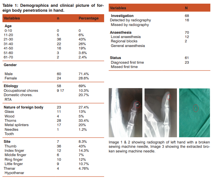
The mean age among the 84 patients was 33.03 with standard deviation of ± 10.98 years, ranging from 14 years to 64 ears. Most of them were between the age of 21 and 30 years (43%). About 71.4% (60) of the injured were males. Etiological assessment of foreign body penetration revealed, that the most of them were injured as a result of occupational accidents (69%), second most common mode of injury was road traffic accident (20.3%) and 9% had sustained injuries during domestic chores. A variety of substances were isolated from the site of injury; they included metal splinters(33.4%),broken glass( 27.4%),broken needles(20%),wood splinters(13%) thorns(5%) and tooth( 1%). In all these cases two views roentgenogram was taken. The two view roentgenograms were able to identify 81% (68) of the cases with foreign body. In sixty one (73%) cases the diagnosis was made at the first consultation with the doctor, either at our department or somewhere else and subsequently referred to our hospital for removal of foreign body. About in 21% (17) of cases the diagnosis of foreign body penetration was never made during the first consultation. All of them had presented to our hospital between three weeks to about four months from initial trauma. In all these cases there was history of some medical treatment at the time of initial trauma. Roentgenograms were taken first time at our hospital in all these 17 cases. Eleven (47.8%) cases among these cases had broken glass inside the wound which were visible in radiographs. In rest of the cases foreign bodies ranged from wooden splinters (13%) broken thorn (8.7%) and metal splinters (26%). The rarest foreign body among these was that of a retained canine tooth which was undetected for two months6 .
DISCUSSION
Foreign body penetrations of the hand wrist usually present as emergency cases, but patients with embedded objects presenting to the outpatient department are not uncommon2 . Embedded Foreign bodies can also be removed from patients who are unaware or uncertain of foreign body entry7 .Even our study revealed a similar picture with regard to presentation, 27% (23) of the patients had presented with embedded foreign bodies for duration ranging from three weeks to four months and none of them were aware of the embedded foreign body. Some centres include the fluoroscopy as routine component of foreign body removal surgeries8 . Similarly all cases were subjected to routine two view plain radiograph, including those with visible foreign bodies. The plane radiographs were able to pick up the foreign body in 81% (68) of the cases. Wood splinters and broken thorns were most common foreign bodies missed in radiographs. It has been stated that the two-view radiographs have been shown to be equivalent to the three-view radiographs in detecting glass foreign bodies9 . In another study, when only plain films were utilized, wood and glass FBs were missed in 93% and 25% of the cases10. In majority of cases the foreign bodies were of metallic origin (53.4%), while there were also broken glass (27.4%), wooden splinters (13%), thorns (5%) and one case of canine tooth6 .Metallic origin foreign bodies were usually broken industrial sewing machine needles or metal splinters from fabrication units. Such high incidence of metallic foreign bodies may attributed to the fact that majority of patients attending our hospital are industrial workers. Case reports of embedded organic foreign bodies such as splinters of plants, wood and fish fin fragments demonstrate the typical clinical picture of inflammatory reaction that develops in days or weeks11. In this sense, metal objects are less risky than the organic ones12. The foreign bodies like broken glasses, metal splinters, broken sewing needles etc were all picked up by the radiographs. Additional investigation used for the detection of foreign body alone was ultra sonogram, used in fifteen patients (18%). The ultra sonogram picked up wooden pieces and broken thorns. The literature shows strong support for radiographic detection of glass and metal foreign bodies, albeit with wooden and gravel foreign bodies detected at lower rates10, 13, 14, 15. Additional investigations in the form of a computer tomography were utilised for understanding the location of foreign body penetration and for planning its extraction in about 10% of the cases. The type of anaesthesia is determined by considering the location of the FB, the depth of penetration, the most likely injured structures, the age and the predicted duration of the operation. Similar to other studies16, 17, 18, 19, the vast majority of foreign body removal in our population was performed in the under local anaesthesia. In seventy cases(83.3%) local anaesthesia was enough for the extraction of the foreign body;12.3%(12) and 2.4%(2) required regional blocks or general anaesthesia respectively,dictated mostly due to the depth of penetration by the foreign body and proximity to the vital structures.
CONCLUSION
We in this study have tried to present the demographics, clinical presentation and treatment aspect of the simple entity of foreign body penetration. The study being a prospective analysis has its limitations and being conducted in a single institute may not exactly reflect the demographics of the entire population. Inspite of its limitations, the study helps us to understand about the demographics, nature of foreign bodies routinely encountered, high degree of clinical suspicion and utilization of all available investigative modalities required for diagnosis and management of foreign body penetration in a better way.
ACKNOWLEDGMENTS
Authors acknowledge the immense help received from the scholars whose articles are cited and included in references of this manuscript. The authors are also grateful to authors / editors / publishers of all those articles, journals and books from where the literature for this article has been reviewed and discussed. Source of funding: None Conflict of interest: None Declared
References:
1. Vishnu C Potini, Ramces Francisco, Benhoor Shamian, Virak Tan. Sequelae of foreign bodies in the wrist and hand. Hand 2013; 8:77–81
2. Emre Hocao?lu, Samet Vasfi Kuvat, Burhan Özalp, Anvar Akhmedov, Yunus Do?an, Erol Kozano?lu, Fethi Sarper Mete, Metin Erer. Foreign body penetrations of hand and wrist: a retrospective study. Turkish Journal of Trauma and Emergency Surgery 2013; 19 (1):58-64
3. Han KJ, Lee YS, Kim JH. Progressive median neuropathy caused by the proximal migration of a retained foreign body (a glass splinter). J Hand Surg Eur 2011; 36:608-609.
4. Talan DA, Abrahamian FM, Moran GJ, Mower WR, Alagappan K, Tiffany BR, et al. Tetanus immunity and physician compliance with tetanus prophylaxis practices among emergency department patients presenting with wounds. Ann Emerg Med 2004; 43:305-314.
5. Banwell H. What is the evidence for tissue regeneration impairment when using a formulation of PVP-I antiseptic on open wounds? Dermatology 2006; 212:66-76.
6. Ranganatha BT, Pawan Kumar KM. Canine tooth in hand - A rare entity. Journal of Clinical Orthopaedics and Trauma 2014; 5: 91-93.
7. Ozsarac M, Demircan A, Sener S. Glass foreign body in soft tissue: possibility of high morbidity due to delayed migration. J Emerg Med 2011; 41:125-128.
8. Tuncer S, Ozcelik IB, Mersa B, Kabakas F, Ozkan T. Evaluation of patients undergoing removal of glass fragments from injured hands: a retrospective study. Ann Plast Surg 2011;67: 114-118
9. Steele MT, Tran LV, Watson WA, Muelleman RL. Retained glass foreign bodies in wounds: predictive value of wound characteristics, patient perception, and wound exploration. Am J Emerg Med 1998; 16:627-630
10. Levine MR, Gorman SM, Young CF, Courtney DM. Clinical characteristics and management of wound foreign bodies in the ED. Am J Emerg Med 2008;26:918-922.
11. Hamnett NT, Tehrani H, McArthur P. Perch fin foreign body in a paediatric hand. J Plast Reconstr Aesthet Surg 2013; 63:2198-2199.
12. Halaas GW. Management of foreign bodies in the skin. Am Fam Physician 2007; 76:683-688.
13. Anderson MA, Newmeyer 3rd WL, Kilgore Jr ES. Diagnosis and treatment of retained foreign bodies in the hand. Am J Surg.1982; 144(1):63–67.
14. De Lacey G, Evans R, Sandin B. Penetrating injuries: how easy is it to see glass (and plastic) on radiographs? Br J Radiol. 1985; 58(685):27–30.
15. Russell RC, Williamson DA, Sullivan JW, Suchy H, Suliman O. Detection of foreign bodies in the hand. J Hand SurgAm. 1991;16 (1):2–11.
16. Blankstein A, Cohen I, Heiman Z, Salai M, Heim M, Chechick A. Localization, detection and guided removal of soft tissue in the hands using sonography. Arch Orthop Trauma Surg. 2000; 120 (9):514–517.
17. Salati SA, Rather A. Missed foreign bodies in the hand: an experience from a center in Kashmir. Libyan J Med 2010; 5:5083.
18. Smoot EC, Robson MC. Acute management of foreign body injuries of the hand. Ann Emerg Med. 1983; 12(7):434– 437.
19. Tuncer S, Ozcelik IB, Mersa B, Kabakas F, Ozkan T. Evaluation of patients undergoing removal of glass fragments from injured hands: a retrospective study. Ann Plast Surg. 2011; 67(2):114–118.

|


 This work is licensed under a Creative Commons Attribution-NonCommercial 4.0 International License