IJCRR - 7(14), July, 2015
Pages: 85-90
Print Article
Download XML Download PDF
STUDY OF VARIATIONS IN THE ORIGIN AND COURSE OF VERTEBRAL ARTERY
Author: Preeti Sonje, Vasanti Arole, Rieshav Anand
Category: Healthcare
Abstract:Arterial variations are always very commonly seen in different arteries of the body and they are very important from surgical and
diagnostic point of view. Aim of this study is to find variations in the origin and course of vertebral artery. The vertebral arteries
are major arteries of the neck. The vertebral arteries and their major branches, i.e. vertebro-basilar system, essentially supply
blood to the upper spinal cord, the brain stem and cerebellum and variable parts of posterior cerebral hemispheres. Different
types of variations in the origin and course of vertebral artery were found .The operative indications for surgery in the cervical
region include spondylosis, a herniated inter-vertebral disc, tumour, infection and trauma. . So, apart from understanding the
clinical implications, the data which is derived from the gross anatomical dissections of cadavers can be a useful guideline to the
surgeons for careful pre-operative planning in cases with an unusual course of vertebral artery and can help them in avoiding
potentially life threatening complications1.
Keywords: Vertebral artery, Foramina transversaria, Aorta
Full Text:
INTRODUCTION
The vertebral arteries are major arteries of the neck. The vertebral arteries and their major branches, i.e. vertebro-basilar system, essentially supply blood to the upper spinal cord, the brain stem and cerebellum and variable parts of posterior cerebral hemispheres. The vertebral arteries are normally derived from the subclavian arteries. After taking origin from the subclavian arteries; the vertebral arteries ascend through the neck in the foramina transversaria from C6 upwards up to C1 vertebrae (i.e. C6- C1) and enter the cranial cavity through the foramen magnum, close to the anterolateral aspect of the medulla. They converge medially as they ascend the medulla and unite to form the midline basilar artery at approximately the level of the junction between the pons and medulla. The course of the vertebral artery is divided into four parts as follows:
First part-The first part extends from its origin from the subclavian artery to reach the transverse process of C6.
Second part-The second part runs upward through the foramen transversarium of the C6 to C1 vertebrae.
The third part lies in the suboccipital triangle. It emerges from the foramen transversarium of C1, winds around the posterior aspect of the lateral mass of atlas, runs medially lying on posterior arch of C1 and enters the vertebral canal. The fourth part extends from the posterior atlanto-occipital membrane to the lower border of pons. In our study we will be dealing with the first and second part of the vertebral artery.
Numerous variations are found in the origin of the vertebral artery as well as in its course i.e. its passage through the foramina transversaria of cervical vertebrae. Normally it enters into the foramen transversarium of C6 and passes upwards but sometimes it may enter into foramen transversarium of any other cervical vertebrae. Also, sometimes the vertebral artery takes its origin directly from the arch of aorta instead of subclavian artery. These type of variations, if present, are very important from the diagnostic as well as surgical point of view.
The operative indications for surgery in the cervical region include spondylosis, a herniated inter-vertebral disc, tumour, infection and trauma. The failure to recognize the anomalies during the preoperative planning can lead to surgical complications. The laceration of the vertebral artery is the most challenging surgical dilemma which is faced during anterior cervical spinal surgery. So, apart from understanding the clinical implications, the data which is derived from the gross anatomical dissections of cadavers can be a useful guideline to the surgeons for careful pre-operative planning in cases with an unusual course of vertebral artery and can help them in avoiding potentially life threatening complications1 .
MATERIAL AND METHODS
Cadavers embalmed in 10% formalin from Department of Anatomy, Dr. D.Y. Patil Medical College formed the armamentarium of the study. 40 cadavers were dissected to study the variations in the origin and course of the vertebral artery. Dissection was carried out in the scaleno-vertebral region (between the scalenus anterior and the longus colli muscles) on both the sides, so a total of 80 vertebral arteries were dissected. The prevertebral segment of the vertebral artery as well as the entrance and course of vertebral artery into foramina transversaria was studied in detail. Variations in the origin as well as course of vertebral artery were noted and photographs were taken.
Observations and Results Numerous variations were found in the origin and course of vertebral artery. These variations were noted down as follows.
Variations in the origin of vertebral artery
Variations in the origin of vertebral artery were found in four cases out of 40 cases dissected.
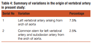
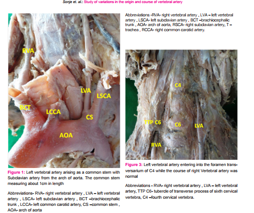
• Left vertebral artery was arising as a common stem with Subclavian artery from the arch of aorta in one case. The common stem was measuring about 1cm in length (Figure 1)
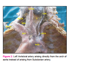
• Left Vertebral artery was arising directly from the arch of aorta instead of arising from Subclavian artery in three cases. In all the three cases the vertebral artery arose as a third branch of arch of aorta, between left common carotid artery and left subclavian artery. (Figure 2)
Variations in the course of the vertebral artery
Variation in the course of vertebral artery were found in four cases.
In two cases the variation in the course of vertebral artery was found to be unilateral.
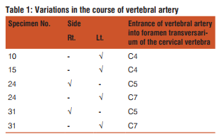
• Left vertebral artery was entering into the foramen transversarium of C4 in two cases .In the same case the course of right Vertebral artery was normal. (Figure 3) Two cases showed the bilateral variation in the course of vertebral artery.
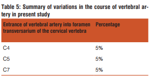
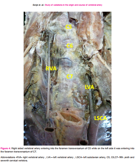
• Right sided vertebral artery was entering into the foramen transversarium of C5 while on the left side it was entering into the foramen transversarium of C7 in both the cases.(Figure 4)
DISCUSSION
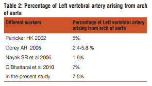
Panicker HK (2002) reported the origin of vertebral artery from arch of aorta in 5 percent of cases, while . Gorey AR (2005) et al stated that the most frequent variant is the left vertebral artery arising directly from arch of aorta between left common carotid and left subclavian arteries which accounts to about 2.4-5.8 percent of cases. Nayak SR et al (2006) reported the classical branching pattern of the aortic arch i.e.the left vertebral artery arising from the arch of aorta in 1.6% of the cases. C Bhattarai et al (2010) had studied 85 cadavers in Nepalese to find out 6 (7%) cases left vertebral artery was arising from arch of aorta. In the present study the vertebral artery originated from the arch of aorta in 7.5% of cases. In this study it was also observed that there was a common stem for left subclavian artery and left vertebral artery, arising from arch of aorta. This type of variation was observed in 2.5% cases.
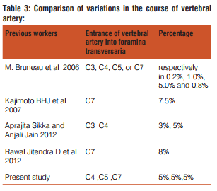
M. Bruneau et al (2006) studied 500 vertebral arteries, they observed an abnormal level of entrance into foramen transversarium in 7% specimens (35 cases), with a level of entrance into the C3, C4, C5, or C7 foramen transversarium respectively in 0.2%, 1.0%, 5.0% and 0.8% of all specimens. Seventeen abnormalities were right sided and 18 were left sided. Thirty-one out of 250 cases had a unilateral anomaly and two had a bilateral anomaly.
Kajimoto BHJ et al (2007) in his study described variations of vertebral artery entering the transverse foramen of seventh cervical vertebra (C7) to be 7.5%.
Aprajita Sikka and Anjali Jain (2012) found the origin of left vertebral artery from the arch of aorta between the left common carotid artery and the left subclavian artery and entered the foramen transversarium of the fourth cervical vertebra .The origin of the right vertebral artery was also variable in the same cadaver. It took origin from the right subclavian artery at the junction of its origin from the Brachiocephalic trunk. It then ascended to enter the foramen transversarium of the third cervical vertebra.
Rawal Jitendra D et al 2012 12 dissected 50 vertebral arteries, and found 46 vertebral arteries entering the transverse foramen of the sixth cervical vertebra (92 %) and 4 of them entering transverse process of seventh cervical vertebra (8%).
In this study also entrance of vertebral artery into different foramina transversaria other than normal i.e. C6 was found. Vertebral artery was entering into the foramen transversarium of C7 was found in 5% of cases. Vertebral artery entering into the foramen transversarium of C5 was observed in 5% of cases. Also we observed the entrance of vertebral artery into foramen transversarium of C4 in 5% cases.
Embryological explanation- At the end of the third week of development, small posterolateral sprouts arise from the dorsal aorta at the cervical through sacral level and grow into spaces between the developing somites. In the cervical region, the intersegmental sprouts form vertical anastomosis with each other, secondarily lose their intersegmental connections to form vertebral arteries.12
Also in the fourth week of development vertebrae are formed from the sclerotomes. Due to resegmentation of the sclerotomes, intersegmental arteries at first lying between the sclerotomes now pass midway over vertebral bodies .When the vertebral arch fuses with body, the vertebral artery, formed from intersegmental anastomosis is enclosed into foramina transversaria of cervical vertebrae. Alteration in this development leads into entry of vertebral artery into different foramina transversaria.13
CONCLUSION
Variations in the origin of left vertebral artery are necessary and beneficial for planning of aortic arch surgery or endovascular interventions, surgical procedures that would necessitate exposure of vertebral artery, include repair of aneurysms, excisions of cranio-cervical junction masses, vertebral endarterectomy, vertebral artery bypass surgery, and bony decompression of the vertebral artery. Also, an anatomical variations in vertebral artery, if missed, can lead to catastrophic sequelae in surgeries like atlanto-axial transarticular screw fixation, anterior cordectomy. Advances in technology have increased our knowledge regarding different variations in our body and an awareness regarding them can help avoid unwanted complications during various interventions 14.
SUMMARY
The vertebral arteries are major arteries of the neck. The vertebral arteries and their major branches, i.e. vertebro-basilar system, essentially supply blood to the upper spinal cord, the brain stem and cerebellum and variable parts of posterior cerebral hemispheres. Numerous variations are found in the origin of the vertebral artery as well as in its course. These are clinically and diagnostically important, were studied. Summary of findings is tabulated in Table no. 4 and Table no. 5
ACKNOWLEDGEMENT
I am very much thankful to Dr Vatsalaswamy, professor and head of Anatomy as well as Dr Neelesh Kanaskar for supporting help for this study.
Authors acknowledge the immense help received from the scholars whose articles are cited and included in references of this manuscript. The authors are also grateful to authors / editors / publishers of all those articles, journals and books from where the literature for this article has been reviewed and discussed
References:
1. Palmar FJ. Origin of the right vertebral artery from the right common carotid artery: agiographic demonstration of three cases. Br J Radiol 1977;50:185-187.
2. Panicker HK, Tarnekar A, Dhawane V, Gosh SK Anomalous origin of left vertebral artery:- Embryological basis and applied aspects-a case report. J Anat Soc India(2004), 51: 234-235.
3. Skandalakis JE The Embryologic and Anatomic Basis of Modern Surgery(2004). Paschalidis Medical Publication Ltd.
4. Gorey AR, Joshi R, Garg A, Merchant S, Yadav B, Maheswari P .Aortic arch variation: a unique case with anomalous origin of both vertebral arteries as additional branches of the aortic arch distal to left subclavian artery. AJNR (2005), 26:93-95.
5. M. Bruneau, J. F. Cornelius, V. Marneffe, M. Triffaux, and B. George .Anatomical variations of the V2 segment of the vertebral artery Neurosurgery.(2005), vol. 59, supplement 1, pp. S20– S24, 2006.
6. Nayak SR, Pai MM, Prabhu LV, D’costa S, Shetty S. Anatomical organization of aortic arch variations in India: embryological basis and review. J Vasc Bras. 2006: 5(2): 95-100
7. Suresh R, Ovchinnikov N, Mcrae A. variations in branching pattern of the aortic arch in three Trinidadians. West Indian Med J. 2006; 55(5):1
8. Ben Hur Junitiro Kajimoto, Renato Luis Dainesi Addeo, Gustavo Constantino de Campos et al Anatomical study of the vertebral artery path in human lower cervical spine. ACTA ORTOP BRAS 2007: 15: vol-2: 84-86.
9. Tsai IC, Tzeng WS, Lee T, Jan SL, Fu YC, Chen MC, et al .Vertebral and carotid artery anomalies in patients with aberrant right subclavian arteries. Pediatr Radiol (2007)37: 1007-1012.
10. Bhattarai C, Poudel PP. Study on the variation of branching pattern of arch of aorta in Nepalese. Nepal med Col J. 2010; 12(2): 84-86
11. Aparjita Sikka and Anjali jain. Bilateral variation in the origin and course of vertebral artery. Anatomy Research InternationalHindawi Publishing Corporation (2012):1-3.
12. Rawal Jitendra D, Jadav Hrishikesh R .Anatomical study of variation of vertebral artery entering the foramen transversarium of cervical vertebrae. National journal of medical research. Volume 2 Issue 2 Apr – June 2012 pg 199-201.
13. William J. Larsen Human Embryology .Third edition .Churchill Livingstone Newyork, 1997;p:207-209.
14. Langman’s Medical Embryology. 11th ed. Lippincott Williams and Wilkins; 2009. p. 140-141.
15. Young-Don Kim, Hyung-Tae Yeo, Young-Dae Cho, Anomalous Variations of the Origin and Course of Vertebral Arteries in Patients with Retroesophageal Right Subclavian Artery J Korean Neurosurg Soc 45, 2009 : 297-299.
16. Gray H. The Anatomical Basis of Clinical Practice. 40thed. Susan Standring, Elsevier Churchill Livingstone; 2008. p. 447-449.
17. Yanik B, Conkbayir I, Keyik B, Hekimoglu B. A rare anomalous origin of right vertebral artery: findings on Doppler sonoghraphy. J Clin Ultrasound 2004; 32:211-214.
18. Vikram N., M.B. Patil, Basavaraj B., Y.D. Badiger. ANATOMICAL VARIATION OF THE ORIGIN OF THE LEFT VERTEBRALARTERYA CASE REPORT .International Journal of Current Research and Review, June 2013/ Vol 05 (11);Page 133-136.
19. S.R. Satti, C.A. Cerniglia, R.A. Koenigsberg. Cervical Vertebral Artery Variations: An Anatomic Study AJNR 28, May 2007: page -976-980.
|
 IJCRR
IJCRR 


 This work is licensed under a Creative Commons Attribution-NonCommercial 4.0 International License


