IJCRR - 7(2), January, 2015
Pages: 54-56
Print Article
Download XML Download PDF
BILATERAL ABNORMALITIES IN THE COURSE OF RENAL VEINS AND SUPERNUMERARY LEFT RENAL ARTERY: A CASE REPORT
Author: Preksha Sharma, Sangita Chauhan, Seema Gupta, Shreya Sharma
Category: Healthcare
Abstract:Introduction: A precise knowledge of variations in origin and course of both renal and gonadal vessels is of utmost importance during diagnostic and operative abdominal surgical procedures.
Methods: A variation was found in the gonadal vein and renal artery during routine dissection of a formaline fixed adult male cadaver in the department of Anatomy, SMS Medical college, Jaipur, Rajasthan.
Results: Following variations were found during the routine dissection \? • Double gonadal veins were found on both the sides.
• Three renal arteries were found on the left side arising from the abdominal aorta. Anomaly in other system was not obvious.
Conclusions: In the present case supernumerary renal arteries were found on the left side which took origin from the lateral aspect of abdominal aorta and double gonadal vessels were found on both the sides. The pair of gonadal veins on the left side drained into lateral aspect of inferior vena cava whereas on the right side the pair of gonadal veins drained into Inferior Vena Cava, one on the anterior aspect and the other on its posterior aspect.Very less incidences of anomalous renal arteries and gonadal vessels have been reported so far. Anomalous renal artery has clinical implication in nephrotomy procedure and renal transplants whereas variation in gonadal vessels has shown its importance in the treatment of syndrome of pelviureteral junction.
Keywords: Variations, Left renal artery, Gonadal veins.
Full Text:
INTRODUCTION
During the routine dissection of an adult male cadaver for medical undergraduates, variations in the origin of the left renal artery and course of bilateral gonadal veins were observed in the department of Anatomy, SMS Medical College, Jaipur , Rajasthan .
METHOD
A variation was found in the gonadal veins bilaterally and renal artery on the left side during routine dissection of a formaline fixed adult male cadaver in the department of Anatomy, SMS Medical college, Jaipur, Rajasthan.During dissection other organs were also preserved and specimens were made for the purpose of teaching.
RESULT
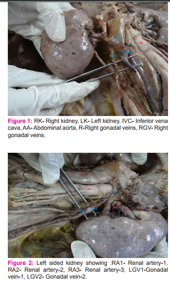
The left kidney received three renal arteries , all of them took origin from the lateral aspect of the abdominal aorta. Two gonadal veins were seen on both the sides. On the right side one gonadal vein was draining on the anterior aspect of inferior vena cava and the other one on its posterior aspect. Similarly both the left gonadal veins were seen draining on the lateral aspect of inferior vena cava, as illustrated in figure-1and figure-2
DISCUSSION
Precise knowledge about variations in renal and gonadal vasculature is mandatory to rule out various anomalies. [1] Soni S et al 2010 reported case of right sided triple renal arteries with double renal arteries on the left side. He also reported variation in testicular arteries and origin of phrenic arteries.[1] Patel S et al 2012 showed a case of unilateral left double renal artery in 54-year-old male cadaver in which first renal artery (RA) arose from aorta at the level of L1 vertebra, whereas 2nd renal artery arose from same 5 cm below to the first one and both RA ran laterally and entered the kidney through the hilum with their anterior and posterior divisions.[2] Similarly a case of additional renal vein was reported by Sharmistha Biswas et al 2006 reported presence of an additional renal vein on the right side draining directly into IVC which was observed during a routine dissection in a middle-aged male cadaver. [3] We found left sided variation in the renal artery but in contradiction to our study, in a study done in thirtyseven formalin-fixed cadavers, the kidneys along with their arteries were explored and the morphological variations of renal arteries were noted during routine abdominal dissection conducted for medical undergraduates. In this study, supernumerary renal arteries were present in 23/37 (62.2%) cases (48.6% of aortic origin and 13.5% of renal origin) on the right side and 21/37 (56.8%) cases (45.9% of aortic origin and 10.8% of renal origin) on the left side.[4].Embryological explanation of such variations has been discussed by Felix.The developing mesonephros, metanephros, gonads and suprarenal glands are supplied by 9 pairs of lateral mesonephric arteries arising from the dorsal aorta and this was seen in an 18mm fetus. These arteries were divided by Felix into three groups- 1st and 2nd arteries were in the cranial group, 3rd to 5th in the middle, and 6th to 9th in the caudal group. Renal arteries were arising from the middle group.[5] Therefore, persistence of more than one artery of the middle group results in multiple renal arteries as seen in our cadaver.
CONCLUSION
Knowledge of variations in renal as well as gonadal artery is mandatory in various renal surgeries, renal transplantations and different radiodiagnostic procedures to prevent serious consequences. Inspite of huge importance there is scarcity of literature in this field.our sudy has tried to fill this gap.
ACKNOWLEDGEMENTS
The authors would like to thank Dr. Sangita Chauhan for her valuable support throughout the study. Authors acknowledge the immense help received from the scholars whose articles are cited and included in references of this manuscript. The authors are also grateful to authors/ editors/ publishers of all those articles, journals, books from where the literature for this article has been reviewed and discussed.
CONFLICTS OF INTEREST: The authors declare that they have no competing interests.
FUNDING: Not Applicable
References:
1. Sony S, Wadhwa A. Multiple variations in the paired arteries of abdominal aorta- Clinical implications. Journal of Clinical and Diagnostic Research. 2010 June ;(4):2622- 2625.
2. PatelS,Wanjari A, Naik A. A case report: double renal arteries. International Journal of Anatomical Variations (2012) 5: 22–24.
3. Sharmistha Biswas, J.C.Chattopadhyay, H. Panicker.Variations In Renal And Testicular Veins –A Case Report. J.Anat. Soc 2006. India 55 (2) 69-71 2006.
4. Virendra Budhiraja, Rakhi Rastogi, Vaibhav Anjankar. “Supernumerary Renal Arteries and Their Embryological and Clinical Correlation: A Cadaveric Study from North India,” ISRN Anatomy 2013. Volume 2013, Article ID 405712, 4 pages.
5. W.Felix. “Mesonephric arteries (aa. Mesonephrica)”, in Manual of Human Embryology, F. Keibel and F.P. Mall, Eds, vol.22, pp.8820-825, Lippincott, Philadelphia, Pa, USA,1912.
|
 IJCRR
IJCRR 


 This work is licensed under a Creative Commons Attribution-NonCommercial 4.0 International License


