IJCRR - 7(10), May, 2015
Pages: 49-50
Print Article
Download XML Download PDF
GIANT CELL TUMOUR OF TENDON SHEATH OF THE ANKLE - A CASE REPORT
Author: Ashish K. Jose, Sandeep MMR , Surendher Kumar R., Krishnagopal R., Ravikumar N.
Category: General Sciences
Abstract:Aim: To study an ankle swelling following a trivial trauma in a young female. Case Report: A 30 year old patient presented with pain and swelling in the right ankle following a trivial injury 4 months back. Radiograph showed soft tissue swelling and USG showed a cystic swelling over the flexor tendon. We did an excision biopsy and the histopathology report was consistent with Giant Cell Tumour of the tendon sheath. Discussion: Giant cell tumour of the tendon sheath is a benign tumour usually arising in the limbs. It occurs more commonly in the hand and rarely in the foot. Patients usually present to the hospital because of pain and swelling. The masses arising from the tendons are of soft tissue density and calcification is uncommon. GCT-TS is histologically similar to pigmented villonodularsynovitis as haemosiderin deposits and numerous macrophages are seen in both. Conclusion: Giant Cell Tumours of tendon sheath are similar to pigmented villo nodular synovitis (PVNS). They are the second most common benign tumor next to ganglions. The treatment is surgical excision of the mass.
Keywords: Giant cell tumour, Histopathological examination, Pigmented villonodularsynovitis (PVNS)
Full Text:
INTRODUCTION
Giant cell tumour of the tendon sheath (GCT-TS) isa benign solitary tumour that occurs more often in the hand and less commonly in the foot (1).There are 2 types: the localized type which is more common and the diffuse type which is rare. The diffuse type is considered to be the localized form of pigmented villonodularsynovitis (PVNS) of tendons. When the pathology is in the joint it is called PVNS. Both are benignproliferative conditions of unknown etiology that tend to affect patients younger than 40 years old. The diffuse form can be aggressive locally and has a high recurrence rate(2). GCT-TS and PVNS are histopathologically similar, but clinically distinct (3). Mononuclear and giant cells histologically resembleosteoclasts and are non-neoplastic (3) . Final diagnosis relies on a pathology specimen from excision although a recent study indicates that a fine-needle aspiration may provide a reliable diagnosis (4). The mass will often be hypervascular on Doppler imaging with ultrasound (5). Ultrasound may confirm the presence of a soft tissue tumor adjacent to a tendon but it can be difficult to differentiate between a solid and a cysticmass accurately (6). MRI appearance of these extra-articular lesions (and PVNS) are distinctive compared to other soft tissue tumors because of the intermediate to low signal intensity areas on both T1- and T2-weighted images. This reflects the hemosiderin deposition that is common to GCT-TS and PVNS tumors (although PVNS tends to have higher hemosiderin content and therefore tends to be darker)(2).
CASE REPORT
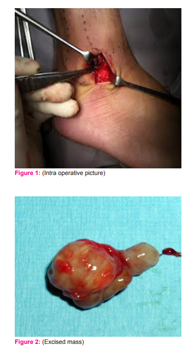
A 30 year female patient named Mrs. Gowri presented to the Orthopaedics OPD of Mahatma Gandhi Medical College and Research Institute Pondicherry, with pain and swelling over her right ankle for 4 months.She gave a history of trivial twisting injury to her ankle 4 months back. Following the injury she developed a small swelling which gradually increased in size.On examination there was a swelling over the anterior aspect of the lateral malleolus measuring 3 x 3 centimeters.Tenderness was present over the swelling. The swelling was firm to hard in consistency, mobile, non pulsatile and the skin over the swelling was pinchable.
Radiograph of right ankle revealed soft tissue swelling below the lateral malleolus.USG showed cystic swelling due to hematoma over the flexor tendon. We planned an excision biopsy. Intra-operativelythe swelling measured 3 x 3 cm and was found over the flexor digitorum tendon (Fig. 1). It was firm in consistency and haemorrhagic. No fluid collection was seen. The excised mass (Fig.2) was sent for histopathological examination. The histopathological changes were typicalof GCT-TS with the presence of numerous fibroblasts, macrophages and scattered giant cells.
DISCUSSION
Patients affected by giant cell tumour usually present to the hospital because of pain and swelling. Radiographic assessment involves both anteroposterior and lateralviews of the affected limb and compared with films of the contralateral side. During the evolution, masses arising from the tendons are of soft tissue density. Calcification is uncommon . Trauma is considered to be one of the etiological factors in GCT. Rodrigues et al (7) reported a history of trauma in 21% cases of GCT-TS of the hand. Our patient also gave a history of twisting injury to her ankle. GCT-TS is histologically similar to pigmented villonodularsynovitis as haemosiderin deposits and numerous macrophages are seen in both.
CONCLUSION
Giant Cell Tumours of tendon sheath are not really tumors but reactive lesions which are similar to PVNS. They can arise from the synovium instead of the tendon sheath. They are the second most common benign tumor next to ganglions. The treatment is surgical excision of the mass.
ACKNOWLEDGEMENT
Authors acknowledge the immense help received from the scholars whose articles are cited and included in references of this manuscript. The authors are also grateful to authors / editors / publishers of all those articles, journals and books from where the literature for this article has been reviewed and discussed.
References:
1. C. L. M. H. Gibbons, H. A. Khwaja, A. S. Cole,P.H. Cooke, N. A. Athanasou Giant-cell tumour of the tendon sheath in the foot and ankle . J Bone Joint Surg [Br] 2002;84-B:1000-3.
2. Annette D. Filiatrault, Joe T. Southerland, William D. Fishco,Giant Cell Tumor of Tendon Sheath: Case Studies.
3. Verheyden, JR, Damron TA. Giant Cell Tumor of Tendon Sheath. Emedecine. Jun26, 2009.
4. Jakowski JD, Mayerson J, Wakely PE Jr. Fine-needle aspirationbiopsy of the distal extremities: a study of 141 cases. Am J ClinPathol 2010;133:224-31.
5. Wan JM, Magarelli N, PehWC, Guglielmi G, Shek TW. Imagingof giant cell tumour or the tendon sheath. Radiol Med .2010;115:141-51.
6. Lee MH, Kim NR, Ryu JA. Cyst-like solid tumors of themusculoskeletal system: an analysis of ultrasound findings. SkeletalRadiol 2010;39:981-6.
7. Rodrigues C, Desai S, Chinoy R. Giant cell tumor of tendon sheath:a retrospective study of 28 cases. J SurgOncol1998;68:100-3.
|
 IJCRR
IJCRR 


 This work is licensed under a Creative Commons Attribution-NonCommercial 4.0 International License


