IJCRR - 7(15), August, 2015
Pages: 17-20
Date of Publication: 11-Aug-2015
Print Article
Download XML Download PDF
ASSESSMENT OF BOHLER'S AND GISSANE'S ANGLES OF THE CALCANEUM IN A GROUP OF SOUTH INDIAN POPULATION - A RADIOLOGICAL STUDY
Author: Rahe Ramachandran, Shailaja Shetty
Category: Healthcare
Abstract:Calcaneus is the largest tarsal bone designed to withstand the daily stresses of weight bearing. Bohler's and Gissane's angles are subtended by the Calcaneus, these angles are used in the assessment of Calcaneal fractures. Objective: Measuring the Bohler's and Gissane's angles from computed ankle radiographs and comparing the results with previous studies. Materials and Methods: This study was done in M.S. Ramaiah teaching hospital and M.S. Ramaiah memorial hospitals, Bangalore, India. Study was conducted on 92 patients older than 17 years. The angles were measured and the variation in range was compared with that of the previous studies. Observation and Results: From this study it was observed that the Bohler's angle ranges between 18.7 \? 46.2 degrees. Gissane's angle ranges between 87.5 \? 137.8 degrees. The normal international reference values are; Bohler's angle 280 to 400 and Gissane's angle 1200 to 1400. The lower limit of Bohler's angle in our study group, like few other studies is found to be much less than the reference values which is used to diagnose Calcaneal fractures. Conclusion: The range of Bohler's and Gissane's angles in the Indian population is much less when compared to the standard reference values, indicating that range of the angles is variable in different population groups. Hence knowledge of the exact range of the angles in a specific population group aids in accurate diagnosis and meticulous treatment of Calcaneal fractures.
Keywords: Calcaneal angle, Measurement, Ankle fractures
Full Text:
INTRODUCTION
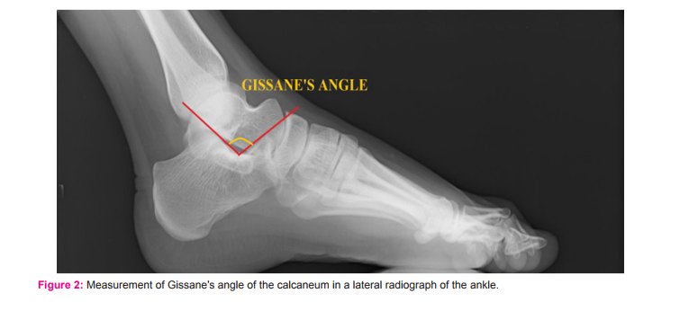
Foot has evolved over many million years to attain the human type. Foot has 28 bones and 31 articulations to support daily biomechanical loads of up to three to seven times the body weight. One of the key bones supporting the body weight is the calcaneus. Fracture of the calcaneus is common in fall from height and slipping from stairways. Imaging plays major role in the diagnosis, treatment and prognosis of Calcaneal fractures. Radiological measurement of Bohler’s angle and Gissane’s angles of the calcaneum are important parameters in Calcaneal fracture diagnosis, management and assessment of prognosis. Bohler’s and Gissane’s angles are subtended by the Calcaneal bone. Bohler’s angle is also known as Calcaneal angle, Tuber joint angle or Salient angle. It is used for assessing the loss of Calcaneal inclination (or ankle dorsiflexion impingement). It is measured at the intersection of a line drawn between the posterior superior aspect of calcaneal tuberosity and the highest point in the posterior talar facet, to another line drawn to the anterior process of the calcaneus. It normally ranges from 280 to 400 1,2 . Fracture through the calcaneus with displacement causes reduction of angle to less than 280 1 . Measurement of Bohler’s angle plays a key role in diagnosis of Calcaneal fractures and its intra operative reduction and fixation. Gissane’s angle or Critical angle of Gissane is the angle subtended between the anterior and posterior talar facets of the calcaneus. The angle normally ranges between 1200 and 1400 2 .
Since the calcaneum is a weight bearing bone the Calcaneal angles can possibly be variable in different races due to variation in built and load bearing. The objective of the study is to measure the Bohler’s and Gissane’s angles of the calcaneum from computed radiographs of lateral view of ankle joint in a group of South Indian population and look for deviations from the present international reference values. The study also includes comparison of results with previous studies conducted in different racial groups. The study was done on radiographs as they are the most common tools used for the measurement of the Calcaneal angles pre-operatively and intra-operatively.
MATERIALS AND METHODS:
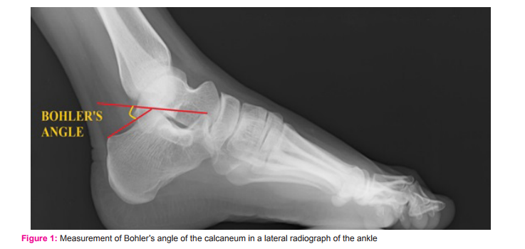
This is a hospital based cross sectional study done in M.S. Ramaiah Medical college hospital, Bangalore from September 2011 to May 2013. By analysing the previous studies the minimum sample size was calculated as 84. In this study computed radiographs of 184 normal ankles of 92 patients, older than 17 years were obtained and analysed . The study population included 61 males and 31 females. Ethical clearance was obtained. Patients referred to the radiology department were randomly included for the study. Patients with ankle deformities, Calcaneal fractures, congenital anomalies and arthritis were excluded. Radiograph of the lateral view of both right and left the ankles were taken in a single exposure. A medio-lateral view was taken. The digital radiograph was obtained and the Bohler’s and Gissane’s angles were measured using the angle tool in software called Dicom Viewer (Fig;1,2). Each angle was measured thrice and the mean was calculated and tabulated.
RESULTS
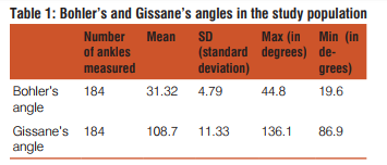
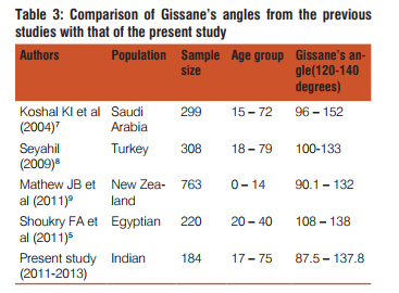
In the present study done in the Indian population, 184 ankle radiographs were taken (118male and 66 female ankles). The mean Bohler’s angle was found to be 31.3±5SD. Minimum and maximum values were 18.7° and 46.2° (Table:1). The lower limit which is diagnostically significant, is lower than that observed in Nigerians (28 to 38 degrees) 3 , Ugandans (20 to 50 degrees) 4 and Egyptians (22 to 40 degrees) 5 and higher than that observed in Americans (14 to 50 degrees)6 and Saudi Arabians (16 to 47 degree)7 (Table:2). The mean Gissane’s angle in the present study was 108.5 degrees. The maximum and minimum values for Gissane angle were 86.3° and 137.8° (Table:1). The minimum angle was found to be lower than that observed in all the previous studies (Table 3).
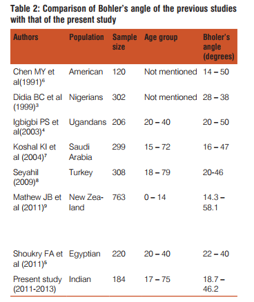
DISCUSSION Calcaneum is the most vulnerable bone of the ankle region. It is the tarsal bone most commonly prone for fractures and accounts for about 2% of the total fractures1 . Various studies have been conducted on Bohler’s and Gissane’s angles on different races. There is no literature suggestive of such a study done in South India. The tuber angle of Bohler ranges from 28 – 40 degrees. A decrease in angle indicates that the posterior facet i.e. the weight bearing surface of the calcaneus has collapsed, this leads to shifting of the weight of the body anteriorly. The reduction of Bohler’s angle indicates mainly the degree of proximal displacement of the Calcaneal tuberosity. Hence the angle is reduced in both intra-articular and extra-articular fractures of the calcaneus 11. The study conducted in 1991 in the American population concluded that if 28 degree is taken as the lower limit of normal for Bohler’s angle, 37 cases (31%) would be false-positive abnormal. The use of 20 degree as the lower limit may decrease the number of false-positive to three cases (2.5%); using 18 degree (mean − 2 SD) reduces the false-positive rate to less than 1% (one case) 6 . in studies conducted in most of the above mentioned races (Table:2,3), the lower limit of Bohler’s angle is less than 28 degrees. The Bohler’s and Gissane’s angles are the most frequently assessed angles in the interpretation of Calcaneal fractures 12. Radiographic evaluation for a suspected Calcaneal fracture should include the 4 views : (i)Antero posterior view / dorsoplantar view, (ii) Oblique view / medial oblique axial projection, (iii) Broden’s view and the lateral view 13,14. Among these the lateral view includes assessment of height loss (using Bohler’s angle) and assessment of rotation of posterior facet (using Gissane’s angle). Bohler’s angle and Gissane’s angle are used also in classification of Calcaneal fractures. Bohler’s angle also plays a role in the surgical management of Calcaneal fractures15. Bohler’s angle of 15 degrees or less is an indication for surgical reduction of the fracture15. During the surgical procedure the displaced fragment is reduced in such a way that the Bohler’s angle is restored16. The initial value of Bohler’s angle at the time of trauma guides the treatment 17. Literature affirms that Bohler’s angle serves as a guide in assessing outcome following surgical or non surgical treatment of Calcaneal fractures 18. The angle escalates towards near normal values with appropriate management.
CONCLUSION
Calcaneus, the weight bearing bone of the lower limb handles the stresses of the upright human posture. Fracture of this bone leads to hindrance in his or her day to day activities. The evaluation of Bohler’s and Gissane’s angles plays an important role in the diagnosis, treatment and prognosis of Calcaneal fractures. The range of Bohler’s and Gissane’s angles in the present study group of south Indian population is much less when compared to the standard reference values, indicating that range of the angles is variable in different population groups. The study of Bohler’s and Gissane’s angles in many such groups in a particular race helps in standardisation of range to that population. Hence this study helps the radiologist and orthopaedic surgeons to achieve accurate diagnosis, deliver apt treatment and in better assessment of prognosis.
References:
1. Sonin AH, Boles CA, Roger LF. Skeletal Trauma. In: Grainger RG, Alleson DJ, Adam A, Dixon AK, editors. Diagnostic Radiology. 4th ed. Vol 3. London, UK: Elsevier Churchill Livingstone; 2001. p. 1813.
2. Yochum TR, Rowe LJ. Essentials Of Skeletal Radiology. 3rd ed. Vol 1. Philadelphia: Lippincott Williams And Wilkins; 1996. p. 241.
3. Didia BC, Dimkpa JN. The calcaneal angle in Nigerians. Relationship to sex, age, and side of the body. J Am Podiatr Med Assoc 1999;89:472-4.
4. Igbigbi PS, Mutesasira AN. Calcaneal angle in Ugandans. Clin Anat 2003;16:328-30.
5. Shoukry FA, Aref YK, Sabry AE. Evaluation of the normal calcaneal angl
6. Chen MY, Bohrer SP, Kelley TF. Boehler’s angle: a reappraisal. Ann Emerg Med 1991;20:122-4.
7. Khoshhal KI, Ibrahim AF, Al-Nakshabandi NA, Zamzam MM, Al-Boukai AA, Zamzami MM. Bohler’s and Gissane’s angles of the calcaneus in the Saudi population. Saudi Med J 2004;25:1967-70.
8. Seyahi A, Uludag S, Koyuncu LO, Atalar AC, Demerhan M. The calcaneal angles in the Turkish population. Acta Orthop Traumatol Turc 2009;43:406-11.
9. Boyle MJ, Walker CG, Crawford HA. The paediatric Bohler’s angle and crucial angle of Gissane: a case series. Journal of Orthopaedic Surgery and Research 2011;6:2-5.
10. Daftary A, Haims AH, Baumgaertner MR. Fractures of the calcaneus: a review with emphasis on CT. Radiographics. 2005;25(5):1215–26.
11. Sanders R. Displaced intra-articular fractures of the calcaneus. The Journal Of Bone And Joint Surgery 2000;82a:225-50.
12. Schepers T, Ginai AZ, Mulder PG, Patka P. Radiographic evaluation of calcaneal fractures; To measure or not to measure. Skeletal Radiol 2007;36(9):847-52.
13. Canale ST. Campbell’s operative orthopaedics, 10th edition. Fractures and dislocations of foot. Murphy GA. Philadelphia, Pennsylvania: Mosby, Elsevier; 2003: 4231- 47.
14. Gupta MK. An outcome evaluation of operative compared with non operative management of intra-articular Calcaneum fractures [unpublished dissertation]. M.S.Ramaiah medical college: Rajiv Gandhi University Of Health Sciences; 2011.
15. Schepers T, Patka P. Intra-articular calcaneal fractures; A review of the literature. Ned Tijdschr Trauma 2008;16(2):40-7.
16. Meraj A, Zahid M, Ahmad S. Management of intra-articular calcaneal fractures by minimally invasive sinus tarsi approachearly results. Malay Orthop J 2012;6:13–17.
17. Bakker B, Halm JA, Van Lieshout EM, Schepers T. The fate of Böhler’s angle in conservatively treated displaced intra-articular calcaneal fractures. Int Orthop 2012 Dec;36(12):2495-9.
18. Schepers T, Vogels LMM, Schipper IB, Patka P. Percutaneous reduction and fixation of intra-articular calcaneal fractures. Oper Orthop Traumatol 2008;20(2):168-175.
|
 IJCRR
IJCRR 


 This work is licensed under a Creative Commons Attribution-NonCommercial 4.0 International License


