IJCRR - 7(16), August, 2015
Pages: 06-08
Date of Publication: 21-Aug-2015
Print Article
Download XML Download PDF
DOUBLE FORAMEN TRANSVERSARIUM-A CASE REPORT
Author: Nilofer Gausmohiyuddin Mulla, Sunil J. Pundge
Category: Healthcare
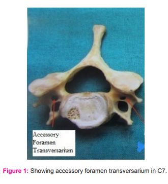
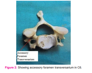
Abstract:Aim: To report a case of bilateral accessory foramen transversarium in lower cervical vertebrae. Case Report: In the present case we observed two abnormal foramina in transverse process of C7 and probably C6 cervical vertebrae. Rest of cervical vertebrae were normal. Discussion: The reasons for the presence of accessory foramen transversarium can be developmental or vascular. The presence of accessory foramen can alter the course of vertebral artery and even lead to compression of vertebral artery. This can lead to symptoms like headache, migraine and fainting attacks. Conclusion: The study of these variations is important to radiologists in interpreting CT and MRI scans.
Keywords: Cervical vertebra, Foramen transversarium, Double
Full Text:
INTRODUCTION
Foramen transversarium (FT) is a unique feature which is present in the transverse process of cervical vertebrae. The transverse process is morphologically composite around the foramen transversarium [1]. It has a dorsal and ventral bar, which terminates laterally as corresponding tubercles. These tubercles are connected, lateral to the foramen, by the costal (or, intertubercular) lamella. In upper six vertebrae foramen transversarium normally transmits vertebral artery, vertebral vein and a branch from cervicothoracic ganglion (vertebral nerve) [1]. In the seventh cervical vertebra it transmits only accessory vertebral veins and each is often divided by a bony spicule [1]. These foramina are known to exhibit variations and their etiology may be related to variations of the course of vertebral artery and is developmental [2]. The foramen transversarium is a result of the special formation of cervical vertebra. It is formed by vestigial coastal element fused to the body and the originally true transverse process of the vertebra. The vertebral vessels and nerve plexus are caught between the bony parts [3]. The deformation and variations of this foramen may affect the anatomical course of vascular and neural structures, and consequently may cause pathological conditions like vertebrobasilar insufficiency [4]. We noted abnormal foramina in the transverse process of C7 and probably C6 vertebrae.
CASE REPORT
During the routine osteology demonstration classes of cervical vertebrae in anatomy department, we notice two abnormal foramina in the transverse process of C7 and probably C6 vertebrae. The foramina were smaller, bilaterally present and posterior to the normal foramen transversarium. The foramina were complete and separated from the main foramen by a thin bar of bone. There was no other abnormality in the bone and remaining cervical vertebrae were normal.
DISCUSSION
Double foramen transversarium may be unilateral or bilateral depending on the course of vertebral artery. The reasons for the presence of accessory F.T can be developmental or vascular. It might be due to double rib bone element on the same side fusing to the original transverse process resulting in unusual number of FT [5].The association of double FTand duplication of vertebral artery is also possible but it is not a rule. The lack of foramen transversarium may indicate bypassing the vertebra by the vertebral artery. These have been confirmed by radiological studies [6]. Taitz et al studied 480 vertebrae from various populations and reported double foramen transversarium in 34 cases i.e.in 7% of vertebrae and only one vertebra manifested three transverse foramina unilaterally [3]. In contrast Das et al has reported duplicated foramen transversarium in two cases out of 132 cases he examined [2]. El Shaaray et al reported accessory foramen transversarium were most common in lower cervical vertebrae (C5, C6 and C7) which goes well with the present case [7]. Murlimanju et al studied 363 vertebrae and reported accessory foramen in 1.6% cases (only in 6 vertebrae) out of which 5 vertebrae showed double foramen and all the foramen were observed in lower vertebrae (C6 and C7) [8]. The vertebral vessels are a factor in the formation of the FT, thus it is assumed that variations in the presence and course of the vessels will be manifested in changes of the FT. Conversely, variations of the FT can be useful for estimating changes or variations of the vessels and accompanying nerve structures. Similar correlation may be suggested for double FT [3].
CONCLUSION
The knowledge of these variations helps in determining a more accurate approach to the removal of osteophytes or spurs compressing the vertebral arteries or in other interventions in the area. The surgical anatomy of foramen transversarium and vertebral artery are important to neurophysicians and radiologists in interpretation of radiographic films, angiograms and CT scans.
ACKNOWLEDGEMENT
Authors acknowledge the immense help received from the scholars whose articles are cited and included in references of this manuscript. The authors are also grateful to authors / editors / publishers of all those articles, journals and books from where the literature for this article has been reviewed and discussed.
References:
1. Standring S, 40th ed. Gray’s anatomy-The anatomical Basis of Clinical Practice. Spain: Churchill Livingstone Elsevier. 2005:718-721.
2. Das S, Suri R, Kapur V. Double foramen transversaria: An osteological study with clinical implications. Int Med J. 2005; 12:311-313.
3. Taitz C, Nathan H, Arensburg B. Anatomical observations of the foramina transversaria. Journal of Neurology, Neurosurgery and Psychiatry 1978; 41:170–176.
4. Mishra GP, Kumari S, Bhatnagar S, Singh B. Sixth cervical vertebra with bilateral double foramen transversarium and nonbifid spine: a rare case. Int J Res Med Sci. 2015; 3(1): 352-353.
5. Veeramani and Shankar. An unusual origin of the right vertebral artery from the thyrocervical trunk- a case report. International Journal of Basic Medical Sciences. (2011); 2(4).
6. Jarostaw Wysocki, Mariusz Bubrowski, Jerzy Reymond, Jan Kwiatkowski. Anatomical variants of the cervical vertebrae and the first thoracic vertebra in man. Via Medica, (2003); 62:357- 363.
7. El Shaarawy EAA, Sabry SM, El Gammaroy T, Nasr LE. Morphology and morphometry of the foramina transversaria of cervical vertebrae: A correlation with the position of the vertebral artery. Kasr El Aini Medical Journal [serial online] June 2010. Accessed December 10, 2010
8. MurlImanju BV, Prabhu LV, Shilpa K, Rai R, Dhananjaya KVN, Jiji PJ. Accessory transverse foramina in the cervical spine: incidence, embryology basis, morphology and surgical importance. Turkish Neurosurgery.2011; 21(3):384-387.
9. Cloward R. B. The anterior approach for removal of ruptured cervical discs. Journal of Neurosurgery. (1958); 15:607-617.
10. Caovilla HH, Gananca MM, Munhoz MS, Silva ML. Sindrome cervical. Quadros Clinicos Otoneurologicos Mais Comuns. Atheneu, Sao Paulo.2000; 3(11):95-100.
|
 IJCRR
IJCRR 


 This work is licensed under a Creative Commons Attribution-NonCommercial 4.0 International License


