IJCRR - 14(13), July, 2022
Pages: 71-76
Date of Publication: 05-Jul-2022
Print Article
Download XML Download PDF
Multivariate Analysis of Human Adult Femur Length by Various Parameters of Neck of Femur
Author: Ganjewar Manjiri, Shroff Gautam, Mandhana Vaishali
Category: Healthcare
Abstract:Introduction: Bones forms the main framework of the human body. Bone structure is different in different individuals depending upon race, sex, height, work, nutrition and geographical area. The femur is the bone that is the longest and strongest in human body and shows highest correlation with stature. Aims: This study has been done to determine the relationship of neck measurements with femur length in Maharashtrian population. Methodology: In the present Study, 54 human femur bones were studied for the following parameters using standard procedures. We took femur length (FL), neck length (NL), neck diameter at the narrowest site (NDN) and neck diameter at widest site (NDW) in mms. Results: The data was analysed through SPSS software. The descriptive statistics was analysed. Univariate and Multivariate Regression equations were derived to estimate Femur Length from various neck parameters. In univariate analysis, FL was 344.06 + 2.856 NL with R value 0.513 and in multivariate analysis, FL was 209.457+ 0.89 NL + 10.85 NDN -2.98 NDW with R value 0.867. Conclusion: It has been concluded that femur neck length has a positive correlation with total length of femur and femur length can be calculated if measurements of one or more neck parameters are available.
Keywords: Stature Correlation, Multivariate Analysis, Femur length (FL), Neck length (NL), Neck diameter narrow (NDN), Neck diameter widest (NDW).
Full Text:
Introduction
Homo sapiens are biped organisms. Due to shift in posture for walking, he needs strong bone structure and well-developed muscles and proper joint structure, especially in lower limbs. The femur is the lower limb bone, bone of the thigh. It is the longest and strongest bone of the body.1 It is involved in two very important joints which have a crucial role in standing and walking i.e. hip joint and knee joint. If femur is fractured, the working of both hip and knee joints will be disrupted. The fractured femur is very difficult to heal. Hence, in case of a hip replacement or implant surgery for a proximal segment of the femur, we need to have accurate measurements of the femur so that we can provide best possible replacement or corrective surgical measures for that clinical condition.
Femur bones have one of the highest correlations with stature and hence regression equations can be derived for the estimation of stature from available femur length (FL).2
Bone structure is different in different persons depending upon race, sex, height, work, nutrition and geographical area.3 Hence, we need to have adequate data related to bone dimensions from local population. Laishram had formulated the linear regression equation for the estimation of FL from various parameters of proximal segment of femur specifically diameter of head of femur.4 This study has been undertaken to determine the relationship between neck measurements and FL in the Maharashtrian population.
Materials and method
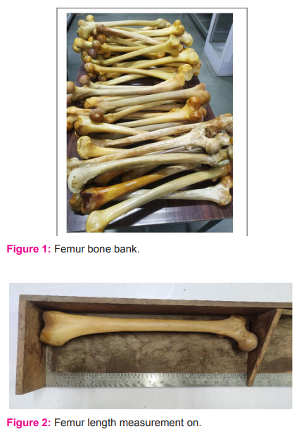
After scientific and ethical committee approval, this study was conducted on 54 dry femurs from Bone bank of the Department of Anatomy, MGM Medical College during the year 2019-20.(Fig. 1) Bones with obvious deformities in shape or size, the bones with visible fracture lines or injuries or surgical corrections were excluded from the study. After the exclusion of the deformed bones, 54 bones irrespective the side were included in study and given tags.
Dry weight of the bones was measured on a weighing scale. Simultaneously, the total length of these bones was also measured on bone osteometric board thrice and the average of all 3 entries was taken as final length for that femur. Both entries were promptly entered in master chart. As this study is aimed to find the femur length from the measurements of proximal segment of femur following measurements were taken in mms. Side of femurs were identified and tabulated in excel sheet.5
1. Femur length (FL) – Femur was placed on an osteometric board with internal rotation and it was measured from highest point of head of femur to the lowest point of medial condyle. (Fig no 2.)
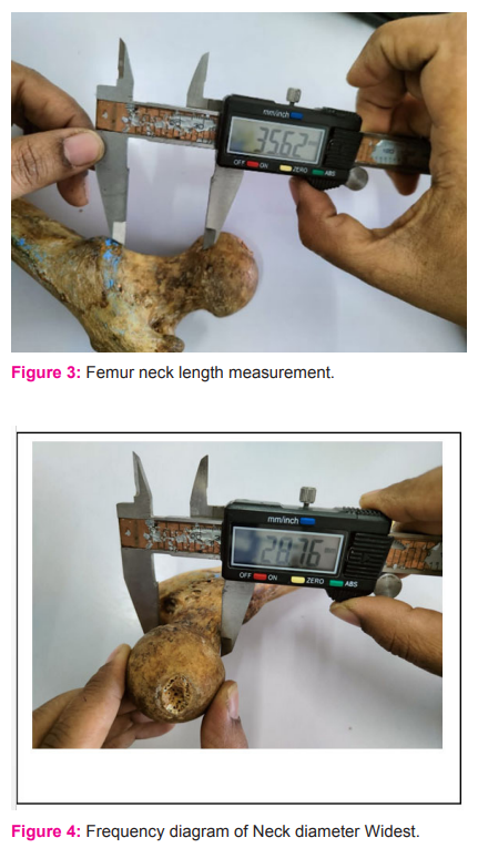
2. Neck length (NL)– It is the distance between the lowest margin of head and lowest point on the intertrochanteric line. It was measured with measuring tape. (Fig no 3)
3. Neck diameter – We have drawn a parallel line to the axis of neck. Diameters were measured at its narrowest (NDN) and at the widest part (NDW) (Fig no 4 and 5)
Measurements were first entered into Microsoft Excel (2010) then transferred to the SPSS version 20 for statistical analysis. After the data met the normality test, descriptive statistics were derived to calculate, mean, standard deviation and proportion of segmental measurements with the MFL (MFL – Mean Femur Length) for each side. Test of significance was applied. Finally, linear regression models were derived for estimating the MFL from different measurements of neck of femur.
Results
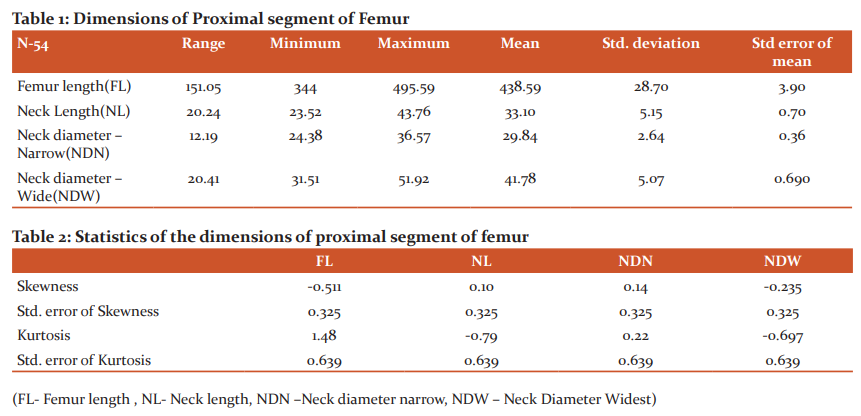
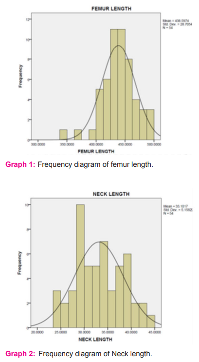
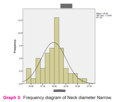
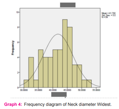
The MFL was 438.59 mm with a ±SD value of 24.38 mm. The mean NL was 33.10 mm with std. deviation of 5.15mm. The mean NDN was 29.84 mm. The NDW was 41.78 mm with a standard deviation of 5.07 mm. MFL in the study was found to be 344 mm, minimum NL was 43.76 mm, NDN was 24.38mm and of NDW was 31.51 mm.(Table no.1) (figure1,2,3,4)
The skewness of all the measurements was within -0.5 to 0.5 and hence it suggests that the data obtained was fairly symmetrical. Kurtosis value for FL is 1.48 and it is less than 3. This distribution is platykurtic. Similarly, kurtosis for the NL is -0.79 which is also platykurtic. NDN and NDW also show the platykurtic distribution. (Table no .2)
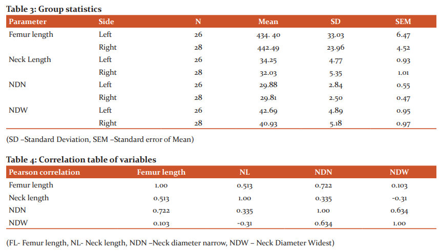
As shown in Table no 3, Out of 54 Femurs 26 bones were left-sided while 28 were right-sided. The MFL of left-sided femurs was 434.40 mm and of right-sided femurs were 442. 49 mm. Similarly, there was a difference between the means of NL of right and left side femurs. The difference in parameters of right and left MFL and right and left femur NL may be related to the dominant use of right side in the majority of the population and needs to be studied further in detail.
Mean NDN was found to be 29.88 mm and 29.81 mm on left and right side respectively. Mean NDN on left and right side were found to be 29.88 mm and 29.81mm whereas mean NDW were 42.69 and 40.93mm respectively. (table no.1)
As shown in Table no 4, the values indicate that there is a positive correlation between FL and NDN and negative correlation between FL and NDW. The univariate equation was derived for estimating FL from measurements of NL.
Femur length = 344.06 + 2.856 NL with R value 0.513.
This formula can be used to predict FL from the available neck length. It is also useful to derive the neck length for the prosthesis in surgeries if femur length is available.
For the calculation of FL from fragments of proximal segment of femur, a regression equation was derived from NL, NDN and NDW. For this, FL was used as a dependent variable and NL, NDN and NDW were used as independent variables. The multivariate was done. Multivariate equations were derived to estimate FL from different neck measurements. As R-value for the multivariate equation is significantly higher than that of univariate equation for the calculation of FL, it is better to predict FL from the multivariate equation when all the relevant dimensions are available.
Femur length = 209.457+ 0.89 NL + 10.85 NDN -2.98 NDW with R-value 0.867.
Discussion
The femur is one of the most important long bone for the erect posture of Homosapiens, and neck of the femur is one of the important factors for weight transmission, stabilization of posture and versatile movements at the hip joint. The measurements of the neck plays important role in femur implants too. Hence, due to scarcity of such data in Indian population, this study has been done to estimate FL from various parameters of femur neck
The MFL is 438.59 mm with std deviation of 24.38 mm. Similar results were seen in study done by Pillai .6 In this study, MFL was 437.4mm.
Micheal studied the linea aspera variations of femur while Shivshankarappa and Shakil et al. both studied the neck shaft angle in the femurs in different populations.7, 8 All these studies guide us to understand the anatomy of femur more precisely and objectively. We wanted to determine the correlation between the proximal segment measurements with femur length. Hence, we have decided to derive equations so as to predict MFL by using the neck measurements. On the literature search, we have found similar such studies.
Ravichandran studied NL and neck diameter (ND)of femur in 578 specimens and mean NL and ND were 31.88 mm and 30.99 mm while in our study NL was 33.10 mm ,NDN was 29.84mm and NDW was 41.mm.9
Bhavna Nath stated in their study, lower limb bone like tibia followed by fibula and then femur have high correlation with stature of the individual. They also derived the regression equation for estimation of stature from FL. S = 77.99 + 2.15 (MFL) ± 3.80 with r value 0.743.10
Seyed Vaghefi et al derived the mean ±SD value of FL in their study and it was 40.31 cm and 43.3 cm in females and males respectively and this difference was found to be significant (P<0.05), MFL in present study was 438.59mm.11
Anil Dwivedi had studied 280 femurs of Maharashtrian population and derived the regression equations for the FL estimation.12
FL= 128.347+ 4.104 (HVD) + 2.959 (HTD) and
FL of left side = 126.767+ 3.985(HVD) +0.963 (HC).
(HVD- Head Vertical diameter, HTD- Head Transverse Diameter, HC- Head Circumference)
In our study, we have derived the regression equations to estimate MFL from neck measurements as below in Maharashtrian Population
FL = 344.06 + 2.856 NL with R value 0.513 and
FL = 209.457+ 0.89 NL + 10.85 NDN -2.98 NDW with R-value 0.867
Conclusion
54 dry bones from bone bank of MGM Medical College, Aurangabad were studied for osteometric measurements. Data was tabulated and analyzed with help of SPSS software. From this study, it can be concluded that FL can be estimated with help of regression equations using measurements of proximal segment (NL, NDN, and NDW). According to our study, we can estimate the FL by both univariate and multivariate equations and R-value for multivariate equation is significant (p value)
FL = 344.06 + 2.856 NL (R value- 0.513)
FL = 209.457+ 0.89 NL + 10.85 NDN -2.98 NDW (R value- 0.867)
These equations will be useful in anthropology, Forensic and Orthopaedics for person identification, estimating the stature and estimating measurement of femur prosthesis. Still there is a scope to expand this study in future with inclusion of different measurements of proximal and distal segments for more accuracy.
Acknowledgement: Authors acknowledge M G M Medical College, Aurangabad for providing the infrastructure to conduct this study.
Conflict of Interest: Author declares that there is no conflict of interest.
Author`s Contribution: The idea generation is by Dr. Shroff Gautam and Dr. Mandhana Vaishali, Dr. Manjiri Ganjewar. Data Collection is by Dr. Ganjewar Manjiri. Data analysis and Tabulation Is done by Dr. Shroff Gautam and Dr. Manjiri. Drafting is done by Dr. Ganjewar Manjiri and Dr. Mandhana Vaishali. Critical revision of the article is done by Dr. Mandhana Vaishali.
.png)
References:
1. Standring S. The Anatomical Basis of Clinical practice. Gray’s Anatomy. 40thed. London: Churchill Livingstone Elsevier 2008.p.1360-1390.
2. Asha KR, Vinaykumar K, Bindurani MK, Kavyashree AN, Subhash L. Reconstruction of femoral length from its proximal fragments and diaphyseal segments in south Indian population. Res J. Pharm. Biol. Chem. Sci. 2014; 5(5):920-25.
3. Singh S, Nair SK, Anjankar V, Bankwar V, Satpathy DK, Malik Y. Regression equation for estimation of femur length in central Indians from the inter-trochanteric crest. J Indian Acad Forensic Med. 2013; 35(3):223-26.
4. Laishram D, Chandrasekaran S, Shastri D. Derivation of regression equation for estimation of stature by using measurement of femur. Asian J Med Sci. Oct 2021; 12(10):147- 151.
5. Khanal L, Shah S, Koirala S. Estimation of Total Length of Femur from its Proximal and Distal Segmental Measurements of Disarticulated Femur Bones of Nepalese Population using Regression Equation Method. J Clin Diagn Res. 2017 Mar; 11(3): HC01–HC05.
6. Pillai TJ, lakshmi Devi CK, Sobha Devi T. Osteometric Studies on Human Femurs. IOSR J. Dent. Sci.(IOSR-JDMS)e-ISSN: 2279-0853, p-ISSN: 2279-0861. Feb. 2014;13(2) I:34-39.
7. Polguj M, Bli?niewska K, J?drzejewski2 K, Majos A, Topol M. Morphological study of linea aspera variations: proposal of classification and sexual dimorphism. Folia Morphol. 72(1) :72–77.
8. Khan SM, Shaik HS. Study on neck-shaft angle and femoral length of south Indian femurs. Int J Anat Res. 2014; 2(4):633-35.
9. Ravichandran D, Muthukumaravel N, Jaikumar R, Das H, Melani R. Proximal femoral geometry in Indians and its clinical applications. J. Anat. Soc. India 60(1) 6-12 (2011):6-12
10. Bhavna, Nath S. Estimation of stature on the basis of measurements of the lower limb. Anthropology Today: Trends, Scope and Applications. 2007;Anthropologist (3): 219-222.
11. Vaghefi SHE, Elyasi L, Amirian SR, Raigan P, Akbari H, Sheikhshoaiee M, Borbor A. Evaluating Anthropometric Dimensions of the Femur Using Direct and Indirect Methods. Anatomical sciences. May 2015; 12(2):89-92
12. Dwivedi AK, Airan N, Bhatnagar R. Estimation of Length of Femur from its Proximal Segment in Maharashtrian Population. Int. J. Anat. Radiol. Surg. 2019, Jan;8(1): AO01-AO05.
|
 IJCRR
IJCRR 


 This work is licensed under a Creative Commons Attribution-NonCommercial 4.0 International License


