IJCRR - 14(2), January, 2022
Pages: 42-46
Date of Publication: 16-Jan-2022
Print Article
Download XML Download PDF
Evaluation Serum Chemerin and Visfatin Levels with Rheumatoid Arthritis: Possible Diagnostic Biomarkers
Author: Maryam Mohammed Jebur, Alaa Hussein J Al-qaisi, Nazar Sattar Harbi
Category: Healthcare
Abstract:Introduction: Rheumatoid Arthritis (RA) is a joint-damaging chronic inflammatory disease that affects the synovium and articular cartilage. RA is characterized by symmetric arthritis that primarily affects the tiny joints of the hands and feet. Adipokines play a role in the etiology of a variety of metabolic, vascular, and inflammatory diseases. The goal of this study is to compare the levels of inflammatory adipocytokines (chemerin and visfatin) and their ratios, as well as certain related biomarkers, in RA patients and healthy controls to see if they can help diagnose Rheumatoid Arthritis. Methods: A total of 70 (25 males and 45 females) RA patients' group with ages ranging from 45-65 years and 30 (10 males and 20 females) as the control group with ages ranging from 40-70 years old were involved in the study. All individuals had their biochemical parameters, demographic profile and serum chemerin and visfatin concentrations analyzed. Results: Our findings indicated that serum levels of Visfatin and Chemerin were significantly higher in individuals with rheumatoid arthritis than in healthy controls. Chemerin had moderate positive correlations with C-reactive protein (CRP) and total cholesterol (TC) while, it had weak negative correlation with high-density lipoprotein cholesterol (HDL). Conclusion: The variations in adipokine levels that we observed could play a role in diagnose of Rheumatoid Arthritis.
Keywords: Rheumatoid arthritis, Chemerin, Visfatin, Adipokines, C-reactive protein, High-density lipoproteins
Full Text:
INTRODUCTION
Rheumatoid arthritis (RA) is a systemic inflammatory disease affecting the joints principally. If left untreated, it is the most prevalent inflammatory disease of the joints, characterized by cartilage and bone degradation, leading to functional deterioration and disability. As a result, increasing cartilage and joint degeneration occurs, as well as impairment.1 According to data, women have a 3:1 chance of developing this condition. Any age group can be affected; however, the onset is most common between the ages of 40 and 60 .2 Rheumatoid arthritis has no recognized etiology. Clinical signs and symptoms can be caused by a mix of genetic predisposition and environmental triggers. RA is characterized by symmetric arthritis that primarily affects the tiny joints of the hands and feet. General discomfort and swelling of joints (often symmetrically), morning stiffness, and movement limitations that last more than one hour and can be eased by mild motions are all common indications and symptoms of RA.
However, RA can affect any organ, with the most prevalent symptoms being interstitial lung disease, renal amyloidosis, skin vasculitis, episcleritis, and poly-neuropathy of numerous mono-neuropathies. Better diagnostic biomarkers for the early detection of RA are always needed.3
Adipose tissue not only serves as a passive energy storage reservoir, but it also serves as an endocrine gland, producing and secreting a variety of bioactive peptides called adipokines.4Adipokines play a role in the etiology of a variety of metabolic, vascular, and inflammatory diseases.5 Adiponectin, resistin, leptin, visfatin, plasminogen activator inhibitor type 1 (PAI-1), tumor necrosis factor alpha (TNF- α), interleukin (IL)-6, and IL-8 are all adipokines .6
One of the future proteins in inflammatory biomarkers might be chemerin, a 16 kDa protein originally discovered in ovarian cancer patients' ascitic fluids and synovial exudates from rheumatoid arthritis patients' synovia as a result of the Tazarotene-induced gene 2 (Tig2).7,8 RA patients have increased levels of Chemerin and ChemR23 expression in their fibroblast-like synoviocytes (FLS) .9
Another pro-inflammatory adipokine that has the potential to help with RA diagnosis is Visfatin was first identified as a cytokine involved in early B-cell development, but it was renamed visfatin since it is mostly released by visceral fat. Visfatin is a peptide that is mostly secreted by the liver. The plasma level of visceral adipose tissue correlates with the amount of visceral fat in humans .10Visfatin levels in the blood are higher in RA, as is its expression in synovial fluids and inflamed synovium. The degree of inflammation, the severity of the disease, and joint destruction were all linked to visfatin serum and synovial fluid levels .10
This study aimed to evaluate RA patients' serum levels of chemerin and visfatin to those of healthy controls.
MATERIALS AND METHODS
Study participants: The current study was conducted in Al-Baghdad teaching hospital, during the period from October 2020 to March 2021. The present study included 70 (25 males and 45 females) RA patients from 45 to 65 years old and 30 (10 males and 20 females) healthy control group ranging from 40-70 years old. This study was approved by the Department of Chemistry, College of Science, Al-Nahrain University, Baghdad, and by the Research Ethics Committee of the Iraqi ministry of health, Iraq, with ethical clearance letter no.3122.
Exclusion criteria: History of hypertension, smoking, heart failure, diabetes mellitus, hypothyroidism, and hepatic or renal disorders, patients taking any medication, and drug used were all eliminated from the current study.
Sample collection: Patients and healthy individuals provided seven milliliters of venous blood placed into gel tubes for 15 minutes to coagulate. Serum was isolated from blood samples by centrifugation at 1840 x g for 15 minutes at room temperature. The serum was separated into aliquots and kept at -70°C until testing.
Measurement of Body Mass Index: BMI was measured by dividing weight (in Kilograms, Kg) by height squared (in meter, m) for each participant.
Biochemical analysis serum levels of chemerin, and visfatin were measured using enzyme-linked immune-sorbent assay (ELISA) provided by (MyBioSource, USA). The photometric method was used to evaluate the serum lipid profile total cholesterol (TC), triglyceride (TG) and high-density lipoprotein cholesterol (HDL) provided by (Linear, Spain). The fluorescence Immunoassay (FIA) method was used to evaluate the CRP level (ichroma, Korea).
Statistical Analysis of Parameters: Demographic and biochemical data in the present study were performed using GraphPad Prism software version 8.0.2 (San Diego, California, USA). T-test unpaired was performed to assess mean ± standard deviation (STD) and significant differences (P-value) among means of the two studied groups. Correlations between parameters in the present study were estimated with Pearson’s correlation coefficient. P ≤ 0.05 was considered statistically significant.
RESULTS
Table (1) shows the demographic data of the two studied groups (Rheumatoid arthritis and control). The results obtained from the preliminary analysis shown in table (1) indicated that there was no significant difference in BMI (P = 0.0993) between RA and control groups. There were no significant differences between RA group and the control group regarding age (P=0.5563).
Table (2) shows laboratory data from the blood analysis among the two groups. Our findings indicate there were significant differences between the RA or control groups regarding serum levels of CRP and TC and there were significantly higher in HDL (P< 0.0001) among two groups
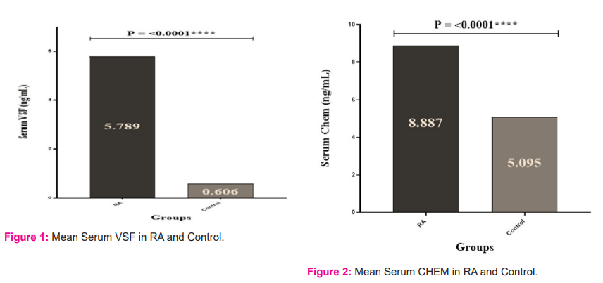
Table (3) shows the different adipokine concentrations among the two groups. Statistical analysis revealed that serum levels of VSF were significantly higher (p< 0.0001) among two groups, as shown in figure (1). Furthermore, serum levels of Chem were significantly higher (P< 0.0001) in RA group compared to controls, as shown in figure (2).
We determined Pearson’s correlation coefficient among the different variables in the study. There were three significant correlations appeared in RA group. Chemerin had moderate positive correlations with CRP (r=0.452, P=0.031) and TC (r=0.504, P=0.014) while, it had weak negative correlation with HDLc(r=-0.245, P=0.041).
DISCUSSION
Rheumatoid arthritis has been linked in several studies to adipokines.11 The majority of prior research have focused on pre-existing atherosclerotic diseases or symptoms to narrow down their patient group. In our research, we looked at the serum levels of Visfatin and Chemerin in patients with RA to see what role adipokines have in the disease's etiology.
In the current study, the RA patients were obese (30.2 ± 3.4), the marked inflammation encountered in them CRP (30.8 ± 3.9) agreed with Jonssonetal.12 who reported that RA patients explained this finding by the increase in inflammatory cytokine production.
Adipocytokines are currently thought to play a role in the etiology of a variety of metabolic and inflammatory diseases, including rheumatoid arthritis. To examine its significance in the pathophysiology of these disorders, we urged researchers to look at the amounts of the adipocytokine in serum samples from patients with inflammatory and non-inflammatory rheumatic diseases, as well as healthy controls .13
serum levels of chemerin is significantly higher (P value<0.0001) in RA patients compared to the healthy individuals, which are similar to findings from a previous study .14
Chemerin was originally recognized in its precursor low biological active state, which was implicated in innate and adaptive immunological responses.15 Once activated, it directed dendritic cells and macrophages to wounded tissues and inflammatory locations, triggering fast defenses throughout the body. Obesity, diabetes, lipid profile components, and early vascular inflammation have all been linked to serum adipokine concentrations.16Chemerin and its receptor CMKLR1 create a complex that is engaged in immune response modulation and can play a role in the start and resolution of acute inflammation. Increased chemerin levels can promote inflammation by attracting immune system cells. Chemerin also elevated inflammatory mediator expression and production in the affected area. Increased levels of this adipokine in adipose tissue stimulate immune cell recruitment, which leads to increased expression of inflammatory mediators such as CRP, interleukine-6 (IL-6), and tumor necrosis factor alpha (TNF-α), which leads to inflammation exacerbation.17
Many substantial correlations between the measured parameters can be found in the correlation research. Chemerin had a positive correlation with total cholesterol TC (r=0.504, P=0.014) and a negative correlation with HDLc (r=-0.245, P=0.041) in the current study.
Chemerin is thought to regulate lipid metabolism enzymes by lowering cyclic adenosine monophosphate (cAMP) buildup and stimulating calcium release in adipocytes .18 These associations could be related to chemerin's multiple effects on various biological systems involved in RA disease, which grew stronger as the disease progressed. Chemerin is a protein that affects adipogenesis, angiogenesis, and inflammation 19, and its levels have been found to rise with the duration of RA. Furthermore, serum levels of this adipokine are linked to metabolic syndrome components such as higher BMI and plasma TG level.
In this study, another correlation was found between chemerin with CRP (r=0.452, P=0.031), indicating a relationship with systemic inflammation. This, in part, may be due to the fact that inflammatory cytokines released by adipose tissue stimulate the synthesis of CRP in the liver, which was observed in inflamed tissues, in RA .20
Moreover, serum levels of visfatin were significantly higher (p< 0.0001) in RA patients than in the healthy control group, which is in line with observations from previous work of ?Alkady et al 21 who found that visfatin level was significantly increased in patients with RA.
Visfatin may play a substantial role in the pathophysiology of RA, according to several observations. In certain studies, visfatin was found to be upregulated in active RA in response to proinflammatory stimuli like IL-6. 22 Synovial fluid and serum, the degree of inflammation, the severity of the disease, and joint destruction are all linked to visfatin levels .23
Despite the fact that visfatin is an adipokine, earlier research has found no link between it and obesity. Some research, however, have discovered a link between visfatin and inflammatory markers.24Visfatin was found in invasive synovial tissue in rheumatoid arthritis patients, and its levels were also elevated in synovial fibroblasts. Visfatin can activate IL6, Matrix metalloproteinase (MMP1 and MMP3), and TNF and IL6 in RA synovial fibroblasts, as well as TNF and IL6 in monocytes .25
CONCLUSION
Our findings reveal that chemerin and visfatin levels are significantly higher. Therefore, these alterations in adipokine levels may play a key role in the development of RA-related inflammation.
ACKNOWLEDGEMENT
The authors would like to acknowledge staff in Baghdad teaching hospital for their assistance in this work.
Source of Funding: No financial assistance was obtained from any sources.
Conflict of Interest: The authors declare that they have no known competing financial or personal relationships that could have appeared to influence the work reported in this paper.
Authors’ Contribution: This is a collaborative work among all authors.
Author 1: Designing, data collection, data analysis, article writing
Author 2: Data verification, article editing
Author 3: Data collection, article writing

All values are shown as mean±SD (Standard Deviation). S: p-value <0.05 (Significant),
NS: p-value <0.05 (Non-Significant),HS: p-value <0.0001 (Highly Significant).
 CRP – C-reactive protein, HDLc– high-density lipoprotein cholesterol, TC – total cholesterol, TG – triglyceride


References:
1. Yap H-Y, Tee SZ-Y, Wong MM-T, Chow S-K, Peh S-C, Teow S-Y. Pathogenic role of immune cells in rheumatoid arthritis: implications in clinical treatment and biomarker development. Cells. 2018;7(10):161.
2. Al-Rubaye AF, Kadhim MJ, Hameed IH. Rheumatoid Arthritis: History, Stages, Epidemiology, Pathogenesis, Diagnosis and Treatment. Int J Toxicol Pharmacol Res. 2017;9(2):145–55.
3. Aletaha D, Neogi T, Silman AJ, Funovits J, Felson DT, Bingham III CO et al. 2010 rheumatoid arthritis classification criteria: an American College of Rheumatology/European League Against Rheumatism collaborative initiative. Arthritis Rheum. 2010;62(9):2569–81.
4. Jakovljevi? B, Paunovi? K, Stojanov V. Adipose tissue as an endocrine organ. Srp Arh Celok Lek. 2005;133(9–10):441–5
5. Ouchi N, Parker JL, Lugus JJ, Walsh K. Adipokines in inflammation and metabolic disease. Nat Rev Immunol. 2011;11(2):85–97.
6. Giralt M, Cereijo R, Villarroya F. Adipokines and the endocrine role of adipose tissues. Metab Control. 2015;265–82.
7. Bonomini M, Pandolfi A. Chemerin in renal dysfunction and cardiovascular disease. Vascul Pharmacol. 2016;77:28–34.
8. Ernst MC, Haidl ID, Zúñiga LA, Dranse HJ, Rourke JL, Zabel BA, et al. Disruption of the chemokine-like receptor-1 (CMKLR1) gene is associated with reduced adiposity and glucose intolerance. Endocrinology. 2012;153(2):672–82.
9. Ha Y-J, Kang E-J, Song J-S, Park Y-B, Lee S-K, Choi ST. Plasma chemerin levels in rheumatoid arthritis are correlated with disease activity rather than obesity. Jt Bone Spine. 2013;81(2):189–90.
10. Fukuhara A, Matsuda M, Nishizawa M, Segawa K, Tanaka M, Kishimoto K, et al. Visfatin: a protein secreted by visceral fat that mimics the effects of insulin. Science (80- ). 2005;307(5708):426–30.
11. Del Prete A, Salvi V, Sozzani S. Adipokines as potential biomarkers in rheumatoid arthritis. Mediators Inflamm. 2014;2014.
12. Jonsson MK, Sundlisæter NP, Nordal HH, Hammer HB, Aga A-B, Olsen IC, et al. Calprotectin as a marker of inflammation in patients with early rheumatoid arthritis. Ann Rheum Dis. 2017;76(12):2031–7.
13. Otero M, Logo R, Gomez R, Logo F, Dieguez C, Gómez-Reino JJ, et al. Changes in plasma levels of fat-derived hormones adiponectin, leptin, resistin and visfatin in patients with rheumatoid arthritis. Ann Rheum Dis. 2006;65(9):1198–201.
14. Ali DMM, Al-Fadhel SZ, Al-Ghuraibawi NHA, Al-Hakeim HK. Estimation of Serum Chemerin, Visfatin Levels and Their Ratio as A Possible Diagnostic Parameters of Rheumatoid Arthritis. 2019;
15. Ernst MC, Sinal CJ. Chemerin: at the crossroads of inflammation and obesity. Trends Endocrinol Metab. 2010;21(11):660–7.
16. Bozaoglu K, Curran JE, Stocker CJ, Zaibi MS, Segal D, Konstantopoulos N, et al. Chemerin, a novel adipokine in the regulation of angiogenesis. J Clin Endocrinol Metab. 2010;95(5):2476–85.
17. Fontes VS, Neves FS, Cândido APC. Chemerin and factors related to cardiovascular risk in children and adolescents: a systematic review. Rev Paul Pediatr. 2018;36:221–9.
18. Maghsoudi Z, Kelishadi R, Hosseinzadeh-Attar MJ. Association of chemerin levels with anthropometric indexes and C-reactive protein in obese and non-obese adolescents. ARYA Atheroscler. 2015;11(Suppl 1):102.
19. Lu B, Zhao M, Jiang W, Ma J, Yang C, Shao J, et al. Independent association of circulating level of chemerin with functional and early morphological vascular changes in newly diagnosed type 2 diabetic patients. Medicine (Baltimore). 2015;94(47).
20. Ali DMM, Al-Fadhel SZ, Al-Ghuraibawi NHA, Al-Hakeim HK. Serum chemerin and visfatin levels and their ratio as possible diagnostic parameters of rheumatoid arthritis. Reumatologia. 2020;58(2):67.
21. Alkady EAM, Ahmed HM, Tag L, Abdou MA. Serum and synovial adiponectin, resistin, and visfatin levels in rheumatoid arthritis patients. Z Rheumatol. 2011;70(7):602.
22. Sglunda O, Mann H, Hulejová H, Kuklová M, Pecha O, Pleštilová L, et al. Decreased circulating visfatin is associated with improved disease activity in early rheumatoid arthritis: data from the PERAC cohort. PLoS One. 2014;9(7):e103495.
23. Institutet K, Hambardzumyan K. Predictive Biomarkers in Rheumatoid Arthritis. 2018.
24. Arogbodo , J. O. ., Faluyi, O. B. ., &Igbe, F. O. . (2021). In vitro Antimicrobial Activity of Ethanolic Leaf Extracts of Hibiscus Asper Hook. F. and Hibiscus Sabdariffa L. on some Pathogenic Bacteria. Journal of Scientific Research in Medical and Biological Sciences, 2(3), 1-12. https://doi.org/10.47631/jsrmbs.v2i3.304
25. Brentano F, Schorr O, Ospelt C, Stanczyk J, Gay RE, Gay S, et al. Pre–B cell colony?enhancing factor/visfatin, a new marker of inflammation in rheumatoid arthritis with proinflammatory and matrix?degrading activities. Arthritis Rheum Off J Am Coll Rheumatol. 2007;56(9):2829–39.
26. Mirfeizi Z, Noubakht Z, Rezaie AE, Jokar MH, Sarabi ZS. Plasma levels of leptin and visfatin in rheumatoid arthritis patients; is there any relationship with joint damage? Iran J Basic Med Sci. 2014;17(9):662–27. Makhlouf, A.-M. A. ., Mahmoud, A. M. ., Ibrahim, R. G. ., & Abdel Aziz, Y. S. . (2021). Effects of Vitamin D and Simvastatin on Inflammatory and Oxidative Stress Markers of High-Fat Diet-Induced Obese Rats. Journal of Scientific Research in Medical and Biological Sciences, 2(3), 39-50. https://doi.org/10.47631/jsrmbs.v2i3.297
|


 This work is licensed under a Creative Commons Attribution-NonCommercial 4.0 International License