IJCRR - 13(21), November, 2021
Pages: 48-52
Date of Publication: 09-Nov-2021
Print Article
Download XML Download PDF
Comparative Evaluation of Four Different Anterior Reference Points & Their Effect on the Protrusive Condylar Guidance Angle Obtained in Semi Adjustable Articulator
Author: Bora Neelam, Jagtap Amit, Dange Bhagyashree, Bulbule Nilesh, Gandage Dhananjay, Swarup Siddharth
Category: Healthcare
Abstract:Introduction: An anterior reference point is a physical requirement for orienting casts in three-dimensional space in an articula�tor. Different anterior points of reference for different articulators have been proposed. Aims: To evaluate four different anterior reference points and their effect on the protrusive condylar guidance angle obtained in a semi-adjustable articulator. Methodology: A total of 20 subjects were selected with Class I dental relationship. Face bow records were taken for orbitale and orbitale minus 7mm as the anterior points of reference. Another face bow record was taken without considering any point on the face and orienting the face bow on the articulator with the superior annular notch and inferior annular notch on the incisal guide pin as an anterior point of reference. Protrusive bite registration was made with the help of an anterior jig. Programming of the articulator was done with the protrusive bite record to evaluate condylar guidance angle. Two lateral cephalograms were made in maximum intercuspation and protrusive position with the help of an anterior jig. Cephalograms were traced and superimposed to obtain condylar guidance angle. Descriptive statistics were performed to obtain the means and standard deviation of all the sample sizes. Paired 't' test was performed between radiographic readings and condylar guidance angle obtained. Result: This study showed that the protrusive condylar guidance values registered for inferior annular notch and Orbitale as anterior points of reference were statistically significantly closer to the radiographic values. Conclusion: The orbital & inferior annular notch mounts the maxillary cast more accurately to the Frankfort Horizontal Plane when they are used as the anterior points of reference.
Keywords: Anterior point of reference, Orientation jaw relation, Articulator, Lateral cephalograms, Orbitale, Orbitale minus 7 mm
Full Text:
INTRODUCTION
The form, function, & patho-function of the dynamic masticatory system comprises one of the most fascinating, basic, and important areas of study in dentistry.1 No speciality in dentistry can be effectively practised at the highest level of competence without an understanding of how the teeth relate to the rest of the masticatory system, including the temporomandibular joints. It is correctly said that dentists who ignore the relationship of the occlusion to the position and condition of the temporomandibular joints can only guess at diagnosing a myriad of problems that are seen in every general practice.2
The use of 2 posterior points and an anterior point of reference for orienting a maxillary cast to an articulator has long been advocated.3 Anterior reference point is any point located on the midface that, together with two posterior reference points, establishes a reference plane.4 Different anterior points of reference for different articulators have been proposed which include the orbitale, orbitale minus 7 mm, nasion, ala of the nose, superior annular notch and inferior annular notch on the incisal guide pin of the Hanau wide vue articulator. Out of these points, the orbitale, orbitale minus 7 mm, superior annular notch & inferior annular notch on the incisal guide pin of the articulator are used in this study as anterior points of reference for Hanau Wide Vue articulators. A variation in the supero-inferior position of casts on the articulator can alter the protrusive condylar guidance making the reliability of these anterior reference points questionable.
With this background of uncertainty, a study was planned to evaluate four different anterior reference points and their effect on the protrusive condylar guidance angle obtained in semi adjustable articulator.
METHODOLOGY
Twenty Students from Dr. D.Y Patil Dental College and Hospital, Pimpri were selected for the study. Irreversible hydrocolloid impressions were made for the maxillary & mandibular arch. One set of the stone cast (maxillary & mandibular) was obtained for each student after making the impressions. Bases were poured for the casts & then a spilt cast was made for the maxillary cast.
Four different anterior reference points were selected for mounting the casts on a Hanau Wide Vue articulator. The orbitale of the patient, orbitale minus 7 on the patient, superior annular notch on the incisal guide pin of the articulator (37 mm below the orbital plane), the inferior annular notch on the incisal guide pin of the articulator (54 mm below the orbital plane) were the anterior reference points used in the study. The maxillary teeth impression was recorded on the bite fork with impression compound.
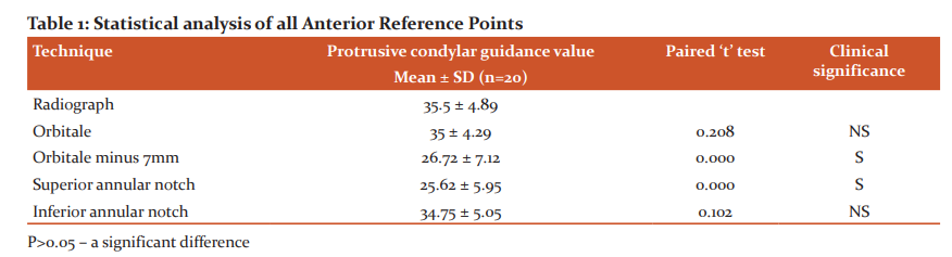
The first anterior point of reference was selected as orbitale. The position of the orbitale was confirmed, which is located on the notch present in the lower rim of the orbit in line with the pupil of the eye & was marked with an indelible pencil.5 (Fig.1) This marking was used as the anterior point of reference for the facebow transfer using a facebow (Hanau spring Bow, Teledyne Water Pik, Fort Collins, Colorado, USA). The external auditory meatus was used as posterior determinants for recording the orientation of the maxilla to the cranium. The facebow record was made followed by transfer of the facebow record onto the Hanau Wide Vue semi-adjustable articulator and indirect mounting of the maxillary cast was done.6 Then the mandibular cast was mounted in maximum intercuspation with the maxillary cast.7
After completion of mounting a pencil mark was made on the buccal cusp tip of the maxillary 1st premolar on both sides of the cast. Another marking was made 6 mm distal to the previous mark on the mandibular tooth on both sides of the cast. Then, the centric locks were opened & the mandibular cast was protruded so that the two lines marked previously coincided. An anterior jig was then fabricated with an impression compound with the mandibular protrusion of 6 mm. This jig was then placed intraorally & checked. This was done to keep a constant of 6 mm protrusion for all the subjects.
Three sets of protrusive interocclusal records were made intraorally using Addition silicone bite registration material with the jig placed between the anterior teeth. The protrusive bite records were used to program the articulator.8 Then the average of the three readings was calculated for both the right & left sides. The condylar guidance angle was tabulated for both the right & left sides.
The casts were then demounted and used again for the mounting with the second anterior point of reference. A second mark was made 7 mm below the orbital marking and was used for the second facebow transfer. (Fig. 2)
Then the casts were mounted similarly as done previously for orbitale as the anterior point of reference. Programming of the articulator was done & the condylar guidance angles were tabulated for both sides.
The third & fourth anterior point of reference i.e. the superior annular notch on the incisal guide pin of the articulator & the inferior annular notch on the incisal guide pin of the articulator was used for mounting the maxillary cast. A facebow record was made without considering the anterior reference point on the patient. (Fig.3)
To locate the incisal edges of the maxillary casts at the level of superior & inferior markings on the incisal guide pin respectively, the facebow was adjusted by using an anterior elevator (no. 010358-000, Teledyne Water Pik, Fort Collins, Colorado, USA). The casts were mounted using the new facebow record. The maxillary cast was mounted followed by mounting of the mandibular cast as done previously. Programming of the articulator was done & the condylar guidance angles were tabulated for both the sides for both the superior & inferior annular notch as the anterior points of reference.
A radiographic marker was then placed in the same position on the patient’s face where the orbitale was marked for orbitale as the anterior point of reference. This was done to trace the same marking of orbitale on the lateral cephalogram as on the face. The two lateral cephalometric radiographs were made using a Broadbent cephalostat to standardize the head positions in the Department of Oral Medicine Diagnosis and Radiology. One lateral cephalometric radiograph was made in maximum intercuspation. The second lateral cephalometric radiograph was made in a protrusive position by placing the anterior jig intraorally.
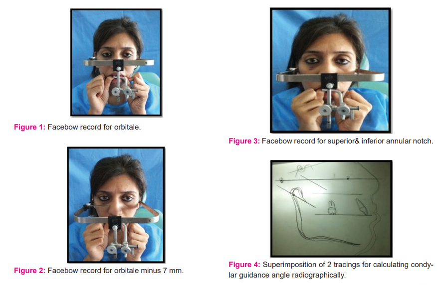
The lateral cephalograms were then traced and superimposed. The following points were traced on each lateral cephalogram: sella, nasion, radiographic orbitale, porion.9 The condyle border was traced for both sides and their mean condyle border was marked. A posterior-most point was marked on the posterior slope of the condyle. Sella & nasion were connected & this line was used for overlapping the two lateral cephalograms. Frankfort horizontal plane was drawn by connecting the radiographic orbital & porion. By joining the posterior-most points on the posterior slope of the condyles in maximum intercuspation and the protrusive position, the protrusive condylar path was gained. The angle between the condylar path and the Frankfort horizontal plane was determined. (Fig.4)
METHOD OF DATA ANALYSIS:
The Paired ‘t’ test was used to analyse the values gained for protrusive condylar guidance in 4 different mountings and the radiographic tracings.
RESULTS:
Comparison of protrusive condylar guidance angle values obtained from 4 different anterior reference points and radiographic tracings. (Table 1)
DISCUSSION
In this study, four different anterior reference points were used i.e. Orbitale, orbitale minus 7 mm, Superior annular notch and inferior annular notch present on the incisal pin of the Hanau wide vue articular.
The values obtained for protrusive condylar guidance in 4 different mountings and the radiographic tracings were analysed using paired t-test & correlation test. The analysis of the data obtained from the study did not support the null hypothesis that all anterior points of reference register the same condylar guidance on both the articulators. The analysis showed that the four different anterior points of reference used in the study recorded different condylar guidance values.
The mean protrusive condylar guidance values registered for mountings with Orbitale as the first anterior point of reference was 35 ± 4.29 degrees, with Orbitale minus 7 mm as the second anterior point of reference was 26.72 ± 7.12 degrees, with the superior annular notch as the third anterior point of reference was 25.62 ± 5.95 degrees, with the inferior annular notch as the fourth anterior point of reference was 34.75 ± 5.05 degrees. The mean protrusive condylar guidance values registered radiographically was 35.5 ± 4.89 degrees.
The protrusive condylar guidance values registered for mountings with Orbitale minus 7 mm (p = 0.000) and superior annular notch (p = 0.000) as anterior points of reference were significantly different (p <0.05), and those for Orbitale (p = 0.208) and inferior annular notch (p= 0.102) as anterior points of reference were not significantly different from those of radiographic tracings.
The difference between condylar guidance angle by orbitale & inferior annular notch as the anterior points of reference and the radiographic condylar guidance angle was 0.5 degrees and 0.75 degrees respectively. This difference of condylar guidance angle values suggests that the orbital & inferior annular notch mounts the maxillary cast more accurately to the Frankfort Horizontal Plane when they are used as the anterior points of reference. Between these two points, the orbital is the one that mounts the maxillary cast closest to the anatomic position.
The difference between condylar guidance angle by orbitale minus 7mm and superior annular notch as the anterior points of reference and the radiographic condylar guidance angle was 8.78 degrees and 9.88 degrees respectively. This difference of condylar guidance angle values suggests that the orbitale minus 7 mm and superior annular notch mounts the maxillary cast less accurately to the Frankfort Horizontal Plane when they are used as the anterior points of reference.
According to the Correlation test between lateral cephalograms and 4 different anterior reference points in the study group, the R-value for orbitale (0.938) & inferior annular notch (0.924) was closest to the radiographic values. The R-value for orbitale minus 7 mm (0.394) & superior annular notch (0.492) was not significant. This test also suggests that the orbital & inferior annular notch are highly significant anterior points of reference taken into consideration.
The mountings with the inferior annular notch present on the incisal pin of the articulator registered the condylar guidance values closer to the radiographic values than did those with the superior annular notch present on the incisal pin of the articulator was used as the third point of reference. This comparison supports the findings of Lauciello and Appelbaum.10
According to Weinberg, a 0.2-mm reduction in the non-working cusp height was observed with a 9-degree decrease in the condylar path inclination. Hence, a variation of 2 to 5 degrees would be clinically inconsequential.11 Hence, mounting the cast with orbitale & inferior annular notch as anterior points of reference can be advocated.
Gonzales and Kingeryobserved the lack of parallelism between the Frankfort horizontal plane and the axis-orbital plane.12 To rectify this, the 7 mm correction was suggested by the authors. Nevertheless, in the newer Hanau articulators, the orbital pointer is placed 7 mm above the level of the condylar plane. Even though, the protrusive condylar guidance values enumerated on the mountings with inferior annular notch exhibited a wide range of variation from individual to individual, the mean of these values was closer to the mean of radiographic values. The orbital reference point was the next closest point of reference. Moreover, when related to the inferior annular notch reference, the range of variation was found to be lesser from individual to individual. Hence, when Hanau Wide Vue articulator is used in practice, the Orbitale can be considered to be the better anterior point of reference for the facebow records. The results of this study supported the findings of Nooji & Sajjan.13
The sequence of the accurate third point of reference used in this study can be given as follows, the orbitale as the anterior point of reference mounts the casts to the closest anatomic position, the second is the inferior annular notch present on the incisal pin of the Hanau Wide Vue articulator which also mounts the maxillary casts closer to the anatomic position. The superior annular notch present on the incisal pin of the articulator & orbitale minus7 mm as the anterior point of reference gave a wide range of differences with anatomic values, they are the least accurate to mount the casts to the anatomic position. So, the use of superior annular notch & orbitale minus 7 mm as the anterior point of reference is questionable.
CONCLUSION-
The most crucial and operator-dependent step in prosthodontic dentistry is the orientation of the maxillary cast in an articulator. This technique-sensitive step acts as a baseline from occlusal rehabilitation of the patient is carried out.14 An error in selecting and transferring the occlusal plane to the articulator may result in instability of complete dentures and/or decreased chewing efficiency of the patient. It can also cause a substantial error in the final occlusal scheme of the prosthesis and/or poor aesthetics.15
The mean protrusive condylar guidance values registered for mountings with Orbitale as the first anterior point of reference was 35 ± 4.29 degrees, with Orbitale minus 7 mm as the second anterior point of reference was 26.72 ± 7.12 degrees, with the superior annular notch as the third anterior point of reference was 25.62 ± 5.95 degrees, with the inferior annular notch as the fourth anterior point of reference was 34.75 ± 5.05 degrees. The mean protrusive condylar guidance values registered radiographically was 35.5 ± 4.89 degrees.
The protrusive condylar guidance values registered for inferior annular notch and orbitale as anterior points of reference were significantly closer (P < 0.05) to the radiographic values. The orbitale (350) and inferior annular notch (34.750) references registered the highest protrusive condylar guidance values. However, the values did not differ significantly from the radiographic values.
Within the limitations of this study, it can be concluded that the orbital & inferior annular notch mounts the maxillary cast more accurately to the Frankfort Horizontal Plane when they are used as the anterior points of reference for mounting casts on Hanau Wide Vue articulators with Hanau Spring Bow. Between these two points, the orbital is the one that mounts the maxillary cast closest to the anatomic position.
Acknowledgements- Authors acknowledge the immense help received from the scholars whose articles are cited and included in references of this manuscript. The authors are also grateful to authors/editors/publishers of all those articles, journals and books from where the literature for this article has been reviewed and discussed.
Author’s contribution-
Bora Neelam- Chief investigator
Jagtap Amit- Methodology and study design
Dange Bhagyashree- Manuscript preparation
Bulbule Nilesh- Methodology and study design
Gandage Dhananjay- Manuscript preparation
Swarup Siddharth- Methodology and study design
Conflict of interest: None
Funding source: None
References:
-
Koolstra, Harm J. Dynamics of the Human Masticatory System. Critical reviews in oral biology and medicine: an official publication of the American Association of Oral Biologists. 2002;13,366-76. DOI:10.1177/154411130201300406.
-
Dawson PE. Functional Occlusion: From TMJ to Smile Design. 2nd Edition. The United Kingdom, Elsevier Health Sci. 2006; ix-x.
-
Bailey JO Jr, Nowlin TP. Evaluation of the third point of reference for mounting maxillary casts on the Hanau articulator. J Prosthet Dent. 1984 Feb;51(2):199-201. doi: 10.1016/0022-3913(84)90260-9. PMID: 6583397.
-
The glossary of prosthodontic terms. J Prosthet Dent. 2005;94:10-92
-
Salzmann JA. Orthodontic practice and techniques. Philadelphia: JB Lippincott Co; 1957. p. 139.
-
Anderson JD. Biological & clinical considerations in making jaw relation records & transferring records from the patient to the articulator. Zarb Bolender. Prosthodontic treatment for edentulous patients.12th edition. St. Louis: Mosby; 2009. p. 268-97.
-
Shillingburg HT. Interocclusal records. Fundamentals of fixed prosthodontics. 3rd ed. Chicago: Quintessence Publications. 1997;35-45
-
Shillingburg HT. Articulation of casts. Fundamentals of fixed prosthodontics. 3rd ed. Chicago: Quintessence Publications. 1997;47-72
-
Gilboa I, CardCash HS, Kaffe I, Gross MD. Condylar guidance: correlation between articular morphology and panoramic radiographic images in dry human skulls. J Prosthet Dent. 2008 Jun;99(6):477-82. doi: 10.1016/S0022-3913(08)60112-2. PMID: 18514670.
-
Lauciello FR, Appelbaum M. Anatomic comparison to arbitrary reference notch on Hanau articulators. J Prosthet Dent. 1978 Dec;40(6):676-81. doi: 10.1016/0022-3913(78)90068-9. PMID: 281520.
-
Lawrence A, Weinberg A.B. An evaluation of the face bow mounting. J Prosthet Dent 1961;11(1):32-42. 10.1016/0022-3913(61)90107-X
-
Gonzalez JB, Kingery RH. Evaluation of plane of reference for orienting maxillary casts on articulators. J Am Dent Assoc. 1968;76:329-336.
-
Nooji D, Sajjan SM. The third point of reference and its effect on the protrusive condylar guidance angles obtained in the semi-adjustable articulator. J Indian Prosthodont Soc 2008;8:71-77.
-
Anusha CV, Singh AA, Sam G, Sangwan B, Shilpa M, Kamath AG. Evaluation of Two Facebow/Semi-adjustable Articulator Systems for Orienting Maxillary Cast on Articulators: A Pilot Study. J Contemp Dent Pract. 2016 Apr 1;17(4):327-30. doi: 10.5005/jp-journals-10024-1849. PMID: 27340168.
-
Shetty S, Shenoy KK, Sabu A. Evaluation of the accuracy of transfer of the maxillary occlusal cant of two articulators using two facebow/semi-adjustable articulator systems: An in vivo study. J Indian Prosthodont Soc. 2016 Jul-Sep;16(3):248-52. doi: 10.4103/0972-4052.176525. PMID: 27621543; PMCID: PMC5000565.
|
 IJCRR
IJCRR 


 This work is licensed under a Creative Commons Attribution-NonCommercial 4.0 International License


