IJCRR - 13(20), October, 2021
Pages: 52-56
Date of Publication: 24-Oct-2021
Print Article
Download XML Download PDF
A Novel Dataset 'Aravind' for Retinal Image Analysis
Author: M. Vijaya Maheswari, G. Murugeswari
Category: Healthcare
Abstract:Introduction: Diabetes is a disorder or a metabolic disease that increases the level of sugar in the blood. Untreated diabetes can cause serious issues to the eye. There are quite a several research works carried out in retina image analysis. For retinal image analysis, there are multifarious datasets available freely for research. Aim: The objective of the article is to introduce a new dataset namely 'ARAVIND' for retina image analysis for research purposes. Method: This dataset incorporates 98 images altogether which helps in pinpointing different types of eye defects like hemorrhages, microaneurysms, hard and soft exudates. Conclusion: The 'ARAVIND' dataset is freely available online that can be used by computer researchers in the field of medical image analysis which bridges the gap between the field of technology and science to a greater extent.
Keywords: Dataset, Exudates, Hemorrhages, Micro aneurysm and Retina
Full Text:
INTRODUCTION
Diabetes leads to various metabolic disorders and physiologic abnormalities in the body 1. The rapid growth of diabetes is a key indicator for the prevalence of various microvascular complications2. The number of people with diabetes is increasing year by year. Diabetes is increasing more in well-developed countries like the United States than in developing countries. Following the International Diabetes Federation (IDF), the threatening fact about diabetes is that 51% of the world population will likely have diabetes by the year 20453. The overriding reason for the cause of diabetes is the insalubrious lifestyle. Early detection and proper treatment of diabetes is a life-saving measure. The cost of treatment for diabetes varies from country to country. The consequences of diabetes may cause damage to the indispensable organs like the heart, retina etc.
Prolonged diabetes may affect the retina to a greater extent. Fundus/clinical images of the retina play a vital role after the diagnosis of diabetes. A proper eye examination is mandatory for patients with diabetes. Retinal abnormalities can lead to serious vascular diseases which do not show the earlier symptoms to the patients. In such cases, a high - quality fundus images provide finer details about the retina. The high-quality images help in detecting the abnormalities accurately.
Various datasets for retinal image analysis are available online which can be freely downloaded for detecting the defects in the human eye. Different algorithms were applied by the researchers or by the team of experts to extract the basic and finer details of the retina for the accurate identification of the abnormalities. The accuracy or efficiency of a code depends not only on the algorithm but also on the quality of the image. Hence, high-quality images are imperative for the researchers to prove their contribution to their research.
Diabetic Retinopathy
Diabetes is a metabolic disorder that heightens the level of sugar in the blood. The retina can be affected badly in persons who suffer from diabetes for a quite long period. This may lead to a disease called Diabetic Retinopathy (DR). This particular disease can be treated at the earlier stage whereas, in the advanced stage it might lead to blindness4. There are four different stages of diabetic retinopathy ranging from mild, moderate, severe non-proliferative to proliferative5. In the severe stage of this disease, the blood vessels get a leak and thereby leading to blindness. When a person is affected by this disease, ophthalmologists check for the presence of microaneurysms, haemorrhages, hard exudates and soft exudates. Fundus images are those images that are acquired using a high-quality camera in analyzing the retina. This fundus image vividly projects all the components of an eye. For the diagnosis of different retinal defects, fundus images are used. Accurate screening is necessary to identify other abnormalities in diabetic retinopathy patients. Figure 1 represents the internal structure of the human eye.
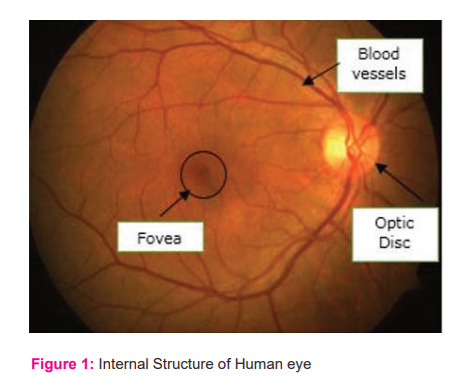
Retinal blood vessels comprise arteries and veins. Arteries of blood vessels bifurcate into branches of vessels. Retinal blood vessels are capable of carrying oxygen to different parts of the eye. The abnormalities of the retina that seems to appear in diabetic patients are elucidated in the following sections.
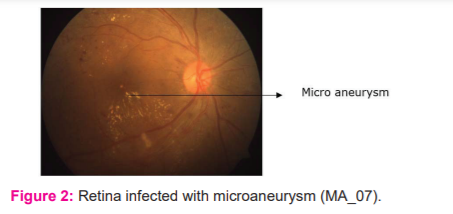
Micro Aneurysm
Micro aneurysms are the tiny blood spots in the capillaries of blood vessels of the retina. The presence of microaneurysm indicates the earlier stage of diabetic retinopathy in diabetic patients6. These abnormalities are always seen as groups or clusters. Sometimes it may occur in isolation also. The presence of microaneurysm can be identified by an eye dilation followed by screening, using fundus images. Figure 2 represents the retina image with microaneurysm which appears as white spots in the fundus images.
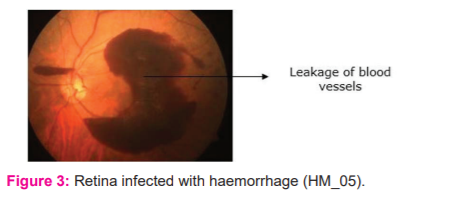
Hemorrhages
Haemorrhages are serious disorder that causes bleeding in the retina. In the advanced stage of diabetic retinopathy, haemorrhages might transpire which lead to the growth of abnormal new blood vessels. The new abnormal blood vessels may leak and the blood may occupy the central portion of the retina. Excessive blood leakage may cause retinal detachment which leads to the loss of vision. Figure 3 shows a retina image with a haemorrhage or leakage of blood.
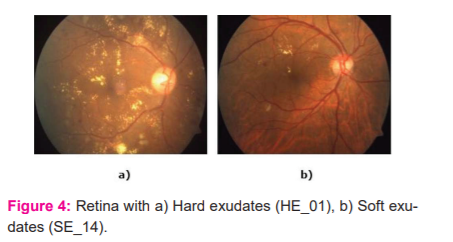
Hard and Soft Exudates
Hard exudates are lipids in the pale yellowish colour that leaks from the capillaries of abnormal retinal blood vessels. This condition is referred to as chronic leakage in diabetic retinopathy patients7. Soft exudates are otherwise called cotton wool spots which appear as a whitish fluffy fibre layer in the abnormal vessels of the retina.8 The presence of hard and soft exudates is more common in Diabetic Retinopathy patients. Figure 4 (a) and (b) represents retina with hard exudates and soft exudates respectively.
ABOUT THE HOSPITAL
Aravind eye hospital9is the renowned hospital established in the year 1976 in Madurai, Tamil Nadu, India. This hospital works as a network of hospitals in various districts in the state of Tamil Nadu. This hospital has performed phenomenal work in treating millions of people with eye defects including surgeries. Aravind eye hospital also does outreach programs in remote villages by conducting free eye camps.
Privacy Policy
For some security and privacy reasons, the information about the patient’s details is not divulged anywhere. All images in the dataset are compressed lossless and are stored in a repository in Github10.
DATA SET
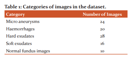
The images in this dataset are procured from a famous hospital in Tirunelveli city, South Tamil Nadu, India. The screening is done on patients between the ages of 30 and 70. This data set is divided into 5 different categories like (i) hard exudates, (ii) soft exudates, (iii) haemorrhages, (iv) microaneurysms and (v) retina with no defect. Each category comprises raw images obtained using the fundus camera. All the images are of high quality which includes colour images and few grayscale images. These images were acquired using an FF450 plus fundus camera with a 50ofield of view. The field of view of every image is circular. Each image is compressed in the format of JPEG. This dataset can be used for identifying different retinal diseases like hard, soft exudates, haemorrhages and detecting microaneurysms in diabetic patients. All the images in this dataset are used for clinical diagnosis. These images can be used alone as a standalone image or they can be used with the combination of other retinal images for comparative study. These images in this dataset can be used for testing image processing techniques such as enhancement, segmentation, etc. The number of images in each category is as follows: Table 1 recapitulates the categories of images in the dataset.
The description of the images is given in Table 2, including the name and size of the images in each category.
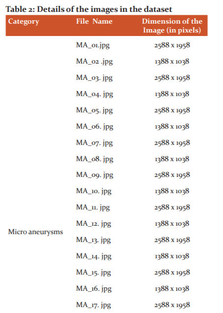
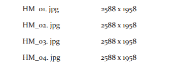
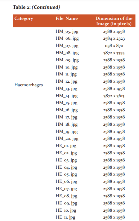
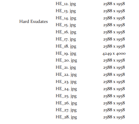
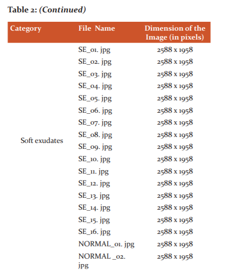

All the images in the dataset are compressed in JPEG format. The dimension of HM_14 (Hemorrhage category) is comparatively high because the retina is highly infected by the leakage of blood vessels. Similarly, an image in the hard exudates (HE_19) category also has a higher dimension due to the presence of a greater number of hard exudates. The images can be downloaded from the following URL:
https://github.com/ARAVINDDATASET/ARAVIND.git
SCOPE FOR COMPUTER RESEARCHERS
Using this dataset, the computer researchers can execute their research in the areas such as detection of abnormalities in the retina, segmentation of the blood vessels, identification of hard and soft exudates, localization of optic disc in the retina, classification, feature extraction, detection of haemorrhages and microaneurysm and grading the level of disease in the case of diabetic retinopathy patients.
DISCUSSION
Retina image analysis is an emerging research area for which many researchers are contributing by proposing new techniques for automatic diagnosis. At present, only very few Indian datasets are available for retina image analysis. There are hundreds of patients visiting the Aravind eye hospital, Tirunelveli every day for eye checkups and eye treatment. We have collected the retina images of various patients from the hospital and created a dataset that contains images of different categories like microaneurysm, haemorrhages, soft exudates, hard exudates and images with no defect. To motivate the research on retina image analysis, we would like to publish the database so that it could be used by the upcoming young researchers in the field of medical imaging. The dataset contains 98 retina images. This dataset is open to researchers for image analysis purposes.
CONCLUSION
The dataset “ARAVIND” is an image database that contains raw images of infected retina and retina with no defect. The images in this dataset can be used for image processing techniques and machine learning techniques to diagnose retinal disease. Using these images, the researchers can propose new algorithms for medical image analysis and thereby the usage of technology can be transferred to the field of medicine.
ACKNOWLEDGMENTS
We would like to acknowledge Aravind eye hospital, Tirunelveli, South Tamil Nadu, India for providing us with the dataset of retinal images. This dataset will help computer researchers to a greater extent to research in the field of medical imaging. We also acknowledge the immense help received from the scholars whose articles are cited and included in references of this manuscript. We are also grateful to authors, editors, and publishers of all those articles, journals and books from where the literature for this article has been reviewed and discussed.
Conflict of Interest: The authors declare there is no conflict of interest.
Funding Source: None
Ethical Clearance: There was no direct human intervention involved in this research.
Author’s Contribution: The images that are stored in the repository are real-time images that have been collected from Aravind Eye Hospital. Ground truth images are generated with the help of experts which can be used for evaluation purposes.
References:
1. Tang J, Kern T. Inflammation in diabetic retinopathy. Progress in Retinal and Eye Research. 2011; 30(5):343-358.
2. Zhang X, Saaddine J, Chou C, Cotch M, Cheng Y, Geiss L et al. Prevalence of Diabetic Retinopathy in the United States, 2005-2008. JAMA. 2010; 304(6):649.
3. Research E, Atlas I. IDF Diabetes Atlas [Internet]. Idf.org. 2021. Available from: https://www.idf.org/e-library/epidemiology-research/diabetes-atlas.html
4. Diabetic retinopathy - Symptoms and causes [Internet]. Mayo Clinic. 2021. Available from: https://www.mayoclinic.org/diseases-conditions/diabetic-retinopathy/symptoms-causes/syc-20371611.
5.[Internet].https://www.griswoldhomecare.com.2021.Availablefrom:https://www.griswoldhomecare.com/blog/2015/January/the-4-stages-of-diabetic-retinopathy-what-you-ca/
6. Tags C, Watch J, A, Glycosmedia A, Team E, questions F et al. Diabetic Retinopathy – Features of Diabetes: Microaneurysms [Internet]. Glycosmedia. 2021. Available from: https://www.glycosmedia.com/education/diabetic-retinopathy/diabetic-retinopathy-features-of-diabetes-microaneurysms/
7. Jaya T, Dheeba J, Singh N. Detection of Hard Exudates in Colour Fundus Images Using Fuzzy Support Vector Machine-Based Expert System. Journal of Digital Imaging. 2015; 28 (6):761-768.
8. Kavitha M, Palani S. Hierarchical classifier for soft and hard exudates detection of retinal fundus images. Journal of Intelligent & Fuzzy Systems. 2014; 27(5):2511-2528.
9. Home - Aravind Eye Care System [Internet]. Aravind Eye Care System. 2021 [cited 14 May 2021]. Available from: https://aravind.org
10.GitHub: Where the world builds software [Internet]. GitHub. 2021. Available from: https://github.com
|
 IJCRR
IJCRR 


 This work is licensed under a Creative Commons Attribution-NonCommercial 4.0 International License


