IJCRR - 13(20), October, 2021
Pages: 28-35
Date of Publication: 24-Oct-2021
Print Article
Download XML Download PDF
Role of High-Resolution Computed Tomography Temporal Bone in Evaluation of Patients with Chronic Suppurative Otitis Media (CSOM)
Author: Karegowda H Lakshmikanth, Singhal Rahul Kumar, Vinay Raju
Category: Healthcare
Abstract:Introduction: CSOM is chronic inflammatory changes of the tympanic cavity and one of the leading causes of chronic aural discharge which has potential for intracranial extension and associated middle ear complications. High-Resolution Computed Tomography (HRCT) is needed for the assessment of disease localisation, ossicular dignity, bony walls status and post-surgical assessment. Objective: To study the HRCT findings of the temporal bone in patients with CSOM and also to determine the efficacy of HRCT in patients with CSOM by comparing the preoperative findings with intraoperative findings (gold standard), whenever available. Method: Prospective observational study conducted over 14 months in 167 ears diagnosed with CSOM who underwent HRCT temporal bone, using Philips Incisive 128-slice MDCT scanner. The radiological findings were further compared with the intraoperative findings to calculate the sensitivity, specificity, negative predictive value, and positive predictive value(PPV) for diagnostic accuracy and further overall accuracy were calculated. Results: From a total of 167 ears, surgical and radiological findings showed a high level of sensitivity and specificity for location and extent of soft tissue density, ossicular status and tegmen erosion, tympanic membrane status, mastoid pneumatisation, scutum erosion with a p < 0.01 but less sensitive for facial canal erosion. Conclusion: HRCT scan is an excellent preoperative imaging modality for the otologist to predict disease during the surgery. Early identification of complications enables a better surgical approach and treatment plan thereby making high resolution computed tomography an essential workup diagnostic tool.
Keywords: High Resolution, CSOM, Otitis, Media
Full Text:
Introduction:
CSOM is a common condition seen in patients visiting the otolaryngology department. Cholesteatoma is a serious condition that can progressively enlarge and can be associated with complications because of erosion into surrounding structures like meninges, brain, internal carotid artery, jugular bulb and facial nerve.1-7
The diagnosis of aural cholesteatoma is made radiologically by Imaging methods, such as HRCT and MRI.HRCT findings suggesting cholesteatoma include scutum erosion, aditusadantrum widening, ossicles erosion, facial nerve canal erosion, tegmen erosion, mastoid destruction (automastoidectomy), sigmoid plate dehiscence, and bony external auditory canal erosion.8,9,10 Prior knowledge of the extension and the complications of cholesteatoma will help clinicians in surgical approach and treatment plan.
The study aimed to assess the role of HRCT temporal bone in the evaluation of patients with chronic suppurative otitis media (CSOM) patients with objectives being to study the HRCT findings of the temporal bone in patients with CSOM and also to determine the efficacy and role of HRCT temporal bone in patients with CSOM by comparing the preoperative findings with intraoperative findings (gold standard), whenever available.
Methods and design:
This prospective study was conducted at the Department of Radiodiagnosis and Imaging over 14 months (Aug. 2019 – November 2020). A total of 167 patients who were referred to the Department of Radiodiagnosis and clinically suspected of having CSOM, was the included subjects. Exclusion criteria were trauma or neoplastic growth involving the middle ear, previous middle ear operation, acute otitis media and granulomatous or residual or recurrent disease.
197 ears in 167 patients, clinically diagnosed with chronic suppurative otitis media were evaluated. Patients were referred to the Department of Radiodiagnosis for HRCT temporal bone which was performed using multi-slice detector Philips Incisive 128-slice CT scanner with serial 0.67 mm thin sections. Axial projections were obtained, Coronal and Sagittal reformatted images were obtained. For ossicular evaluation, the V reconstruction method was used.
Parameters of comparison were:
-
Soft tissue extension in the attic, aditus ad antrum, hypotympanum, mesotympanum, Prussak’s space, mastoid antrum, facial recess and sinus tympani.
-
Ossicular chain status.
-
Bony erosions involving scutum, facial canal, tegmen tympani, sigmoid plate, facial canal and lateral semi-circular canal.
-
Presence of cholesteatoma or granulation tissue.
-
Tympanum changes – Perforation/thickening/retraction.
-
Complications of cholesteatoma
Following surgery, these parameters were individually compared with surgical findings. These intra-operational results were regarded as the gold standard and correlated with radiological observations.
STATISTICAL ANALYSIS:
Comparative parameters between pre-operative HRCT and intraoperative findings were determined using Sensitivity(Sn), Specificity(Sp), negative predictive value(NPV) and positive predictive value(PPV) for diagnostic accuracy and overall accuracy were calculated and tabulated, taking surgery as the gold standard.
The association between radiological and intra-operative findings was tested using the Chi-square test to find out any statistically significant difference. A value of <0.001 was considered significant statistically. Data were analyzed using “Statistical Package for Social Sciences(SPSS) 22 version software. To acquire graphs such as bar diagrams, pie diagrams and scatter plots, MS Excel and MS word were used.
RESULTS:
197 ears in 167 patients, clinically diagnosed with chronic suppurative otitis media were evaluated; of which 58 ears underwent surgery, 101 ears were medically managed and 38 ears lost to follow up.
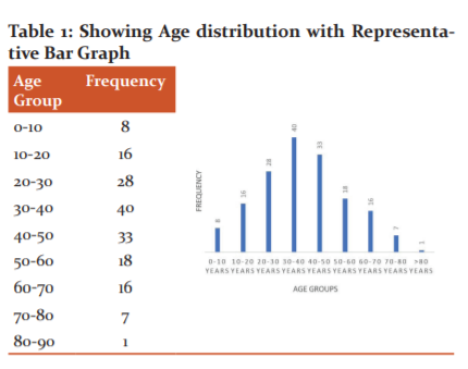
Most of the cases 47/197 (24%) in the study belong to the age group of 30 to 40yrs
SEX DISTRIBUTION:
Total of 105 males and 62 females were included in the study. Most of the affected population in the study were males 62.8%.
CLINICAL SYMPTOMS DISTRIBUTION:
In 197 ears, there were overlapping of different clinical symptoms and also some cases presenting with isolated symptoms, overall the most common presenting clinical symptom was chronic ear discharge seen in 149 ears followed by loss of hearing in 93 ears. Least common clinical manifestation was tinnitus seen in 25 ears.
Ear involvement:
Most common ears involved were found to be on the left side – 94 ears (48%), 73 right ears (39%) and 23 bilateral ears were involved
LOCATION AND EXTENSION OF SOFT TISSUE WITHIN TYMPANIC CAVITY:
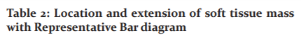
There was overlap in the extent of involvement of the soft tissue in the middle ear, in the form of holotympanic involvement or isolated attic/mesotympanum/hypotympanum involvement.
In the mastoid, soft tissue was seen extending to the aditus, aditus-ad antrum or antrum , with some cases showing whole mastoid involvement and also isolated aditus/antrum involvement. Overall, most common location of soft tissue density(STd) within the tympanic cavity on CT, was seen in the attic (88.8%), followed by mastoid antrum ~ 68% and aditus (~63.9%) whereas hypotympanum was least commonly involved location (~40.6%).
Soft tissue density(STd) extending into the external auditory canal involvement was seen in 66 ears.
TYMPANUM CHANGES:
Tympanum changes in chronic suppurative otitis media was observed as perforation seen in 88 ears, followed by thickening seen in 22 ears and retraction seen in 11 ears.
Tympanum perforation along with thickening was observed in 30 ears and perforation along with retraction in 8 ears.
Whereas tympanum retraction along with thickening was seen in 16 ears. Overall in toto, tymapnum perforation was the most common finding in 126 ears, followed by thickening in 68 ears and retraction in 35 ears.
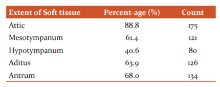
OSSICULAR EROSIONS:
Ossicular erosion was seen in found in 122 ears. Complete ossicular erosion was seen in 15 ears, both malleus and incus erosion in 12 ears, both incus and stapes erosion in 6 ears. Only incus was seen eroded in 11 ears, stapes in 8 ears and malleus in 7 ears. Overall in toto, Incus was the most frequently involved ossicle (36%) followed by Malleus erosion (28%) and then stapes (24%).
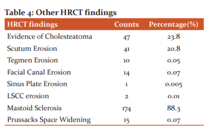
Among other HRCT findings, observation was made of the isolated complications and overlapping of different complications in many cases. Overall the most frequent finding was mastoid sclerosis obtained in 174 ears (88.3%). Presence of choleasteatoma was the second most common finding on HRCT seen in 47 ears (23.8%). Scutum erosion of was found in 41 ears (20.8%).Tegmen and facial canal erosion(FCE) was established in 10 and 14 cases respectively. Lateral semicircular canal(LSCC) erosion was found inn only 2 ears and the least common finding was Sinus plate erosion which was seen in only 1 case. Prussack’s Space widening due to the presence of soft tissue density(STd) in attic was seen in 15 ears.
OTHER COMPLICATIONS:
Among the cases of cholesteatoma, mastoid cortex erosion was found was in 7 cases, out of which 3 cases were confirmed intraoperatively, automastoid cavity formation was seen in 3 cases, all 3 cases were confirmed intra-operatively and thrombosis of sigmoid sinus was seen in 2 cases. Other complication like jugular bulb dehiscence was seen in 2 cases.
COMPARISION OF HRCT FINDINGS AND INTRA-OPERATIVE OBSERVATIONS:
The comparision was performed under following broad headings:
-
Tympanic membrane changes.
-
Soft tissue density in tymanic cavity.
-
Ossicular changes.
-
Prsence of Cholesteatoma
-
Other findings
“Chi square test was performed for all variables with degrees of freedom = 1, p<0.001. All parameters were found statistically significant.”
IMAGE GALLERY
CASE I:
A 43-year-old male came to ENT OPD with complaint of reduced hearing and right ear pain for 3 months. Clinically diagnosed as a c/o congenital cholesteatoma. He was sent to the Radiodiagnosis and Imaging department for HRCT of temporal bone(TB) prior to the surgery.
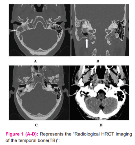
HRCT images - Axial (A.) & Coronal (B.) plane showed lesion of soft tissue density(STd) filling the right mastoid antrum and mastoid apex causing sigmoid sinus plate erosion and medial aspect of mastoid cortex. Contrast enhanced CT scan axial images (D) of brain showed lack of enhancement of right sigmoid sinus which was suggestive of sigmoid sinus thrombosis. Axial (C) HRCT image shows the presence of cholesteatoma in the right petrous apex.
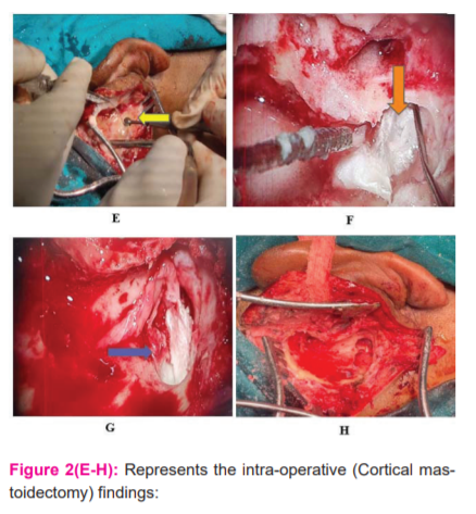
Image(E-H): Intraoperative imaging during Right Cortical Mastoidectomy(E) shows pearly white cholesteatoma (yellow arrow) in the right mastoid antrum and mastoid apex (F) (orange arrow) and right petrous apex (blue arrow - G). Image H shows post cortical mastoidectomy status after removal of cholesteatoma.
CASE II:
A 43-year-old male came to ENT OPD with complaint of reduced hearing and right ear pain for 3 months. Clinically diagnosed as unsafe variety of chronic suppurative otitis media. He was sent to the Radiodiagnosis and Imaging Department for HRCT of temporal bone(TB)
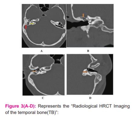
Axial (A) HRCT images showed right mastoid sclerosis and non-dependent soft tissue lesion (cholesteatoma) filling right mastoid aditus-ad-antrum and antrum. HRCT images in Coronal (B) and Axial (C) planes showed cholesteatoma surrounding the ossicles (malleus along with long process of incus). V reconstruction axial image (D) showed cholesteatoma surrounding the lenticular process of incus and supra-structure stapes leading to its improper visualization.
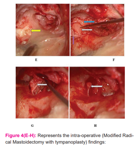
Image(E-H): Intraoperative imaging during shows pearly white cholesteatoma sac (yellow arrow) in the right mastoid antrum and image F shows cholesteatoma (purple arrow) enveloping incus (white arrow) and malleus (blue arrow) causing their erosion. In image G (white arrow) depicts cholesteatoma surrounding the stapes supra-structure. Image H depicts the viable stapes after removal of cholesteatoma.
CASE III:
Male patient, 35-year-old, arrived to ENT OPD by complaints of reduced hearing and ear pain on left side for 7 months. Clinically diagnosed as “cholesteatomatousattico-antral chronic suppurative otitis media.” He was sent to the Radiodiagnosis and Imaging Department for HRCT of temporal bone(TB)
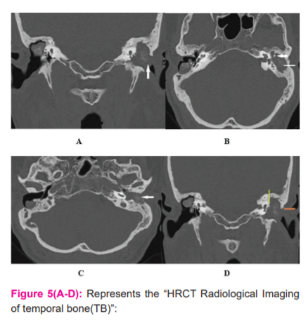
68years old male patient who had undergone previously modified radical mastoidectomy of the right ear with ball-shaped granulation tissue in a post-operative mastoid cavity. The Coronal (A, D) & axial (B., C.) HRCT scan demonstrated mixed density soft tissue lesion noted in the post-operative right mastoid cavity. CT mixed density with “cholesteatomatous” lesion seen in the left middle ear (Arrow in A) along with an “automastoid cavity” (thin arrow in B), and complete ossicular erosion (thick arrow in B) extending into the external auditory canal and causing erosion of its posterior wall (Arrow in Image C) with scutum erosion (orange arrow in Image D) and lateral semicircular canal erosion (yellow arrow in Image D)
DISCUSSION
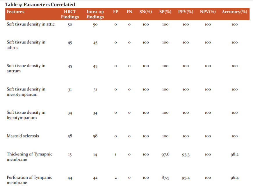
In our study, on radio-surgical correlation, HRCT proved to be 100% sensitive and accurate for the identification of the extent and location of soft tissue density in the tympanic cavity and mastoid, with zero false positive/negative cases. Almost similar results were demonstrated by Chatterjee et al.,2 Sreedhar et al.3 and Kanotra et al.,4
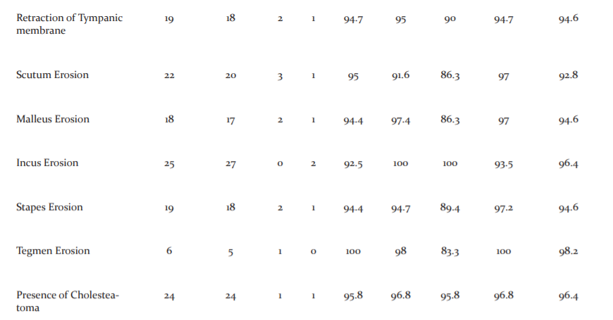
On radio-surgical correlation, HRCT proved to be 100% sensitive for thickening and perforation of the tympanum, however slightly less specific (87.5%) for perforation of the tympanum. For retraction of the tympanum also proved highly sensitive and specific (94.5% and 97.5% respectively).
In our study, HRCT accurately projected 92.5% of cases of incus erosion which was the most commonly eroded ossicle in our study, this result is almost similar to information by Chatterjee et al.2 and Sreedhar et al.3 HRCT was found to be 92.5% sensitive and 100% specific for incus, which was comparable to findings by Chatterjee et al.2 and who found it 98.3% sensitive and 100% specific, and Prakash MD5 et al. who found 95.8% sensitivity and 100% specificity.
Malleus was the second most common eroded ossicle, HRCT showed high sensitivity of 94.4% and specificity of 97.4% for malleus erosion, with 2 false-positive cases and 1 false-negative case. Chatterjee et al.2 have reported almost similar findings in their study, showing 93.6% sensitivity and a specificity of 100%.
Stapes was the least eroded, with the sensitivity of ~94.4% and specificity of 94.7%, with 2 false positives and one false negative case which was higher as compared to studies conducted by Chatterjee, et al2 which showed the sensitivity of 85.6% and specificity of 89.8% and Prakash MD, et al5 which showed the sensitivity of 81.8% and 88.8%, which can be attributed to the “V” special ossicular reconstruction used in our study.
The sensitivity(Sn) and specificity(Sp) obtained for the presence of cholesteatoma on HRCT was high, 95.8% and 96.8% respectively, with 1 false-positive and 1 false-negative case. This aligns with a study conducted by Chatterjee et al.,2 which displayed a sensitivity of and specificity of 93.6% and 100% respectively, and Chidambaram et al.,6 which showed a sensitivity of 94% and specificity of 90%.
For mastoid sclerosis, HRCT demonstrated a very high sensitivity and specificity of 100%.
In our study, high-resolution CT of temporal bone presented with the erosion of tegmen tympani in 6 patients, out of which 5 cases were confirmed intraoperatively, with 1 false-positive case, with a high sensitivity of 100% and 98% specificity. Comparable outcomes were demonstrated by Chatterjee et al., and Prakash MD et al., in their study.
In our study, high-resolution CT of temporal bone presented with the erosion of scutum in 22 patients, with 3 false-positive and 1 false negative, thus overall sensitivity of 95.0% and specificity of 91.3% was achieved, which were by results demonstrated by Sreedhar et al., and Chidambaram et al.,
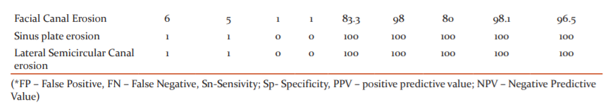
HRCT showed facial canal erosion in 5 cases, out of which in 4 patients findings were confirmed intraoperatively, with one false negative case and 1 false-positive case. Therefore, we got sensitivity(Sn) of 80.1% and specificity(Sp) of 98% for facial canal erosion by HRCT. The low sensitivity and relatively high specificity which our study showed, is following a similar study conducted by Prakash MD et al.,5 which demonstrated sensitivity(Sn) and specificity(Sp) of 80% and 100% respectively. Therefore, this indicates the limited role of HRCT to diagnose facial canal erosion, however, an important feature to note is that HRCT can exclude the absence of facial canal erosion with high accuracy.
Sinus plate erosion and lateral semi-circular canal erosion was found in 1 case with 100% sensitivity and specificity, by a study conducted by Chidambaram et al., which demonstrated sensitivity(Sn) of 100% and specificity(Sp) of 99.2% respectively.
Facial recess and sinus tympani changes could not be correlated as it was surgically a blindspot.
The limitations of the study were
-
The sample size was limited due to the time-bound nature of the study.
-
In distinguishing a cholesteatoma mass from granulation tissue, the HRCT scan is less sensitive.
CONCLUSION
In our study, the correlation between radiological and post-surgical findings concerning the extension of soft tissue, bony boundaries of tympanic cavity, pneumatisation status of mastoid, tegmen erosion, ossicular chain(OC) status and lateral semi-circular canal(LSCC) fistula showed high sensitivity and specificity. It was less sensitive, however to facial canal erosion.
HRCT is the best imaging tool for the learning of suppurative diseases of the tympanic cavity and thus have a vital role in pre-surgical assessment and management advice.
Acknowledgements: Dr VinayRaju, Dr Rajagopal KV, Dr PrakashiniKoteshwar
Competing interests: No competing interests exist.
Authors: Dr Rahul Kumar Singhal1, Dr Lakshmikanth H K2
1Post graduate student, Department of Radiodiagnosis and Imaging, Kasturba Medical College, Manipal University, Karnataka 576104, India
2Associate Professor, Department of Radiodiagnosis and Imaging, Kasturba Medical College, Manipal University, Karnataka 576104, India
Funding: Nil
Disclaimer: The views expressed in the submitted article are my own and not an official position of the institution.
Authors Contribution:
1st n Corresponding author H Karegowda H Lakshmikanth – Principal Investigator
2nd author Dr Singhal Rahul Kumar – Lead investigator
3rd author Dr VinayRaju – Statistics and methodology.
References:
-
Lane JI, Lindell EP, Witte RJ, DeLone DR, Driscoll CLJR. Middle and inner ear: improved depiction with the multiplanar reconstruction of volumetric CT data. 2006;26(1):115-24.
-
Chatterjee P, Khanna S, Talukdar RJIJoO, Head, Surgery N. Role of high resolution computed tomography of mastoids in planning surgery for chronic suppurative otitis media. 2015;67(3):275-80.
-
Sreedhar S, Pujari K, Agarwal AC, Balakrishnan RJIJoO. Role of high-resolution computed tomography scan in the evaluation of cholesteatoma: A correlation of high-resolution computed tomography with intra-operative findings. 2015;21(2):103.
-
Kanotra S, Gupta R, Gupta N, Sharma R, Gupta S, Kotwal SJIJoO. Correlation of high-resolution computed tomography temporal bone (TB) findings with intraoperative findings in patients with cholesteatoma. 2015;21(4):280.
-
Prakash M, Tarannum AJIJoO, Head, Surgery N. Role of high resolution computed tomography of the temporal bone(TB) in preoperative evaluation of chronic suppurative otitis media. 2018;4(5):1287.
-
Karpagam B. High-Resolution CT Imaging in Pathologies of Temporal bone(TB).
-
Shah CP, Shah PC, Shah SD. Role of HRCT temporal bone in the pre-operative evaluation of cholesteatoma. Int J Med Sci Public Health 2014;3:69-72
-
Niveditha J, Chidananda R. Clinical study of the correlation between preoperative findings of HRCT with intra-operative findings of cholesteatoma in cases of CSOM. Indian J Anatomy Surgery Head, Neck Brain 2017;3(1):1-5.
-
Zeifer B. Radiology of the temporal bone. In: Thomas R, Staecker H, editors. Otolaryngology, basic science and clinical review. 1st edition. Thieme Medical Publishers; 2006: 431–442.
-
Mohammadi G, Naderpour M, Mousaviagdas M. Ossicular Erosion in Patients Requiring Surgery for Cholesteatoma. Iranian J Otorhinolaryngol. 2012;24(68):125–8.
|
 IJCRR
IJCRR 


 This work is licensed under a Creative Commons Attribution-NonCommercial 4.0 International License


