IJCRR - 7(20), October, 2015
Pages: 09-11
Date of Publication: 20-Oct-2015
Print Article
Download XML Download PDF
A STUDY OF FORAMEN OF ARCUALE IN ATLAS VERTEBRA: INCIDENCE AND CLINICAL CORRELATIONS
Author: Patel Dinesh K., Shinde Amol A., Roy Nilanjan, Manvikar Purushottam R., Bharambe Vaishaly
Category: Healthcare
Abstract:Introduction: Atlas is the first cervical vertebra. Foramen tranversarium transmits vertebral vessels and sympathetic plexus. An additional foramen on the transverse process has been named foramen of Arcuale or Arcuate foramen. Aim: To find incidence and clinical correlation of foramen of Arcuale. Methodology: 32 atlas vertebrae from various colleges of Maharashtra were studied in the present study for presence of variant foramen. Results: Foramen of Arcuale was seen bilaterally in 3 atlas vertebrae giving a incidence of 9.37%. There were no other abnormalities in the bones. Conclusion: The course of the vertebral artery may be distorted with this extra foramen. This variation has been a known contraindication for C1 lateral mass screw fixation procedures. Knowledge of this variation will prove helpful for radiologists during CT and MRI and neurosurgeons during cervical surgery.
Keywords: Foramen of Arculae, Arcuate foramen, Atlas, Vertebral artery, Foramen transversarium
Full Text:
INTRODUCTION
Atlas is the first cervical vertebra. It is a ring shaped vertebra with anterior and posterior arch but no body. Normally we see a single foramen transversarium bilaterally on the transverse process. It transmits vertebral artery, vertebral vein and sympathetic plexus. The third part of the vertebral artery leaves the foramen transversarium on its way to the cranial cavity. It grooves the superior surface of the posterior arch of atlas vertebra, just behind the superior articular facet. The groove is called the groove for vertebral artery or sulcus arteria vertebralis. It transmits vertebral artery and suboccipital nerve. Sometimes the groove is converted into a foramen by an osseous bridge called foramen of Arcuale or Arcuate foramen. It has also been named as Kimmerle’s anomaly, foramen sagittale, foramen atlantoideum, foramen retroarticulare superior, canalis vertebralis. [1] [2] The Aim of this study is to investigate the incidence of extra foramina in the Atlas vertebrae and to discuss its clinical importance.
MATERIAL AND METHODS
32 atlas vertebrae from various medical and dental colleges of Maharashtra were studied for variations in foramen transversarium. Any Variation in number and position of foramen transversarium were noted
RESULTS
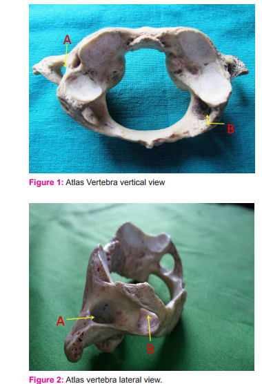
Extra foramen on transverse process were seen bilaterally in 3 atlas vertebrae giving an incidence of 9.37% . No “incomplete canal for vertebral artery” or “retrotransverse foramen” were observed. No other abnormality was seen in each of these bones. The foramina were situated at the lateral part of the posterior arch behind the lateral mass as seen in Figures 1 and 2.

Showing A – Foramen transversarium and B – Foramen of Arcuale
DISCUSSION
When the groove for vertebral artery (while arching over the posterior arch of atlas vertebra), gets converted into an osseous tunnel by formation of a bony arch over it, the so formed foramen through which the artery now traverses is called as arcuate foramen. Such a foramen can be a complete foramen or it can be in form of an incomplete arch. [3,4]. Patel et al [5] in a study of atlas vertebra in Gujarat region give a 13% incidence of foramen of Arcuale which is higher than 9.37% incidence reported by present study. Krisnamurthy A et al [1] give a 8.33% incidence of foramen of Arcuale in atlas vertebra. This incidence coincides with present study. They conclude that this variation should be considered in patients with symptoms of vertebrobasilar insufficiency like shoulder pain vertigo and headache. A study of 60 atlas vertebra by Ozgur Ckmak et al in Turkey give a 11.6 % incidence of arcuate foramen which is more than incidence of our study. They state that patients with arcuate foramen have many complaints which may be due to compression of the sympathetic plexus around the vertebral artery. So presence of arcuate foramen should be considered in patients of vertigo, headache, shoulder pain and neck pain. A cervical spine radiography is indicated in such cases.[6] Brown and Verheyden [7] have described a case of a 1 year old boy who underwent surgery for cleft palate and suffered “posterior fossa infarction” following the surgery. A threeview cervical spine radiographic series showed no fracture or subluxation, with the disk spaces appearing normal. A questionable calcific density was however observed superior to the posterior arch of C1. This could be because of possible presence of a arcuate foramen. They stated that some surgeons prefer to operate on cleft palate with the neck in full extension. In this position it is possible that the extension of the neck in presence of an arcuate foramen, could lead to bilateral occlusion of the vertebral arteries resulting in “posterior fossa infarction”. They have therefore suggested that during such surgeries the neck be extended only as far as is necessary to perform the procedure, rather than extending it more to make the repair easier on the surgeon. Cushing K et al [8] give a relation between tethering of vertebral artery in the foramen of arcuale and dissection of the artery from repetitive trauma during neck movements. P Potaliya et al [9] gave a 13.33 % incidence of accessory foramen transversarium .They state that this finding could be helpful to orthopedic surgeons, neurosurgeons and radiologists to avoid misdiagnosis in their clinical practice. A qualitative analysis of atlas vertebra by C. Gupta et al gives a Incidence of 5.7%. Presence of bridges to form foramen on the groove may indicate ossification of posterior atlanto occipital membrane. This variation may predispose to vertebrobasilar insufficiency and cervicogenic syndromes on strenuous neck movements.[10] S.S. Baeesa et al in a study of Saudi population gave a 16 % incidence of foramen of Arcuale. They attributed various cases of stroke, subarachnoid bleeds, vertebrobasilar insufficiency to presence of foramen of Arcuale. They state that theories on formation of the foramen are controversial and that some authors have reported presence of this foramen even in fetuses and children where it was present in cartilaginous form indicating a congenital origin. Another theory stated that the foramen could develop as a result of degenerative calcification of atlanto-occipital membrane. The vertebral artery accompanied by venous plexus, periarterial sympathetic plexus, suboccipital and 1st cervical nerves passes through the bony foramen arcuale. The canal can therefore compress any or all of these structures as the neck rotates. So radiographic study is indicated in cases of spine surgery and screw fixation in the atlas vertebra.[3] Michael J Huang and John Glaser state that C1 lateral mass screw fixation is contraindicated in patients with foramen of arcuale. Preoperative radiographs are indicated to avoid endangering the vertebral artery during screw fixation. Also the foramen of arcuale is known to be an associated factor in cases of migraines and vertebrobasilar artery stroke.[11] In a study of south Indian population, R Agrawal et al give an incidence of 10.7 % for foramen of Arculae. This incidence coincides with present study (9.37%). They opine that this abnormal foramen can cause compression of vertebral artery resulting in compromised blood flow.[12] Kwaiatkowska et al did a morphometric study of foramen transversarium in Poland. Vertebral and basal arteries supply blood directly to the brain and the spinal arteries. Any abnormality in course of vertebral arteries may hence lead to deficient blood supply to the cerebellum and the brainstem posing a threat to the vascular-cerebral system.[13]
CONCLUSION
Vertebral artery passes through the foramen transversarium of atlas before entering foramen magnum. An extra foramen will cause additional external pressure on the artery. Presence of foramen of Arcuale have been noted by studies in various countries giving a incidence from 8 to 16 %. We found a incidence of 9.37% .Presence of foramen of Arcuale should be considered in patients of vertigo, headache, migraine, shoulder and neck pain. Cervical spine radiography is indicated in cases of C1 lateral mass screw fixation and other spine surgeries to avoid damage to the vertebral artery. Excess extension of neck should also be avoided during surgery to avoid compression and resulting tethering of 3rd part of vertebral artery in case of presence of foramen of Arcuale.
ACKNOWLEDGEMENT
The Authors acknowledge the immense help received from the scholars whose articles are cited and included in references of this manuscript. The authors are also grateful to the authors/editors/ publishers of all those articles, journals and books from where the literature for this article has been reviewed and discussed.
Source of funding: As this study was carried out in the dissection hall of our Department, there was no separate financial aid provided for it.
Conflict of interest: There is no conflict of interest
References:
1. A. Krishnamurthy, S. R. Nayak, S. Khan, Latha V. Prabhu,Lakshmi A. Ramanathan, C. Ganesh Kumar, Abhishek Prasad Sinha arcuate foramen of atlas: incidence, phylogenetic and clinical significance Romanian Journal of Morphology and Embryology 2007, 48(3):263–266.
2. Abduelmenem Alashkman and Roger Soames, Bilateral foramina on the posterior arch of atlas, Rev Arg de Anat Clin,2014,6(2): 90-94.
3. S.S. Baeesa, F. Rakan . Bokhari, M. Khalid, Bajunaid, J. Mohammad, Al-Sayyad, Prevalence of the foramen arcuale of the atlas in a Saudi population, Neurosciences 2012’ 17 (4): 345-351
4. G. Paraskevas, B. Papazioga, A Tzaveas, K Natsis, S Spanidou, P. Kitsoulis, Morphological parameters of the superior articular facets of the atlas and potential clinical significance. Surg Radiol Anat. 2008 Nov;30(8):611-7.
5. Z. Patel, A Zalawadia, C.A. Pensi C A . Study of Arcuate Foramen in Atlas Vertebrae in Gujarat Region. NJIRM. 2012; 3(2): 73-75.
6. O. Cakmak, E. Gurdal, G. Ekinci, E. Yildiz, S. Cavdar, Arcuate foramen and its clinical significance, Saudi Med J, 2005, 26(9):1409-1413.
7. M. Brown and C. Verheyden. Posterior Fossa Infarction following Cleft Palate Repair and the Arcuate Foramen. Plastic and Reconstructive Surgery. 11/2009; 124(5):237-9.
8. K.E. Cushing, V. Ramesh , D. Gardner-Medwin, N.V.Todd,A. Gholkar, P. Baxter., P.D. Griffiths, Tethering of the vertebral artery in the congenital arcuate foramen of the atlas vertebra: a possible cause of vertebral artery dissection in children, Dev Med Child Neurol, 2001,43(7):491-496.
9. P. Potaliya, A. Dadhich, D.S. Chawdhary, Accessory foramen transversarium and it’s incidence in atlas vertebrae, Indian Journal of Clinical Anatomy and Physiology,2014,1(1):8-9.
10. C. Gupta, P. Radhakrishnan, V. Palimar, A.S. D’souza, N.L. Kiruba, A quantitative analysis of atlas vertebrae and its abnormalities, J. Morphol. Sci., 2013, vol. 30, no. 2, p. 77-81.
11. M.J. Huang and J.A. Glaser, Complete Arcuate Foramen Precluding C1 Lateral Mass Screw Fixation in a Patient with Rheumatoid Arthritis: Case Report, Iowa Orthop J. 2003; 23: 96-99.
12. R. Agrawal, S Sanathi, S. Agrawal, K. Usha. Posterior arch of atlas with abnormal foramina in south Indians.JASI, 2012,61(1):30-32.
13. B. Kwiatkowska, J. Szczurowski, D. Nowalkowski, Variation in foramina transversaria of human cervical vertebrae in the medieval population from Sypniewo (Poland), Anthropological Review’2014 ,77 (2), 175-188.
|


 This work is licensed under a Creative Commons Attribution-NonCommercial 4.0 International License