IJCRR - 13(19), October, 2021
Pages: 76-79
Date of Publication: 11-Oct-2021
Print Article
Download XML Download PDF
Cytological Patterns of Cervical Smear Seen at Federal Medical Centre Asaba, Delta State in Nigeria
Author: Anibor Ese, Obaroefe Martins, Jaiyeoba-Ojigho Efe Jennifer, Maidoh Maryjoy Anene
Category: Healthcare
Abstract:Introduction: The cervical smear test is the most commonly carried out cytology test for women and has been proven to help identify the emergence of cancer in the cervix. Aim: This study is aimed at evaluating various cytological patterns as well as their relationship with age. Methodology: This study employed a retrospective study design. Purposive sampling was used to collect the histological results of 82 patients seen for 4 years at the Federal Medical Centre (FMC) Asaba, Delta State in Nigeria. Ethical approval was sought from the Research and Ethics Committee of the Department of Human Anatomy and Cell Biology, Delta State University, Abraka in Nigeria. Data collected were analyzed via the Statistical Package for the Social Sciences (SPSS version 23), Chi-square test was adopted to help determine the association between observed variables. Results: Findings from this study showed patients with the ages of 31-40 years (42.7%) being the most frequently affected with cytological cervical disorders while the least was those within the ages of 41-50 years (28.0%). The preponderance of cervical smear of normal cervical lesions (61.0%) ranked the highest which was followed by Inflammatory cells (20.7%) with the least been squamous cells (1.2%). It was also evaluated from the findings that no significant association existed between age and cervical lesion patterns (p=0.732). Conclusion: From this study, it can be concluded that patients within the ages of 31-40 years of age (42.7%) have a high susceptibility to cytological cervical disorders compared to other age groups. The inflammatory cell pattern was the most commonly observed cytological disorder of the cervix while the least was squamous cell carcinoma. The pattern of cervical smear was not notably associated with the age of the patients.
Keywords: Asaba, Cytological, Cervical, Patterns, Smear
Full Text:
Introduction
The cervix as an anatomical organ is made up of tissues that are primarily composed of cells; such tissues include the endocervical mucosa, a conglomerate of single-layer columnar mucous cells. These cells have the necessary amount of mucus required for lubrication of cervical walls during copulation.1 The ectocervix is also another tissue present in the cervix which consists of non-keratinized squamous epithelium which joins with the vaginal wall, this tissue helps prevent erosion of cells in the ectocervical region of the cervix. 1, 2 The cervix has fibrous tissue such as elastin (which provides the elastic properties of the cervix), collagen (which provide the structural rigidity and strength of the cervix).1
The cervix is structurally the link/passage between the uterus and vagina.2 Its neurovascular supply entails the uterine artery, uterine vein, external iliac lymph nodes and lastly the pelvic splanchnic nerve.3 It functions as a pathway for sperm entry to the uterus during copulation and also in childbirth.2 The cervix like all structures of the human body is also predisposed to abnormal cell growth of which cervical cancer is the most common condition as regards the cytological disorders of the cervix.4
Examples of such cytological disorders of the cervix are as follows: atypical glandular cell, (glandular cell of cervix deviation from the normal cytological structure/pattern of glandular cell in cervix, studies conducted by several researchers it was observed that atypical glandular cell had an incidence of 0.1-2.1% among cervical lesions),5 Atypical squamous cell, (this simply refers to inflammatory, reactive abnormal cells lining the ectocervix, they are usually associated with infections such as papillomavirus, yeast infection etc and also hormonal imbalance and benign growth of cervix. In a study conducted by Mahira and Elmir, it was observed that in an examination of 1784 female patients in Bosnia and Herzegovina only 1.7% had atypical squamous cell as a cervical cytological abnormality.6, 7 High and low grade squamous intraepithelial lesions (these refer to moderate to severe or fewer changes of cervical cells), this cytological aberration in the cervix constituted 1.3 and 1.5% in researches conducted by Mahira and Elmir also that of Tamboli respectively. 6, 8
Squamous cell carcinoma is a deviant of the normal cervical cytology which arises as a result of abnormal cell growth and changes of cervical cells such as metaplasia of cervical squamous cell, hyperplasia and abnormal production of the cervical cells. 9, 10 According to WHO, about 80% of all known cases of cervical cancer worldwide are associated with developing or low-resources countries, which can be attributed to the lack of awareness and difficulty in running cytological based screening. 11 This condition is a major health problem worldwide with a significant level of morbidity and mortality, in a recent note there has been a decline in the prevalence level of this condition in developed countries.12 Cervical Cancer ranks third (3rd) globally among female associated malignancies which are predominant in developing countries. 13
Studies have shown those below 40 years of age with the possibility of developing cervical cancer as 2%. It has also been observed that an estimate of 500,000 new cases appears each year globally, with it being the second rank most usual cause of death in the world associated with the female gender with breast cancer ranking first (1st).13 Over the years, researches have been conducted on the means and methods of identifying this condition; one of such methods is the commonly conducted Cervical smear test also known as Papanicolaou (Pap) test, a simple, non-invasive screening test conducted by examining cells of the cervix to help evaluate if there are any cytological abnormalities in the cell/tissue structure of the cervix.14 This method was introduced by George Papanicolaou in the year 1914, aimed at examining the cells of the cervix histologically to help early discovering abnormal cell development in the cervix.15 This study aimed at evaluating the various cytological patterns of cervical smear with relation to age at the Federal Medical Centre, Asaba, Delta State in Nigeria.
Material and Method:
Ethical Consideration: The ethical clearance used was issued by the Research and Ethics Committee Human Anatomy Department, Faculty of Basic Medical Sciences, Delta State University, Abraka. The ethical approval number is DELSU/CHS/ANA/2020/34.
Study Design: The study design adopted for this research was a retrospective cross-sectional study design.
Sampling Technique and Sample Size: Purposive sampling was employed for sample collection. The study population comprised 82 patients who came for cervical smear screening at the Federal Medical Centre, Asaba, Delta State, Nigeria within a period of 4 years (2016-2019).
Data Collection: Data were obtained from the medical records unit of the Pathology Department, and Gynaecology Clinic registers. Data on the age of patients and cytology results of their pap smear were recorded.
Data Analysis: Data were analyzed and the results were presented in tables and charts. A Chi-square test was used to evaluate the association between age and patterns of cervical smear observed with a probability acceptable at less than 0.05.
Results
Figure 1 showed that the most affected age group with cervical lesions are those with the ages of 31-40 years with a frequency of 42.7%, which was closely followed by those within the age group of 20-30 years with a frequency of 29.3%, the least prevalent age with this condition were those within the ages of 41-50 years of age (28.0%).
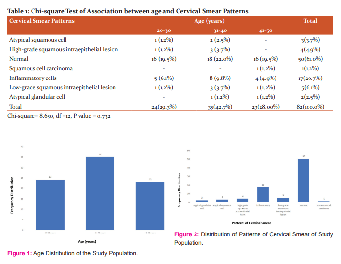
Figure 2 revealed the distribution of the various patterns of cervical smear observed in the study. Normal pap smear had a frequency of 61.0%, Inflammatory pattern was the most predominant cervical lesion with a frequency of 20.7%. The rest observed patterns are arranged as follows in order of preponderance; Low-grade squamous intraepithelial lesion, High-grade squamous intraepithelial lesion, Atypical squamous cell, atypical glandular cell and the least common being Squamous cell carcinoma with their respective frequencies as follows 6.1%, 4.9%, 3.7%, 2.4% and 1.2%.
Table 1 depicts that there was no significant association (p<0.05) between age and cytological pattern of cervical smear observed.
Discussion
The cervix like all structures of the human body is also predisposed to abnormal cell growth of which cervical cancer is the most common condition as regards the cytological disorders of the cervix.2
Findings from this study as regards age and cytological disorders of the cervix are in line with a study conducted Researchers who reported females of 30-45years age bracket to be predominant with cervical disorders after cytological examination of female patients at the Obstetrics and Gynaecology Department of the National Institute of Unani Medicine, Bangalore, India.14 Our finding is also in tandem with the submissions of Nepalese which indicated that most patients who had undergone cytological examination in the Tertiary Hospital of Nepal were those in the age group of 31-40years.16 But these findings do not conform with studies conducted by Nigerians who reported ages of 41-50 years to be the most prevalent age group with high susceptibility to abnormal cervical cytology.17
Results from this study on evaluation of the preponderance of the cervical lesion patterns were in agreement with results obtained from studies conducted by Nigerians which showed Inflammatory smear as the most common and squamous cell carcinoma as the least lesion.17 Similar results were also observed in studies conducted by Indians, Nigerians and a Saudi Arabian.9,17,18 However, results obtained from studies conducted by Asians are contradictory when compared with findings from this study, the Asians observed that atypical squamous cell was the most common cervical cytological abnormality.19 Also, other studies conducted by Americans, Tunisians, Northern Nigerians and Western Nigerians depicted squamous cell carcinoma as the most common lesion with respective frequencies of 92%, 95%, 93%, 85.7%. 4,20,21,22
Findings from this study as regards the test of association between age and patterns of cervical smear was similar to result obtained from studies conducted by Indians, Nigerians and a Saudi Arabian who submitted that there was no significant (p>0.05) association between age and patterns of cervical smear.9, 17, 18 However, findings from studies conducted by a South African as well as Nigerians showed that age and pattern of cervical smear had a significant (p<0.05) association, hence their submissions do not conform with the findings of this current study.23, 24 Racial and geographical differences could be attributed to these observed variations from our study.
Conclusion
The patterns of the cervical smear of patients seen at Federal Medical Centre, Asaba has been studied. The inflammatory cell pattern was the most commonly observed cytological disorder of the cervix while the least pervasive was squamous cell carcinoma. The finding from the study revealed no association between the pattern of cervical smear as regards the ages of the patients.
Conflict of Interest
The authors declare no conflict of interest
Source of Funding
Nil
Author’s Contribution
Anibor Ese: Concept of the study design, Manuscript drafting and preparation
Obaroefe Martins: Data collection, Data analysis, Manuscript drafting and preparation
Jaiyeoba-Ojigho Efe Jennifer: Data collection and manuscript preparation
Maidoh Maryjoy Anene: Data collection and Data analysis.
References:
-
Gray H. Gray's Anatomy (38th ed.). Churchill Livingstone; 2008, pp. 1870–73.
-
Guyton, AC and Hall JE. Textbook of Medical Physiology (11th ed.). Philadelphia, PA: W.B. Saunders, 2005;1027.
-
Drake RL, Vogl W and Tibbitts AWM. Gray's Anatomy for Students. Philadelphia, PA: Elsevier/Churchill Livingstone. 2005; 415, 423.
-
Chris-Ozoko LE, Barovbe MU, Oyem JC and Ekanem VJ. 5-Year Retrospective Study of the Prevalence of Cervical Lesions at the University of Benin Teaching Hospital, Nigeria. Acta Sci Anat. 2020; 1(4): 235-243.
-
Marques JP, Costa LB, Pinto AP, Lima AF, Duarte ME, and Barbosa AP. Atypical glandular cell and cervical cancer: Sys Rev. 2011; 57:234-8
-
Mahira J and Elmir J. Diagnostic Approach to patients with Atypical Squamous Cells of Undetermined Significance cytological findings on the cervix. Med Arch. 2016; 70(4):296-298.
-
Toews HA. The Abnormal Pap Smear: A Rationale for Follow Up. Can Fam Physician. 1983; 29:759-62
-
Tamboli GD. Accuracy of Cytological Findings in Abnormal Cervical Smear by Cyto-histological Comparison. J Med Edu Res. 2013; 3(2):19-24.
-
Pushp LS, Meenakshi S, Munna LP, and Rekha S. A Study on Cervical Cancer Screening Using Pap Smear Test and Clinical Correlation. Asia Pac J Oncol Nurs. 2018; 5:337-41.
-
Patel MM, Pandya AN, and Modi J. Cervical Pap Smear Study and its Utility in Cancer Screening, to Specify the Strategy for Cervical Cancer Control. Natl J Community Med. 2011; 2:49?51.
-
Ferlay J, Soerjomataram I, Dikshit R, Eser S, Mathers C and Rebelo M. Cancer Incidence and Mortality Worldwide: Sources, Methods and Major Patterns in GLOBOCAN 2012. Int J Cancer. 2015; 136:359?86.
-
Juneja, A., Sehgal, A., Sharma, S.and Pandey, A. Cervical Cancer Screening in India: Strategies Revisited. Indian J Med Sci. 2007; 61:34-47.
-
Gyawali B, Keeling JJ, Teijlingen E, Dhakal L and Aro AR. Cervical Cancer Screening in Nepal: Ethical Considerations. Medico-legal Bioethics. 2015; 1-4.
-
Bangalore NR and Arshiya S. Cytopathological Study of Cervical Smear: A Hospital-Based Retrospective Study. Med J Islamic World Aca of Sci. 2014; 22(1): 42-49.
-
Padubidri V and Daftary SN. Gynaecological Diagnosis. In: Howkins and Bourne Shaw’s textbook of Gynecology. 13th ed. Noida: Elsevier India Private Ltd; 2004; 77-187.
-
Pragya GG, Durga BCR, Kavita S, Kamar J. and Richa S. Spectrum of Cytological Patterns in Cervical PAP Smears in a Tertiary Care Center of Western Region of Nepal. Nepal J Med Sci. 2019; 4 (1): 2-8.
-
Adepiti CA, Ajenifuja KO, Okunola O, Omoniyi-Esan GO. and Uche O. Age and Pattern of Pap smear Abnormalities: Implications for Cervical Cancer Control in a Developing Country. J Cytol; 2017; 34 (4): 208-211.
-
Fadwa JA. Cervical Cancer Screening with Pattern of Pap Smear. Review of Multicenter Studies. Saudi Med J. 2006; 27 (10): 1498-1502.
-
Bhagya LA, Prasad UMS. and Satish K. Cytological Patterns of Cervical Pap Smear with Histopathological Correlation. Int J Res Med Sci. 2015; 3 (8): 1911-1916.
-
Missaoui N, Trabelsi A, Landolsi H, Jaidaine L, Mokni M. and Korbi S. Cervical adenocarcinoma and squamous cell carcinoma incidence trends among Tunisian women. Asian Pac J Cancer Prev. 2010; 11:777?780.
-
Mohammed A, Ahmed SA, Oluwole OP. and Avidime S. Malignant tumours of the female genital tract in Zaria, Nigeria. Ann Afr Med. 2006; 5:93?96.
-
Babarinsa A, Akang EE. and Adewole IF Pattern of gynaecological malignancies at the Ibadan cancer registry (1976?1995). Nig Q J Hosp Med.; 1998, 8:103?106.
-
Denny L. Cervical Cancer: Prevention and Treatment. Discov Med. 2012; 14(75):125-31.
-
Mosuro AO, Ajayi I, Ademola AO, Adeniji OA and Oluwasola O. Prevalence of Cervical Dysplasia and associated Risk factors among women presenting at a primary care clinic in Nigeria. J of Basic & Clin Rep Sci. 2017; 4(2): 70-79.
|
 IJCRR
IJCRR 


 This work is licensed under a Creative Commons Attribution-NonCommercial 4.0 International License


