IJCRR - 13(16), August, 2021
Pages: 84-87
Date of Publication: 30-Aug-2021
Print Article
Download XML Download PDF
Customized Gingival Retraction Cord using Additive Manufacturing: A Novel Technique
Author: A. Hemavardhini, V. Vidyashree Nandini, Mathivathani S P, Shiney Boruah
Category: Healthcare
Abstract:Introduction: In Prosthodontics, impressions are required to produce indirect restorations with accurate fit. The main objective of impressions for fixed dental prostheses is to displace the gingival tissue and register the finish lines and prepared abutments accurately. To acquire preventive therapeutic, and aesthetic properties, the margins of restoration are accurately positioned in the prepared finish line of the abutment. In tooth-supported and implant-supported fixed prostheses, impression making requires an accurate record of the prepared finish line area, particularly where the finish line is located at the same level as gingiva or sub-gingiva. The Gingival retraction can cause tissue injury, bleeding, apical recession when proper positioning and minimal force application are not followed. Ideally, the retraction technique choice must be simple, quick, and inexpensive and should not cause damage to periodontal tissues. This article is an attempt to present the technique that involves the fabrication of a customized gingival cuff for abutment tooth by 3D printing technology using thermoplastic polyurethane material. Aim: The primary goal of this technique is to reversibly displace the gingival tissues such that an adequate amount of low�viscosity impression material can be introduced into the widened sulcus and records the marginal detail. Result: The customized 3D printed gingival cuff was packed into the gingival sulcus using a cord packer facilitated easy gingival retraction without damaging the surrounding gingival tissue. Conclusion: This technique greatly reduces the marginal discrepancy of the final restoration with minimal trauma to the gingival tissues and achieve a desirable emergence profile of the restoration especially when the finish line is at, or just within, the gingival sulcus
Keywords: Impression making, Finish line, Marginal discrepancy, Gingival retraction, 3D printing, Polyurethane material
Full Text:
INTRODUCTION:
The success of fixed dental prostheses depends on various factors like selection of material, ideal design, the technique of impression and maintenance of proper oral hygiene. The accurate finish line geometry plays a major role in the survival of the restoration. This will be accomplished by proper retraction of the gingival tissue surrounding the prepared tooth. The retraction of gingiva is a method of deflecting the gingival tissues away from the tooth and helps in proper recording of finish line of the abutment. Retraction of the gingiva will produce horizontal and vertical space around the prepared finish line facilitating ample of space for the introduction of impression material.1 The precise recording of the finish line geometry will achieve good marginal adaptation along with better durability as well as the success of the restoration. The gingival displacement technique has been categorized as, chemical, mechanical, surgical and a combination of the above.2 Most commonly employed technique is a chemico-mechanical method, in which retraction cords are used with hemostatic agent2,3In mechanical or chemical-mechanical method the implementation of retraction cord on the prepared tooth is well known in practice compared with rotary gingival curettage and electrosurgery because of the irrelative predictability, success, and safety.4 In order to achieve effective gingival displacement, a cord of adequate thickness can be used to retract the gingiva so that sufficient amount of impression material can be introduced in to the gingival sulcus. The larger diameter cord that causes minimal trauma while placing in the gingival sulcus should be used. A common error of inexperienced dental practitioners is using a small diameter gingival cord. Although these cords cause less trauma, they do not provide sufficient gingival displacement of the gingival tissues.5Redesigning the retraction cord and application of the minimum force can easily over the drawbacks of using the retraction cord technique.
The main aim of the technique is to fabricate a customized mechanical gingival retraction cord for abutment tooth by 3D printing technology which acts as an alternative to the conventional mechanical gingival retraction method. Various dental products are fabricated by Additive manufacturing which acts as a potentially useful technique. Of the various 3D printing technology available, the Fused deposition modeling printer is a robotic glue gun; an extruder moves over a stationary platform or vise versa.6 The software slices objects into various layers and transfers the coordinates to the printer. Thermoplastic materials must be used. Biodegradable polymer polylactic acid is the most commonly used material. Similar materials have also been used as scaffold structures for ‘bioprinting.7Thermoplastic polyurethane is widely made available as biomaterials in the field of medical science for fabricating various medical devices.
The Gingival cuff designed here is a customized 3D printed one using thermoplastic polyurethane material to facilitate a simple, inexpensive and easy gingival retraction method without damaging the surrounding gingival tissue.
The proposed technique was approved by the Institutional Scientific and Ethical Review Board and prior approval was procured before the commencement of the study (IEC approval:1475/IEC/2018).
TECHNIQUE:
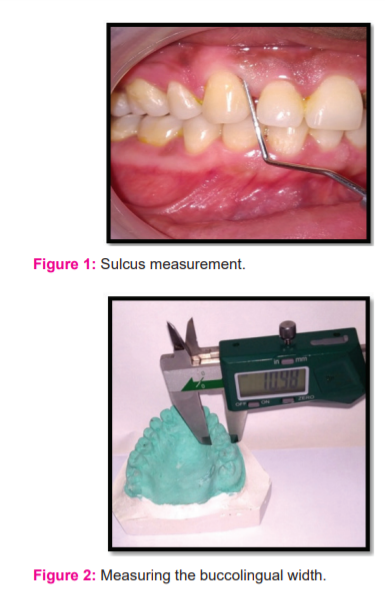
1. Measure and record the dimension of the gingival sulcus of the tooth to be prepared using William’s Periodontal Probe by inserting it parallel to the long axis of the tooth and move along the tooth. The sulcus depth is measured from the deepest point of penetration to the free gingival margin. (FIG-1)
2. Obtain upper and lower diagnostic impression.
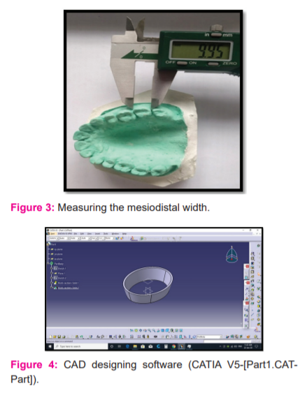
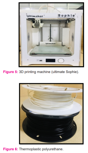
3. Measure the bucco-lingual and mesio-distal width of the tooth to be prepared from diagnostic cast of the patient using digital Vernier calliper.(FIG-2,3)
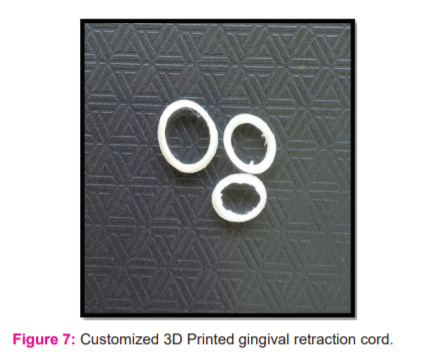
4. The recorded measurements are loaded in the CAD designing software (CATIA V5-[Part1.CATPart]) (FIG-4).The CAD designing of the gingival cuff is done with the following measurement :
-
-
-
The Inner diameter of the cuff: Mesiodistal and labiolingual diameter of the tooth
-
The height of the cuff: Gingival sulcular depth of respective tooth
-
The angulation for the outside slanter: 2o
Finally, which makes it a shape sketch design.
5. Print the retraction cord in the 3D printing machine (Ultimakersophie, Model no. ULTIMAKER 3P03, Brand-IMIK, Utrecht Netherlands.) using 0.2mm nozzle size and material used being thermoplastic polyurethane material. (FIG-5,6)
6. After tooth preparation, insert the customized gingival retraction cord using a cord packer, tack the cord in the sulcus from the mesial side of the tooth to distal crevice and retain it in the sulcus for 3-4min.(FIG-7)
7.The 3D printed cord is removed from the gingival sulcus and an elastomeric impression is obtained.
In comparison with the existing commercial gingival retraction cords this customized gingival retraction cord by 3d printing technology is advantageous in terms of time, manipulation of gingival tissue, controlling of bleeding and is very cost-effective. It even controls the thickness of the cord, so that it can deliver a constant amount of force on to the gingival tissue.
Discussion:
The purpose of this technique was to analyse the impact of the gingival displacement on the marginal fit of a restoration. A properly fit restoration prevents the damage of the periodontium.3Gingival tissues are sensitive to mechanical or chemical trauma.8 The main drawback of the chemical-mechanical method is the rapid collapse of sulcus after removal which prevent accurate impression making, Trauma to epithelial attachment, is Time-consuming, Risk of sulcus contamination, Painful.1In the conventional method, the amount of displacement takes time and most of the time it leads to trauma of the gingival tissue because the operator is not aware of the sulcular anatomy. Many studies have described the use of knitted or braided, impregnated or unimpregnated strings or fibers of different materials as a mechanical gingival displacement method.4The unique knitted design of the retraction cord can exert a gentle, constant outward force following placement, and as the knitted loops try to open, it dilates the free gingival margin.9Some of the drawbacks reported can be listed as the traumatic injury to the gingival tissue by slippage of the instrument or excessive packing force during packing eventually resulting in gingival recession. Mechanical compression built up in the sulcular area limits the GCF flow but once the cord is removed rebound increase in GCF flow can occur compromising moisture control. In this technique the sulcular anatomy is pre-measured and based on the readings the 3D cuff is designed digitally and fabricated to occupy the exact form and shape of the gingival sulcus. This will prevent compression and trauma to the gingival tissue thereby helps in achieving the desired displacement aiding in finish line exposure. Additive manufacturing or 3d printing according to the American Society for Testing and Materials (ASTM) is the process of joining materials to make objects from 3D model data, usually layer upon layer, as opposed to subtractive manufacturing methodologies.6The material used thermoplastic polyurethane, is a biocompatible material with good abrasion resistance and tear resistance, so the chances of tearing during placing it into the sulcus is less and it is cost effective.10 Thermoplastic polyurethane (TPU) are linear, segmented block copolymers with excellent properties such as durability, flexibility, elasticity, biostability fatigue resistant, and insulating properties. These polyurethanes have a vital role in developing many medical devices such as catheters, endotracheal tubes, blood bags, drug delivery vehicles, heart pacemakers connectors, orthopedic splints, vascular grafts and patches.11 Because of non-thrombogenic characteristics polyurethanes are used in cardiovascular areas.12 TPU comprises of hard and soft segments.13Diisocyanide and short-chain extender molecules like diols or diamines are used to manufacture the hard segments. The high interchain interactiveness of hard segments is due to the hydrogen bonds and even acts as reinforcing fillers for the soft matrix. On the contrary, soft segments which have long, linear flexible polyether or polyester facilitates the interconnecting of two hard segments. Hard segments differ from soft segments in terms of rigidity and polarity.14 TPU is a biocompatible material with good abrasion performance; high tear propagation resistance; high damping power; and high resistance to oils, fats, and many solvents.15
Limitations of the study:
-
The amount of gingival retraction obtained is not quantified.
-
It requires an in-office 3D printer.
-
The incorporation of hemostatic agents can be employed in future studies.
Conclusion :
This article described a customize gingival retraction cord fabricated using 3D printing technology which aids as an alternative to the conventional gingival retraction method with improved properties in term of feasibility and compatibility. The gingival retraction technique is easy and safe to use. Thus, the newly advanced technique have been found to be cost-effective and even controls the thickness of the cord, so that it can deliver a constant amount of force onto the gingival tissue.
Conflict of interest:
No potential conflict of interest with respect to the research, authorship and/or publication of this article.
Funding:
No financial support received for the research, authorship and/or publication of this article.
Acknowledgement:
The authors acknowledge the support provided by Mr. Kalaiselvan for printing the retraction cords


References:
-
Safari S, Vossoghisheshkalani Ma ,Vossoghi Sheshkalani Mi, Hoseinni Ghavam F, Hamedi M. Gingival Retraction Methods for Fabrication of Fixed Partial Denture: Literature Review. J Dent Biomater. 2016;3(2):205-213.
-
Phatale S, Marawar PP, Byakod G, Lagdive SB, Kalburge JV. Effect of retraction materials on gingival health: A histopathological study. J Indian SocPeriodontol. 2010;14(1):35-39.
-
Wostmann B, Rehmann P, Trost D, Balkenhol M. Effect of different retraction and impression techniques on the marginal fit of crowns. J Dent. 2008;36(7):508-512.
-
Anupam P, Namratha N, Vibha S, Anandakrishna GN, Shally K, Singh A. Efficacy of two gingival retraction systems on lateral gingival displacement: A prospective clinical study. J Oral BiolCraniofac Res. 2013;3(2):68-72.
-
Donovan TE, Chee WW. Current concepts in gingival displacement. Dent Clin North Am. 2004;48(2): 433-444.
-
GaliSivaranjani, Sirsi Sharad. 3d printing: the future technology in prosthodontics. J Dental and Oro-facial Res. 2015; 11(1):37-40.
-
Dawood A, Marti Marti B, Sauret-Jackson V, Darwood A. 3D printing in dentistry. Br Dent J. 2015;219(11):521-529.
-
Prasad KD, Hegde C, Agrawal G, Shetty M. Gingival displacement in prosthodontics: A critical review of existing methods. J Interdiscip Dentistry 2011;1:80-86.
-
Huang C, Somar M, Li K, Mohadeb JVN. Efficiency of Cordless Versus Cord Techniques of Gingival Retraction: A Systematic Review. J Prosthodont. 2017;26(3):177-185.
-
Akindoyo JO, Beg MDH, Ghazali S, Islam MR, Jeyaratnam N, Yuvaraj AR. Polyurethane types, synthesis and applications – a review. RSC Adv. 2016;6(115):114453-114482.
-
Joseph J, Patel R.M, Wenham A, Smith J.R. Biomedical applications of polyurethane materials and coatings. Trans IMF.2018; 96(3):121-129.
-
Burke A, Hasirci N. Polyurethanes in biomedical applications. Adv Exp Med Biol. 2004;553:83-101.
-
OsmanAzlinFazlina, Martin Darren James. Thermoplastic Polyurethane (TPU) / Organo- fluoromicaNanocomposites for Biomedical Applications: In Vitro Fatigue Properties, IOP Conf. Series: Materials Science and Engineering. 2019;701-709.
-
Dicesare P, Fox WM, Hill MJ, Krishnan GR, Yang S, Sarkar D. Cell-material interactions on biphasic polyurethane matrix. J Biomed Mater Res A. 2013;101(8):2151-2163.
-
Pucci A.Smart and Modern Thermoplastic Polymer Materials. Polymers (Basel). 2018;10(11):1211-1213.
|


 This work is licensed under a Creative Commons Attribution-NonCommercial 4.0 International License