IJCRR - 13(14), July, 2021
Pages: 170-176
Date of Publication: 20-Jul-2021
Print Article
Download XML Download PDF
A Study of Anthropometric Measurements of Anterior Cruciate Ligament and Corresponding Femoral Intercondylar Notch in Human Cadavers in Indian Population
Author: Bharambe Vaishaly Kishore, Gangani Seherish Karim, Anand Shubhangi, Vanshikha Gupta
Category: Healthcare
Abstract:Introduction: Damage to the Anterior Cruciate ligament (ACL) is a very frequently occurring injury. There are reports of the possibility of this injury being associated with the narrow intercondylar notch of the femur Aims: To measure and correlate the dimensions of the Anterior Cruciate ligament and intercondylar notch of the femur in the Indian population. Methodology: 25 knee joints in formalin-fixed cadavers were dissected and anterior cruciate ligaments and corresponding intercondylar notches were measured using vernier callipers. Results: The average length and width of ACL were found to be 38.8 and 9.6 mm. The standard deviations were found to be 2.3 and 1.7 respectively with standard errors being 0.46 and 0.34 respectively. The average width of the intercondylar notch was found to be 11.9, 15.8 and 14.3 mm in the lower, middle and upper parts of the notch respectively. Conclusion: The findings were compared with those of other authors and it was found that the length of ACL was found to be more and the width of the femoral intercondylar notch was found to be less in the Indian population than those reported by other authors. In the present study, 52% of the intercondylar notches had a width less than 17mm. A positive correlation was found between the length of ACL and the width of the middle part of the intercondylar notch. The authors state that it is possible that long ACL may compensate for narrow intercondylar notch and that a finding of short ACL and narrow intercondylar notch may be necessary risk factors for ACL tears.
Keywords: Cadaver, Anterior cruciate ligament, The intercondylar notch of the femur, Posterior cruciate ligament, Anthro�pometric measuremen
Full Text:
Introduction:
All ligaments of the knee are crucial for the functioning of the knee joint.1Out of all the ligaments found supporting and acting at the knee joint, the anterior cruciate ligament (ACL) is the one that is most frequently injured. Damage to the ACL is a very frequently occurring sports-related injury. 2 Thus ACL repair surgeries are also becoming very frequent. 3It has been reported that reconstruction of the ACL is the sixth most common surgical procedure performed by sports medicine experts.1The repair of the ACL requires detailed knowledge of its anatomical morphology.4
Several researchers have indicated that a smaller intercondylar notch can lead to contact with the ACL predisposing to an ACL injury during movements at the knee joint. 5, 6 Shepstone et al stated that the morphology of the intercondylar notch is related to the functioning and damage to the ACL. The injured ACL is, in turn, a known predisposing factor for osteoarthritis.7
A cadaveric study was carried out by Wilson and Barhorst.8 It investigated the possibility of intercondylar notch impingement of the anterior cruciate ligament in six cadaveric knees, three female and three male on exposure to impingement force. They found that the impingement occurred more frequently in female than in male knees. However, the authors concluded that the small sample size limited the conclusivity of the results.
Tillman et al studied the intercondylar notch geometry and found that the notch shape index for males, exceeded that for females and the intercondylar notch was less round in the females and could play a role in ACL injury. The authors stressed the necessity to further investigate the geometry of intercondylar notch and ACL.9
The present research attempts to study the morphology of ACL and femoral intercondylar notch and by comparing them both, the study will attempt to give an insight into ACL injuries about intercondylar notch impingement.
Objectives
The objectives of the present study were to dissect the ACL and measure the length and width of the ACL in a state of flexion and to study the intercondylar notch by measuring the width of the intercondylar notch in the upper, middle and lower regions of the notch. The study finally attempted to perform a correlate and analyse between the ACL measurements, the notch size and ACL injuries.
Methodology
The study was carried out after obtaining clearance from the ethical committee. The study is observational descriptive study design.
Sample size: A sample size of 25 lower limbs was calculated. The study was conducted on bodies that were donated after taking the consent of the donor and donor family, for purpose of Anatomical dissection and research.
Inclusion and exclusion criteria: The lower limbs were obtained from formalin-fixed adult cadavers. Inclusion criteria for a limb to be selected for the study were that on examination the limb did not have any damage or injuries in the area to be dissected or any visible abnormality of the knee joint. Limbs with damage or injuries or having knee joint related abnormalities were excluded from the study. Instruments used were gloves, scalpel, blunt and tooth forceps, fine small scissors, digital vernier calliper, goniometer and bone saw. For photography, a 16-megapixel camera was used.
Once selected, each lower limb was labelled separately for identification. Dissection of the lower limb was commenced with a skin incision. The upper incision was taken horizontally midway in the thigh and the lower incision midway in the leg region. The incision almost completely encircled the thigh and leg remaining incomplete posteriorly. The two incisions were joined by a vertical incision anteriorly to complete an “I” shaped incision. The overlying skin was removed in form of two flaps starting from the “I” shaped incision thus exposing the patella and the region above and below it.
The muscles, ligaments, patella and joint capsule were identified and observed for any abnormalities. All the soft tissue around the knee capsule was removed along with the patella. The major ligaments of the knee i.e. the ACL, posterior cruciate ligament (PCL), medial and lateral menisci and medial and lateral collateral ligaments were preserved.
The knee was flexed at around 90° flexion. The synovial membrane covering the ACL was removed. The ACL was dissected in detail till it was well delineated. It was photographed and any variations were noted.
The femoral intercondylar notch was measured. The height of the intercondylar notch was measured. Its width at the upper and lower ends and the width in the middle of the notch was measured.
The findings of measurements of intercondylar notch and ACL were fed in a Microsoft Excel spreadsheet and analyzed statistically.
Results:
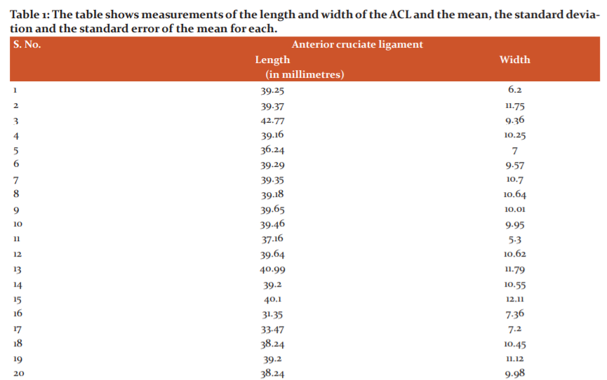
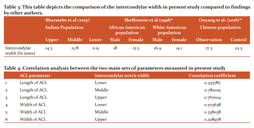
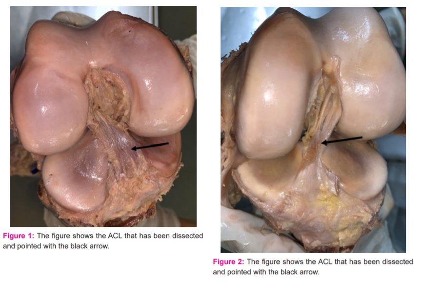
The measurements of the ACL in all 25 cases are given in table 1. The mean, standard deviation and the standard error of the mean are also given in the table. The dissected ACLs can be visualized in Figure 1, 2.
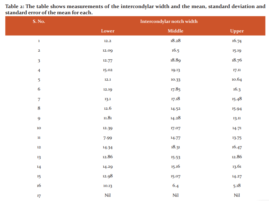
The width of the intercondylar notch is given in table 2 in the upper, middle and lower region of the notch. In each case, the mean, standard deviation of the mean and the standard error of the mean are also given in the table. In the case of specimen number 17, the intercondylar notch had been cut open during the study and hence the intercondylar notch could not be measured reliably.
Discussion:
The number of ACL injuries has been observed to have increased in the last two decades. This has been observed to be especially true in the case of young children and adolescents. This could be because of more number of younger individuals entering into competitive sports.10 However, a smaller diameter of the ACL has been reported to be associated with an ACL injury.11 In their study, Mahajan et al took the measurements of the ACL by use of ultrasonography. Other authors too have corroborated this finding that ACL injured patients often have small-sized ACLs.12, 13 Many authors have based their observations on the fact that there is a difference in the rate of ACL injuries in males and females. Sutton and Bullock reported that females have a three times higher rate of ACL injury compared to males. This was probably because of increased quadriceps angle and increased posterior tibial slope they stated.14 A combination of various pressure influences leads to mechanical loading that leads to ACL rupture. 15,16,17,18
Length and width of ACL:
The present study was a cadaveric study in which the ACLs were measured by the process of dissection. The average length and width of ACL were found to be 38.8 and 9.6 mms. respectively with the standard deviation being 2.31 and 1.74, and the error of the mean being 0.46 and 0.34 respectively.
In a study carried out by Saxena et al. the length and width of ACL was found to be 32.5 and 9.38 respectively.19In a study by Jayagandhi et al. the average length of the ACL was found to be 32.2 mms while in a study by Odenston et al. it was found to be 31 mms.20, 21
Thus in the present study, the length of the ACL was found to be more than the reports by other researchers. Understanding the length of ACL will assist surgeons in ACL reconstructive surgery. Knowing the size of ACL helps the surgeons to select grafts for ACL reconstruction surgery.
Mahajan et al stated that smaller ACL is seen among subjects with ACL injuries.11 The incidence of ACL tears is 2.3 times higher in females than in males.22, 23, 24 Chandrashekhar et al reported that the ACL length was found to be smaller in females than that in males.15As stated, in the present study the length of the ACL was found to be more than the reports by other researchers. Thus it would appear that chances of ACL injuries can be lesser in the present set of knee joints studied.
Width of Intercondylar notch of femur:
In the present study the mean intercondylar width was found to be in the range between 11.94 mm at the lower end of the notch, 15.83 mm in the middle and 14.34 mm at the upper end of the intercondylar notch.
In a study by Shelbourne et al., the middle intercondylar width was found to be 15.5 mm and 18 mm in the case of females and males of the African American part of the study group respectively.6 The similar measurements of the white females and males were found to be 14.1 mm and 16.9 mm respectively.6 The authors noted that there were reports that white female basketball players had a 6.5% higher incidence of ACL tears compared to their African American counterparts.25 In the present case the sex of the cadavers being dissected could not be determined in all cases as some of the lower limbs had been disarticulated from the hip.
The present study shows the average width of the intercondylar notch to be 15.8 mm which is lesser than the width in the case of African Americans as well as white Americans participating in the study carried out by Shelborne and the findings reported by Ouyang et al in the Chinese population.6, 26(Table 3) Femoral intercondylar notch has been often indicated as a possible risk factor in cases of ACL injury. However, it is still not proved that a narrow notch leads to ACL tears. There have been many studies that have reported a narrow notch as being the risk factor for ACL injuries.27, 28Many of these studies have been conducted on X-ray films or CT scan images. While there are studies that implicate the intercondylar notch as a possible risk factor, there are few studies that state that there is no correlation between ACL injury and size of the intercondylar notch (Figure 3).29-32
In the present study, the authors report findings where the intercondylar notch has been measured directly on the femur bone. Lund-Hanssen et al. reported that when the intercondylar notch width was less than 17 mm, chances of ACL injury increased by 6 times compared to those with the wider intercondylar notch.33 In the present study 52% of the intercondylar notches studied had a width less than 17mm.
The ACL measurements in the lower limbs dissected in the present study sample were more than that reported by other researchers and indicating lesser chances of ACL tears which counters the above statement concerning the width of the intercondylar notch. Mahajan et al. have stated the higher incidence of ACL tears is associated with a shorter length of ACL( Figure 4).34
Correlation between the parameters studied i.e. length and width of ACL and the width of the intercondylar notch was analyzed by applying correlation analysis. Table 4 shows the results of the correlation analysis of the length and width of the ACL and the width of intercondylar notches of the femur at all 3 levels i.e. upper, middle and lower applied to present findings. A positive correlation was found between the length of the ACL and the width of the intercondylar notch of the femur as measured in the middle of the notch. No comparative studies in this regard were found in the literature. However, a study by Stijak et al. found a positive correlation between the width of ACL and the intercondylar notch in the males.35,36
The authors in the present study would like to state that it is possible that long ACL may compensate for narrow intercondylar notch and that a finding of short ACL and narrow intercondylar notch may be necessary risk factors for ACL tears. This hypothesis may need further research.
Limitations of the study: The small sample size and inability to differentiate the findings of the sex of the lower limbs being studied are the limitations of this study.
New topics for research: The study puts up a hypothesis wherein there is a need to explore the possibility of correlation of both short ACL and narrow intercondylar notch coexisting in ACL injuries.
Conclusion
In the present study, the length of ACL was found to be more and the width of the femoral intercondylar notch was found to be less in the Indian population than those reported by other authors and 52% of the intercondylar notches had a width less than 17mm. A positive correlation was found between the length of ACL and the width of the middle part of the intercondylar notch. It may be concluded it is possible that long ACL may compensate for narrow intercondylar notch and that a finding of short ACL and narrow intercondylar notch may be necessary risk factors for ACL tears.
Acknowledgement
The authors acknowledge the immense help received from the scholars whose articles are cited and included in the references of this manuscript. The authors are also grateful to authors/ editors/publishers of all those articles, journals and books from where the literature for this article has been reviewed and discussed.
Conflict of interest
The authors declare that there is no conflict of interest
Source of funding:
NIL
Individual Author’s contribution
All the authors have contributed to dissection, collection of data, its analysis and final discussion and manuscript writing.
References:
-
Abulhasan JF, Grey MJ. Anatomy and Physiology of Knee Stability J. Funct. Morphol. Kinesiol. 2017, 2, 34.
-
Moghaddam AB, Torkaman A.A Cadaver Study of the Structures and Positions of the
Anterior Cruciate Ligament in Humans.Int J Prev Med. 2013 Apr; 4(1): 85-9.
-
Tran TD, Tran QL.A Cadaveric study on the anatomy of the anterior cruciate ligament in Vietnamese adults. Asia-Pacific J Sports Med, Arthur, Rehab Techn.2018,14:22-25.
-
Oka S,Schuhmacher P,Brehmer A,Traut U, Kirsch J.Siebold R. Histological analysis of the tibial anterior cruciate ligament insertion.KneeSurg Sports Traumatol Arthrosc 2016; 24:747–753
-
Souryal TO, Freeman TR. Intercondylar notch size and anterior cruciate ligament injuries in athletes. A prospective study.Am J Sports Med. 1993 Jul-Aug;21(4):535-9.
-
Shelbourne K D, Davis TJ, KlootwykTE. The relationship between intercondylar notch width of the femur and the incidence of anterior cruciate ligament tears. A prospective study. Am J Sports Med. 1998 May-Jun;26(3):402-8.
-
Shepstone L, J Rogers, J R Kirwan, B W Silverman.The shape of the intercondylar notch of the human femur: a comparison of osteoarthritic and non-osteoarthritic bones from a skeletal sample.Ann Rheum Dis. 2001;60:968–97.
-
Wilson R, Barhorst A.Intercondylar Notch Impingement of the Anterior Cruciate Ligament: A Cadaveric In Vitro Study Using Robots. J Health Engg. 2018;Article ID 8698167: 27.
-
Tillman MD, Smith KR, Bauer JA, Cauraugh JH, Falsetti AB, PattishallJL.Differences in three intercondylar notch geometry indices between males and females: a cadaver study. J Knee. 2002 Feb;9(1):41-6.
-
LaBella C, HenrickusW, Hewett T. Anterior Cruciate Ligament Injuries: Diagnosis, Treatment, and Prevention.Clin J Sports Med. 2012.22(4):349-55.
-
Mahajan PS, ChandraP, Negi VC, Jayaram AP, Hussein SA. Smaller Anterior Cruciate Ligament Diameter Is a Predictor of Subjects Prone to Ligament Injuries: An Ultrasound Study.Biomed Res Int. 2015;23(4): 71-77..
-
Chaudhari A. M. W., Zelman E. A., Flanigan D. C., Kaeding C. C., Nagaraja H. N. Anterior cruciate ligament-injured subjects have smaller anterior cruciate ligaments than matched controls: a magnetic resonance imaging study. Amer J Sports Med. 2009;37(7):1282–1287. doi: 10.1177/0363546509332256. PMC free articlePubMedCrossRefGoogle Scholar
-
Vaidya S, Dhongade H, Mahajan S, Aneesh MK. Evaluation of Anterior and Posterior Cruciate Ligament Injuries of the Knee on Magnetic Resonance Imaging: A Cross-sectional Study.Int J Curr Res Rev. 2020.12(14):105-108.
-
Sutton KM, Bullock JM.Anterior cruciate ligament rupture: differences between males and females. J Am Acad Orthop Surg. 2013 Jan;21(1):41-50.
-
Chandrashekar N., Slauterbeck J., Hashemi J. Sex-based differences in the anthropometric characteristics of the anterior cruciate ligament and its relation to intercondylar notch geometry: a cadaveric study. Am J Sports Med. 2005;33(10):1492–1498. doi: 10.1177/0363546504274149. PubMedCrossRefGoogle Scholar
-
Hewett T. E., Myer G. D., Ford K. R. Anterior cruciate ligament injuries in female athletes: part 1, mechanisms and risk factors. Amer J Sports Med. 2006;34(2):299–311. doi: 10.1177/0363546505284183. PubMedCrossRefGoogle Scholar
-
Huston L. J., Greenfield M. L., Wojtys E. M. Anterior cruciate ligament injuries in the female athlete: potential risk factors. Clin Orthop Relat Res. 2000;(372):50–63. PubMedGoogle Scholar
-
Davis I., Ireland M. L., Hanaki S. ACL injuries: the gender bias. J Orthop Sports Physical Ther. 2007;37(2):p. A1. doi: 10.2519/jospt.2007.0301. PubMedCrossRefGoogle Scholar
-
Saxena A, Ray B, Rajgopal KV, D’Souza AS,PyrtuhS.Morphometry and magnetic resonance imaging of anterior cruciate ligament and measurement of secondary signs of anterior cruciate ligament tear. Bratisllek Listy.2012;113(1):539-543.
-
JayagandhiS, NimVK, Kumar M, S Senthilkumar.The Morphometric Study Of Anterior Cruciate Ligament: A Cadaveric Study. Int J Anat Res. 2018;6(3.2):5581-86.
-
Odensten M, Gillquist J. Functional anatomy of the anterior cruciate ligament and a rationale for reconstruction. J Bone Joint Surg Am. 1985 Feb; 67(2): 257-262.
-
Oliphant JG, Drawbert JP. Gender Differences in Anterior Cruciate Ligament Injury Rates in Wisconsin Intercollegiate Basketball. J Athl Train. 1996 Jul-Sep; 31(3):245-247.
-
Wirtz PD. High school basketball knee ligament injuries. J Iowa Med Soc. 1982 Mar;72(3):105–106.
-
Zelisko JA, Noble HB, Porter M. A comparison of men's and women's professional basketball injuries. Am J Sports Med. 1982 Sep-Oct;10(5):297–299.
-
Trojian T, Collins S. The Anterior Cruciate Ligament Tear Rate Varies by Race in Professional Women's Basketball. J Iowa Med Soc. 2006. 34(6):895-8.
-
Ouyang X, Yu HW, WangJ, Shi Dong Hong, XinF, Wang L.MRI measurement on intercondylar notch after anterior cruciate ligament rupture and its correlation, Exp Ther Med. 2016.11(4):1275-1278.
-
Houseworth SW, Mauro VJ, Mellon BA, Kieffer DA.The intercondylar notch in acute tears of the anterior cruciate ligament: a computer graphics study. J Iowa Med Soc.1987;15(3):221-224.
-
Anderson AF, Lipscomb AB, Liudahl KJ, Addlestone RB.Analysis of the intercondylar notch by computed tomography. Amer J Sports Med.1987;15(6):547-552.
-
Souryal TO, Freeman TR, and Danie DM. Intercondylarnotch size and anterior cruciate ligament injuries in athletes:a prospective study. Am J Sports Med.1993.21(4):535-539.
-
LaPrade RF, Burnett QM II,D. M. Daniel, “Femoralintercondylar notch stenosis and correlation to anteriorcruciate ligament injuries: a prospective study. Am J Sports Med.1994;22(2):198-203.
-
Ireland ML, Ballantyne BT, Little K, NI. S. McClay.A radiographic analysis of the relationship between the size and shape of the intercondylar notch and anterior cruciate ligament injury. Knee Surgery, Sports Traumatic, Arthrosc. 2001.9(4):200-205.
-
Domzalski M, Grzelak P, GabosP. Risk factors for Anterior Cruciate Ligament injury in skeletally immature patients: analysis of intercondylar notch width using Magnetic Resonance Imaging. IntOrthop. Am J Sports Med .2010;34(5):703-7.
-
Lombardo S, Sethi PM, Starkey C.Intercondylar notch stenosis is not a risk factor for anterior cruciate ligament tears in professional male basketball players: an 11-year prospective study. Am J Sports Med. 2005;33(1):29-34.
|
 IJCRR
IJCRR 


 This work is licensed under a Creative Commons Attribution-NonCommercial 4.0 International License


