IJCRR - 13(10), May, 2021
Pages: 29-35
Date of Publication: 19-May-2021
Print Article
Download XML Download PDF
Morphometric Variations of Mandibular Foramen and Lingula of Mandible with Gender and Age Using Cone Beam Computerised Tomography
Author: Hayagreev D, Kodialbail A, Shetty S, Sujatha S
Category: Healthcare
Abstract:Introduction: Mandibular foramen (MF) is located on the medial surface of mandibular ramus. The inferior alveolar nerve and vessels pass through it and supply the mandibular teeth. Along with the lingula of the mandible, it forms an important bony landmark during oral and maxillofacial surgical procedures and for identifying the site for injection of local anaesthetics. Objective: To study the morphometric variation in the position of the mandibular foramen and lingula and to compare the above measurements in males and females and age groups of < 20 years and > 20 years. Methods: 100 cone beam computerised tomography (CBCT) scans of full skull of both the genders and age group were studied in Ramaiah dental hospital and the location of the mandibular foramen and lingual were measured from the reference points: Base of the mandible, anterior and posterior borders of the ramus of the mandible, sigmoid notch and third molar. Results: The parameter that showed significant variation was; the mandibular foramen and lingula in females was more towards the base than males and among the age group of below 20 years of and adults above 20 years of age, children have their mandibular foramen nearer to the mandibular notch and the posterior border; indicating that the foramen moves downwards and anteriorly with growth. Conclusion: The position of both the mandibular foramen and lingula will vary with age and sex hence the surgeons should consider the age and sex of the patient during osteotomies and nerve block.
Keywords: Osteotomies, Mandibular nerve block, Neurovascular bundle, Cone Beam Computerised tomography, Shape of lingula, Oromaxillofacial surgeries
Full Text:
INTRODUCTION
Several dental procedures such as apical curettage of mandibular premolars, amalgam filling, implant treatment, mandibular osteotomies and periodontal surgeries, dental extractions and correction of various jaw abnormalities require the use of local anesthesia during the surgeries. The most common route of local anesthesia is by giving a nerve block to the inferior alveolar nerve which provides anesthesia of teeth, jaw, lip, gingiva and mucous membrane.1
The mandibular foramen (MF) is located on the medial side of the ramus of the mandible through which the neurovascular bundle containing the inferior alveolar nerve and vessels passes. It forms an anterior loop in the mandibular canal of the lower jaw and leaves the canal after splitting into mental and incisive nerves from the mental foramen in the anterior wall of the alveolar bone.2,3 The failure rate of the anaesthesia is reported to be 20 to 25% and the cause being inaccurate positioning of the mandibular foramen.4,5
The lingula is a tongue-shaped bony projection covering the mandibular foramen. Lingula and MF act as important landmark for the correction of deformities like prognathic, orthognathic and retrognathia by conducting osteotomy procedures such as bilateral sagittal split ramus and intraoral vertical- sagittal ramus surgeries. In the above procedures, the excision of bone should be at the level of the lingula to avoid injury to an inferior alveolar nerve.6
Thus locating these anatomical positions accurately is critical to achieve more successful nerve block and prevent common complications like paraesthesia, skin necrosis and haemorrhage during orthognathic surgery.4,7,8 Variations concerning the individual, gender, age, race, assessing technique and degree of edentulous alveolar bone atrophy has also been noted in the mandibular incisive canal, mental foramen and associated neurovascular bundles.9
In children, a variation in the difference between the distance from the mandibular foramen to the anterior border and the posterior border is observed. This variation is caused by regional growth in different directions.10,11 Hence in the above-mentioned procedures, the exact site of operation and anaesthesia will differ in children and adults.
Radiographic images are obtained clinically to identify the position of the mandibular foramen. However, it has the disadvantage of being less accurate. Computed tomography (CT) scans are accurate but expensive and have a high dose of radiation exposure to the patients. On the other hand, Cone-beam computed tomography (CBCT) scans are less expensive and have low radiation exposure doses12 hence is more beneficial to the patients with accurate results.13-15
Moreover, studies conducted on dry bone cannot determine the age and sex of the individual which was the main aim of this study. Hence CBCT scan was used to study the morphometry and variations in the mandibular foramen and lingula in two age group and males and females. This study aimed to compare the position of the mandibular foramen and lingual in both the sexes and in age groups of below 20 years and above 20 years.
MATERIALS AND METHODS
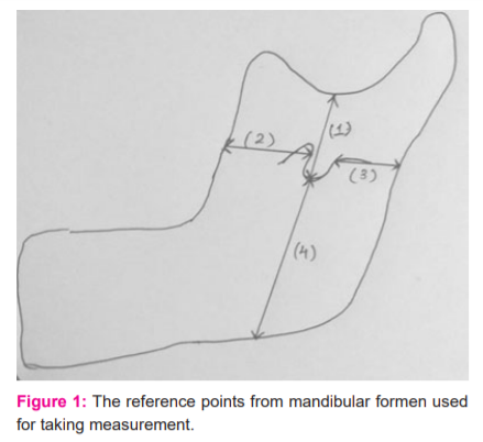
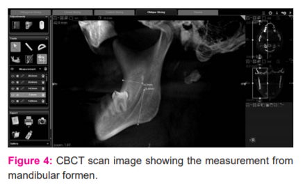
100 CBCT scans of full skull of both genders were taken from the Department of oral medicine and radiology in Ramaiah dental hospital in the period 2014-2017. 50 were of below 20 years and 50 were after 20 years of age. Scans were taken using a Carestream CS 9300 machine at 90 kVp and 6mA with 17*13.5cm field of view. The age, name and gender of the patient were noted. The scan was viewed in cross-sectional slices of 25.5 mm thickness in the sagittal plane. The mandibular foramen on the medial side of the ramus was identified. The shape of the lingula was noted. The presence of a third molar was noted. The location of the foramen and the lingula was assessed by measuring the distances using DIACOM Software. The distance of MF and lingula were measured from the reference points such as base of the ramus, mandibular notch, anterior border of the ramus, posterior border of the ramus, and third molar tooth. The mean of three measurements of each parameter was taken by the same observer to avoid interobserver and intraobserver variations.
Ethical clearance number- DRP/IFP/171- 2/8/2017
Variations in the shape of lingual16, 17 were also noted and were classified into:
-
-
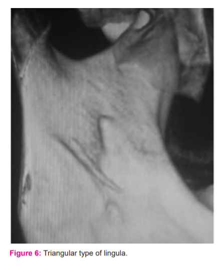
Triangular (with a wide base and a narrow rounded or pointed apex, rounded or pointed apex)
-
Truncated(lingula with somewhat quadrangular bony projection on its top)
-
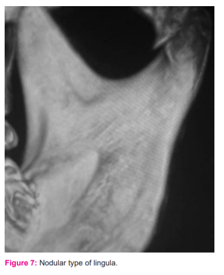
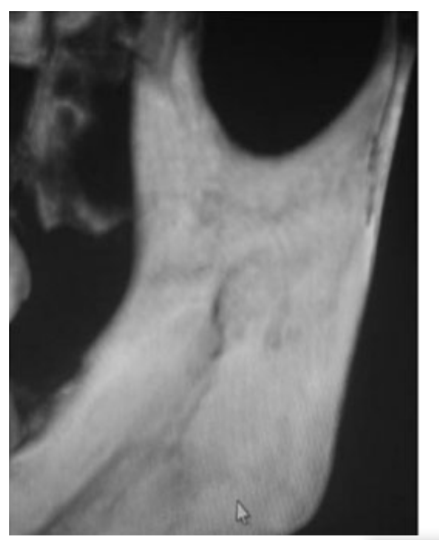
Nodular(lingula is nodular and of variable size, almost the entire lingula except for its apex which was merged into the ramus)
-
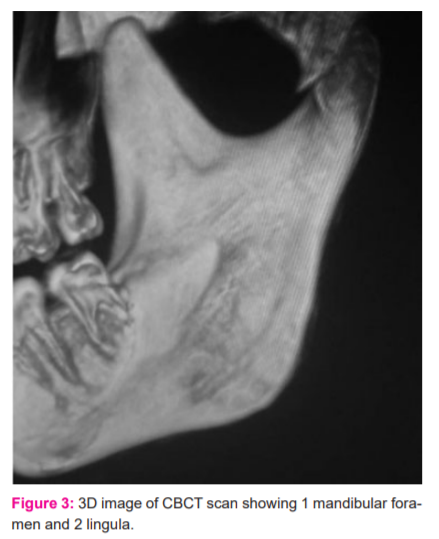
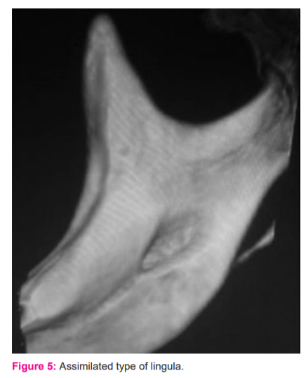
Assimilated (lingula is completely incorporated into the ramus) (Figure 1- 4)
RESULTS
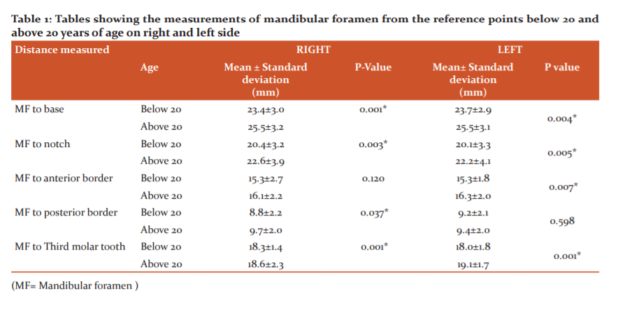
The study showed the mean distance of mandibular foramen from the base of the ramus was 25.55±3.35 mm in the males and 23.6±2.75 mm in females, from the mandibular notch, was 22.25±4.35 mm in males and 20.4±2.85 mm in females, anterior border of the ramus of mandible was recorded as 15.5±2.5 mm in males and 16.00±1.9 mm in females, from the posterior border was recorded as 9.4±2.25 mm in males and 9.15±1.85 mm in females, from third molar tooth 19.1±1.9 in males and 18.25±1.9 in females.
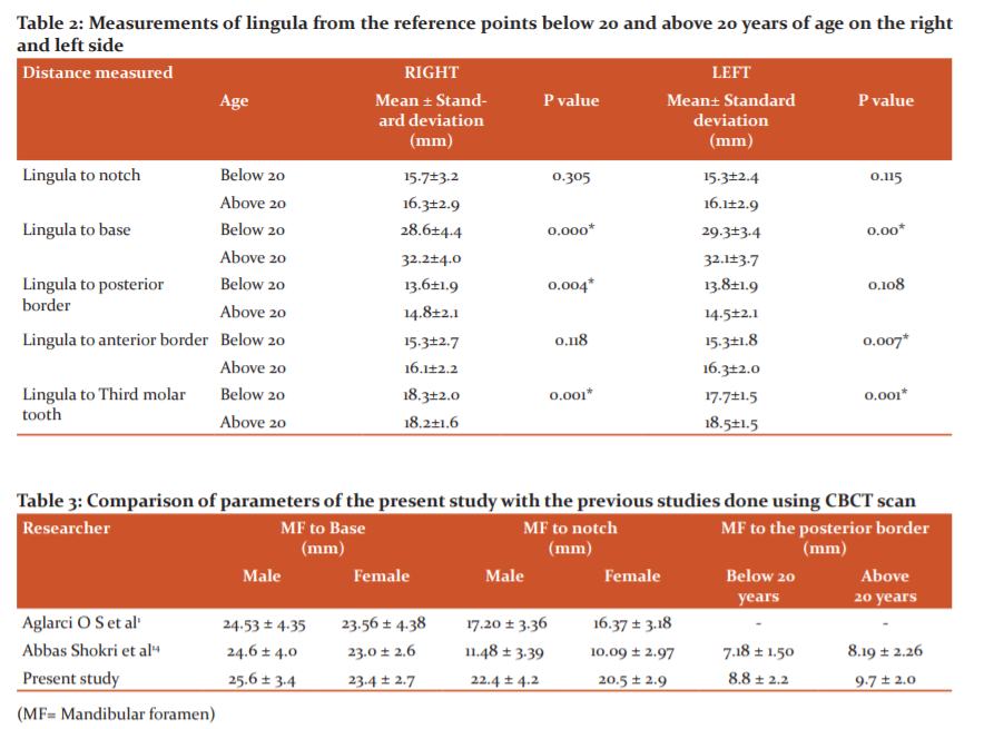
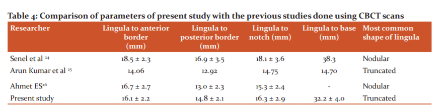
The mean distance of lingula from the base of the ramus was 32.05±3.95 mm in the males and 29.1±3.9 mm in females, from mandibular notch was 16.15±3.05 mm in males and 15.6±2.6 mm in females, anterior border of the ramus of mandible was recorded as 15.5±2.5 mm in males and 16.00±1.9 mm in females, from the posterior border was recorded as 14.55±2.1 mm in males and 13.85±2.0 mm in females, from third molar tooth 18.8±1.4 in males and 17.75±1.65 in females.
The shape of lingula was found to be truncated in 37% on the right side and 34% on the left side, nodular on 22% on the right side and 28% on the left side, assimilated in 25% on the right side and 20% on the left side and triangular in 16% on the right side and 18% on the left side (Figure 5-8).
The measurements from mandibular foramen to the base and mandibular notch and also the distance from lingual to the base of the mandible showed significant variations in males and females with a p-value of 0.001, 0.011 and 0.0 respectively.
The measurements that showed significant variations in the age groups below 20 years and above 20 years with p-value <0.05 are the distance of mandibular foramen to the base, mandibular notch, posterior and anterior borders and the distance of lingula to the base, posterior and anterior border (Table 1 and 2).
The study and its statistical analysis showed that among males and females, females have their mandibular foramen and lingula more towards the base than in males. Among the age groups i.e. group below 20 years of age and adults above 20 years of age, children have their mandibular foramen nearer to the mandibular notch and the posterior border; indicating that the foramen moves downwards and anteriorly with growth.
DISCUSSION
The mandibular ramus and the body are developed from intramembranous ossification. A groove containing nerve and blood vessels develops during the 24th week of the embryonic stage. Initially, 3 inferior alveolar nerves are formed in the embryo innervating each of the 3 groups of mandibular teeth. The fusion of these 3 nerves will result in the formation of a single inferior alveolar nerve. The failure of fusion of any of the two nerves may result in the double mandibular canal.4,13
In 2016, R Shalini et al. in their study found that 21.74±2.74 mm and 21.92±3.33 mm were the distance of mandibular foramen from the mandibular notch on the right and the left side respectively, while 22.33±3.32 mm and 25.35±4.5 mm were the distance from the mandibular foramen to the base of the ramus on the right and left side respectively. The mean distance of mandibular foramen from the anterior border of the ramus of the mandible was 17.11±2.74 mm on the right side and 17.41±3.05 mm on the left side, while from the posterior border was 10.47±2.11 mm on the right side and 9.68±2.03 mm on the left side.4
In 2014, Padmavathi G et al. studied 65 human dry mandibles, wherein lingula was truncated in shape in 33.84%, triangular in 29.23%, nodular 19.23% and assimilated in 17.70% of the mandibles. The mean distance of 21.3 ± 4.12 mm and 19.6 ± 3.30 mm was recorded from the lingula to the anterior and posterior borders of the mandibular ramus respectively. The mean distance of the lingula from the mandibular notch and base of the mandible was 18.6 ± 3.71mm and 36.1 ± 4.12 mm respectively.18
In 2013, according to a study done by Prajna PS et al. on 60 adult human mandibles, the mean distance of mandibular foramen from the posterior border was recorded as 10.47±2.11 mm on the right side and 9.68±2.03 mm on the left side, from the anterior border of the ramus of mandible was recorded as 17.11±2.74 mm on the right side and 17.41±3.05 mm on the left side. The mean distance of mandibular foramen from the base of the ramus was 22.33±3.32 mm on the right side and 25.35±4.5 mm on the left side and from mandibular notch was 21.74±2.74 mm on the right side and 21.92±3.33 mm on the left side. Accessory mandibular foramen was present in 32.36% of mandibles.19
In 2010, 80 dry mandibles were studied by Lopes Paulo Tadeu Campos et al. the triangular shape of the lingula was found on 66 sides (41.3%), the truncated lingula was present on 58 sides (36.3%), and the nodular lingula was seen on 17 sides(10.5%) and the assimilated lingula it was seen on 19 sides (11.9%). Accessory foramina were seen in 11.3 and 3.8% of the right and left mandibular rami respectively.20
A total of 100 mandibular foramina and 18 accessory foramina were observed in the 100 mandibles, in a study held by Gopalakrishna et al. The mean distance of mandibular foramina to the anterior border of ramus was reported to be 14.63±3.16 mm on the right side and 15.31±3.11 mm on the left side. The distance of the mandibular foramen to the posterior border was 12.34±3.10 mm on the right side and 13.51±3.92 mm on the left side. The distances between the foramen and the mandibular notch are 21.23±4.56 mm on the right side and 21.16±3.12 mm on the left side, between the foramen and angle of mandible, was 22.14±3.18 mm on the right side and 22.1±4.12 mm on the left side, while between the foramen and posterior border of 3rd molar tooth was recorded as 14.37±3.16 mm on the right side and 19.26±2.57 mm on the left side.21
O Oguz et al. did a study on 34 Turkish adult male mandibles. The results showed that the distance of the mandibular foramen to the angle of the anterior ramus was 16.9 mm on the right and 16.78 mm on the left. The distance to the posterior side of the ramus was found to be 14.09 mm on the right and 14.37 mm on the left. The distance of the mental foramen to the inferior border of the mandible was found to be 14.61 mm and 14.29 mm on the right and left, respectively. Its distance to the superior border was found to be 13.62 mm on the right and 14.62 mm on the left.22
According to the results of a study done by Jansisyanont on 92 dried mandibles, the truncated lingulae were most frequently found (46.2%) and most appeared to be bilateral (71.7%). Triangular, nodular, and assimilated shapes presented in 29.9%, 19.6%, and 4.3%, respectively. The mean angular height was reported to be 8.2 +/- 2.3 mm. The lingula was located at 20.6 +/- 3.5 mm from the anterior border of the mandibular ramus and 16.6 +/- 2.9 mm from the mandibular notch (Table 3 and 4).
In addition to the above-mentioned measurements, the present study also showed significant variation in the measurements of mandibular foramen to the anterior border, mandibular notch and to the base, and also measurements of lingula to base, posterior and anterior borders of mandible among the age groups of below 20 years and above 20 years.
CONCLUSION
The study and its statistical analysis showed that females have their mandibular foramen and lingula more towards the base than males. Among the age groups, children have their mandibular foramen nearer to the mandibular notch and the posterior border indicating that the foramen moves downwards and anteriorly with growth. The knowledge of the variation of the position of mandibular foramen concerning age and gender is important to improve the efficacy of the nerve block or plane of surgery being performed. Hence the surgeons should consider the age and gender before beginning such procedures to avoid injury to neurovascular bundles.
Acknowledgement: The authors acknowledge the immense help received from the Department of Oral medicine and radiology, Ramaiah Dental College for their support in conducting the study. The authors are also grateful to authors/editors/publishers of all those articles, journals and books from where the literature for this article has been reviewed and discussed.
Disclosure of conflicts of interest: None
Funding: NIL


References:
-
Aglarci O S, Gungor E, Altunsoy M, Nur B, Ok E. Three-Dimensional Analysis of Mandibular Foramen Location: A Cone Beam Computed Tomography Study. OMICS J Radiol 2015; 4 (1): 1-3.
-
Chen Z, Chen D, Tang L, Wang F. Relationship between the position of the mental foramen and the anterior loop of the inferior alveolar nerve as determined by cone-beam computed tomography combined with mimics. J Comput Assist Tomogr 2015; 39(1):86?93.
-
Eren H, Orhan K, Bagis N, Nalcaci R, Misirli M, Hincal E. Cone-beam computed tomography evaluation of mandibular canal anterior loop morphology and volume in a group of Turkish patients. Biotech Equip 2016;30(2):346-53.
-
Shalini R, Ravivarman C, Manaoranjitham R, Veeramuthu M. Morphometric. Study on mandibular foramen and incidence of accessory mandibular foramen in mandibles of south Indian population and its clinical implications in inferior alveolar nerve block. Anat cell Biol 2016;49(4):241-48.
-
Ennes JP, Medeiros RM. Localisation of the mandibular foramen and its clinical impactions. Int J Morp 2009; 27:1305-11.
-
Sophia MM, Alagesan A, Ramchandran K. A Morphometric and Morphological Study of mandibular lingula and its clinical significance. Int J Med Res Rev 2015;3(2):141-48.
-
Castells ET, Albiol JG, Baeza AR, Aytes LB, Escoda CG. Necrosis of the skin of the chin: a possible complication of inferior alveolar nerve block injection. J Am Dent Assoc 2008;139(12):1625?30.
-
Laishram D, Deepti S. Morphometric analysis of mandibular and mental foramen. J Den Med Sci 2015;14(12): 82-86.
-
Juodzbalys G, Wang HL, Sabalys G. Anatomy of Mandibular Vital Structures. Part II: Mandibular incisive canal, Mental foramen and associated neurovascular bundles in relation with dental implantology. J Oral Maxillofac Res 2010;1(1):e3.
-
Ahmet E S, Kenan C, Mustafa A. Cone Beam Computed Tomographic analysis of the shape, height, and location of the Mandibular Lingula in a population of children. BioMed Res Int 2013:1-8.
-
Krishnamurthy NH, Unnikrishnan S, Ramachandra JA, Arali V. Evaluation of the relative position of Mandibular foramen in children as a reference for Inferior alveolar nerve block using Orthopantamograph. J Clin Diagn Res 2017;11(3):71-74.
-
Chau AC, Fung K. Comparison of radiation dose for implant imaging using conventional spiral tomography, computed tomography, and cone-beam computed tomography. Oral Pathol Oral Radiol Endod 2009;107(4):559?65.
-
Park HS, Jae HL. A Comparative Study on the Location of the Mandibular Foramen in CBCT of Normal Occlusion and Skeletal Class II and III Malocclusion. Maxillofac Plast Reconstr Surg 2015;37(1):25
-
Shokri A, Falah-Kooshki S, Poorolajal J, Karimi A, Ostovarrad F. Evaluation of the location of mandibular foramen as an anatomic landmark using CBCT images: a pioneering study in an Iranian population. Braz Dent Sci 2014;17(4):74-81.
-
Al-Shayyab MH. A simple method to locate mandibular foramen with cone-beam computed tomography and its relevance to oral and maxillofacial surgery: a radio-anatomical study. Surg Radiol Anat 2018;40(6):625?34.
-
Tuli A, Choudary R, Choudary S, Raheja S, Agarwal S. Variation in shape of the lingula in the adult human mandible. J Anat 2000;197(2):313-17.
-
Smita Tapas. Variations in the morphological appearance of lingula in dry adult human mandibles. Int J Curr Res Rev 2013;05(24):41-45.
-
Padmavathi G, Varalakshmi K L, Suman T, Roopashree K. A morphological and morphometric study of the lingula in dry adult human mandibles of South Indian origin and its clinical significance. Int J Health Sci Res 2014;4(6):56-61.
-
Prajna PS, Poonam K. Morphometric analysis of mandibular foramen and incidence of accessory mandibular foramen in adult human mandibles of an Indian population. Rev Arg de Anat Clin 2013; 5(2):60-66.
-
Lopes PTC, Pereira GAM and Santos A. Morphological analysis of lingula in dry mandibles of individuals in Southern Brazil. J Morphol Sci 2012;27(3-4):136-38.
-
Gopalakrishna K, Deepalaxmi S, Somashekara SC, Rathna BS. An anatomical study on the position of mandibular foramen in 100 dry mandibles. Int J Anat Res 2016; 4(1):1967- 71.
-
Oguz O, Bzkir MG. Evaluation of the location of the mandibular and the mental foramina in dry, young, adult human male dentulous mandibles. West Indian Med J 2002;51(1):14-16.
-
Sekerci AE, Sisman Y. Cone-beam computed tomography analysis of the shape, height, and location of the mandibular lingula. Surg Radiol Anat 2014;36(2):155?62.
-
Senel B, Ozkan A, Altug H A. Morphological evaluation of the mandibular lingula using cone-beam computed tomography. Folia Morphol 2015;74(4):497–02.
-
Arun Kumar G, Mahantesh C, Bhagyashree MP, Sidra I. The morphology and location of the Mandibular lingula in the south Indian population using cone-beam computed tomography -a retro-sectional study. Int J Curr Res 2016;8(03):28466- 69.
-
Jansisyanont P, Apinhasmit W, Chompoopong S. Shape, height, and location of the lingula for sagittal ramus osteotomy in Thais. Clin Anat 2009;22(7):787?93.
|
 IJCRR
IJCRR 


 This work is licensed under a Creative Commons Attribution-NonCommercial 4.0 International License


