IJCRR - 13(3), February, 2021
Pages: 139-143
Date of Publication: 03-Feb-2021
Print Article
Download XML Download PDF
Does the Quadrant of Location Affect the Prognosis of Breast Lump? A Cytomorphological Study at a Tertiary Care Center
Author: Rani S. S. Sabitha, Vamshidhar I. S.
Category: Healthcare
Abstract:Introduction: The Importance of quadrant location on the prognosis of breast lesions has been investigated for many years. The results are variable. Objective: To determine the prevalence of breast lesions in various quadrants and to assess the nature of breast lumps based on their cytomorphological reports. To determine the prognosis based on their location. Methods: Clinical history, radiological imaging, and physical examination were done noting quadrant of location along with the bilateral examination of nipple, axilla & lymph nodes. FNAC was done with a 22-gauge needle. Cytomorphology evaluation carried out on Hematoxylin and Eosin and Giemsa stained smears. Results: Out of the total n=73 cases n=29 Fibroadenoma cases 58.62% cases were in the Lateral upper Quadrant and 13.79% cases in the Medial Upper Quadrant and 10.34% were found in the Lateral Lower Quadrant and Central area 6.89% cases were detected. In the malignant cases, Invasive duct cell carcinoma (IDCC) was diagnosed in n=10 cases out of which 70% cases were in the Lateral upper quadrant and 30% cases in Medial Upper Quadrant. Conclusion: Among the benign lesions fibroadenoma showed the highest occurrence whereas in malignant lesions, it was IDCC and the Lateral outer quadrant showed the highest involvement for fibroadenoma among the benign lesions and IDCC among the malignant lesions. Therefore, it can be concluded that lesions occurring in the lateral upper quadrant may carry a good prognosis.
Keywords: Breast Lump, Prognosis, cytomorphological study, quadrant location, Fibroadenoma, Invasive duct cell carcinoma
Full Text:
Introduction
This glandular organ such as breasts are under the influence of hormones of females also involved in various lesions and lumps.1 The lesions can be inflammatory to benign to malignant affecting different age groups. The presence of lump and pain is one of the commonest indicators of lesions in the breast. 2 Breast cancer is the most common type of cancer in women across the world and frequency is found to be increasing in countries like India.3,4 It has been estimated that approximately 100,000 new cases are diagnosed every year in India.5 It was previously thought to affect the well affluent population of India. However, recent trends as shown increasing rural populations being affected by breast cancers. As per ICMR-PBCR (Indian Council of Medical Research-Population Based Cancer Registry of India) data, the incidence of breast cancer among women of the urban area such as Delhi, Mumbai, Ahmedabad, Kolkata, and Trivandrum are up to 30% of all cancers of females. 6 It has been found that the reported cases of breast cancer in India 50 – 70% of cases are in advanced stages at the time of diagnosis. If left untreated the mean survival of females is only up to 3-years and a 5-year survival rate is less than 20%. 7 Therefore, early detection and diagnosis are of vital importance to prevent morbidity and mortality. One of the aspects in the study of breast cancers is the importance of quadrant location on the prognosis of breast lesions. It has been investigated for many years. Quadrant location for malignancy has been given importance in (Surveillance Epidemiology End Results) SEER Coding guidelines.8 However prognostic significance of tumour location in breast cancer remains unclear.9 Lymphatic drainage is different for each breast quadrant; therefore, the absence of axillary node positivity could misclassify high-risk lesion to low risk.10,11 Calculated risk for malignant transformation of benign lesions ranges from 3% - 17%. Our study focuses on this range of risk by additional consideration of the breast quadrant utilizing fine needle aspiration cytology (FNAC) as a diagnostic tool.
Material and Methods
Institutional Ethical Committee approval was obtained for the study RC No. IEC/KIMS/Pathology/2018. Written consent was obtained from all the participants of the study. Inclusion criteria: Successive cases with breast lesions, breast lumps referred to the Department of Pathology for cytology were included in the study. Exclusion criteria: History of prior breast lesions and treatment, Secondaries in the breast. A total of n=73 cases were identified as suitable for the study based on the inclusion and exclusion criteria. Clinical history, radiological imaging, and physical examination were done noting quadrant of location along with the bilateral examination of Nipple, Axilla & Lymph Nodes. FNAC was done with a 22-gauge needle. Cytomorphology evaluation carried out on Hematoxylin and Eosin and Giemsa stained smears. Statistics were compiled and p-value calculated using IBM SPSS version 19 software.
Results
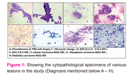
Figure 1 showing stained cytological specimens of patients. A: Fibroadenoma, B: Proliferative Breast Disease with atypia, C: Fibrocystic changes, D: Atypical Ductal Hyperplasia, E: Infiltrating Ductal Cell Carcinoma all A to E are stained with Haematoxylin and Eosin stain [H & E] magnification is 40X. F: Lobar carcinoma, G: Metaplastic Carcinoma, H: Medullary Carcinoma, F to H stained with May-Grunwald Giemsa [MGG] stain.
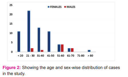
Out of n=73 cases n=64 (87.67%) were females and n=9 (12.33%) were male cases. From the different age groups seen in the study the most commonly involved age group was 21 – 30 years with n=24 (32.87%) cases, followed by 31 – 40 years with n=14 (19.18%) cases the other distribution of cases age-wise is given in figure 2.
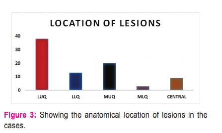
The anatomical location of the lesions was carefully studied, the following observations were made in the study. The Lateral upper quadrant was the location of the highest number of lesions n=38(46.34%) cases. The Lateral Lower Quadrant had n=13(15.85%) of cases, the Medial Upper Quadrant was the location of lesions in n=24(29.27%) cases being the second-highest and Medial Lower Quadrant was involved in n=3(3.66%) least involved quadrant and Central area was involved in n=9(10.97%) cases (figure 3).
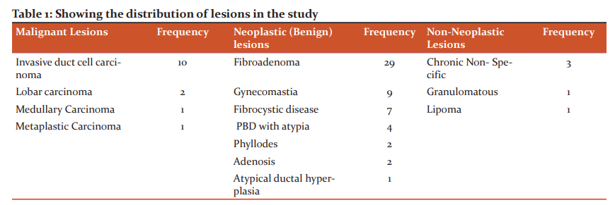
Out of the total n=73 cases, n=14 cases were found to malignant, n=54 cases were benign and n=5 cases were non-neoplastic lesions. Among the malignant lesions diagnosed the Invasive duct cell carcinoma was found in 71.28% of out of the total n = 14 cases of malignancy. In non-neoplastic lesions out of n=54 cases, fibroadenoma was diagnosed in 53.70% cases, followed by gynecomastia in 16.67% cases. In Non-neoplastic lesions, out of n=5 cases, chronic non-specific lesions were found in 60% and 20% each of granulomatous lesions and lipoma cases were diagnosed with details in table 1.
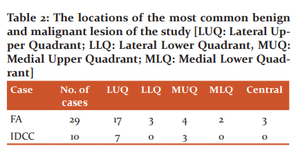
From the total n=29 Fibroadenoma cases 58.62% cases were in the Lateral upper Quadrant and 13.79% cases in the Medial Upper Quadrant and 10.34% were found in the Lateral Lower Quadrant and Central area 6.89% cases were detected. In the malignant cases, Invasive duct cell carcinoma (IDCC) was diagnosed in n=10 cases out of which 70% cases were in the Lateral upper quadrant and 30% cases in Medial Upper Quadrant (Table 2).
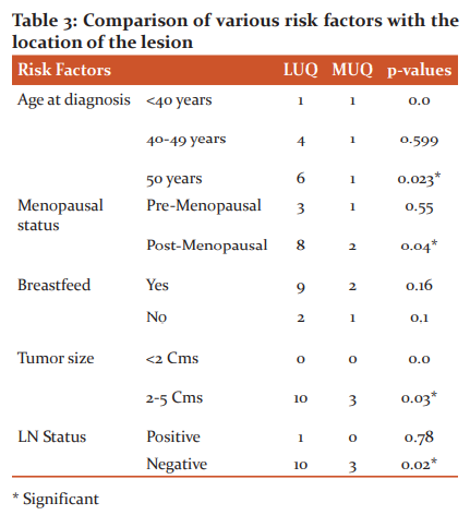
A comparison of the risk factor and the location of lesions was done. The result showed that the presence of a lesion in the Lateral upper quadrant was positively correlated with age and post-menopausal women. It was also found the larger tumour sizes were more often located on the lateral upper quadrant and lymph node-positive status was also significantly related to the presence of lesions in the lateral upper quadrant given in table 3.
The most common location of IDCC in our study was the lateral upper quadrant with SEER coding for IDCC was 2. Nottingham histologic score BR grade to SEER code is shown in table 4.

Discussion
This study was conducted to determine the prevalence of breast lesions in the various quadrant and to assess the quadrant of location as a risk factor for breast lesions. Also, to correlate cytomorphological features of FNAC (Fine Needle Aspiration Cytology) of the spectrum of lesions encountered with the importance of quadrant of location determines the prognosis of the breast lump. The most common age group involved in our study includes females of 3rd decade.12 Haque et al. have reported the 4th decade to be the most common age group of females in their study.13 Sixth decade was the most common age group in males in our study. In the current study, we found the Left breast involvement is more commonly found in n=26 (49.32%) in females The Right side involved in n=28 (38.36%) and bilateral involvement was found in n=9 (12.32%) cases. Prakash HM et al. 14 in their study found more common involvement of the left side. Palpable breast lesions were slightly more common on the left side in agreement with the results of the current study.15-17 However, Chandanwale S et al.18 found the right breast was more frequently involved in their cases. In this study, we found among all the four quadrants the upper and outer quadrants (superolateral) quadrant was involved is most involved n=38(46.34%) cases. The common occurrences of breast lesions in the superolateral quadrants have been also found in other similar studies.16-20 One of the possible explanations for the common occurrence of breast cancer in the upper and outer quadrant is due to the fact the lymphatic drainage of the breast in this region is poor because of inadequate support and a greater amount of target epithelial tissue in this region. In this study, the lump was the important presenting feature reported by 93.15% of patients, the pain was reported by 2.7% patients, and 4.11% reported of discharge. Goyal et al.21 reported that lump was the main presenting symptom in the majority (57.06%) of patients. Kumar et al.22 showed a painless mass in the breast (60.78%) followed by painful mass (13.73%) and associated features like ulceration of the skin, retraction of nipple and nipple discharge. In the current study out of the total n=29 Fibroadenoma cases 58.62% cases were in the Lateral upper Quadrant the calculated risk of fibroadenoma in the study was 3%. Selva Kumaran et al.23 also found the common presentation of fibroadenoma in the lateral upper quadrant agreeing with the results of the present study. The malignant cases Invasive duct cell carcinoma (IDCC) was diagnosed in n=10 cases out of which 70% cases were in the upper Lateral quadrant and 30% cases in Medial Upper Quadrant and the SEER score was 2. Tumour location with the upper outer quadrant has been reported with multiple populations including Chinese, Danish, the United Kingdom, and the USA.24-27 It has also been suggested the tumours in the upper outer quadrant have increased with time and the association of tumour with this location is associated with improved prognosis and data have suggested a trend to reduction in breast cancer mortality. In this study also all the malignant cases were operated, and the excised tissue was then examined histologically for confirmation of diagnosis and SEER grading. The patients were followed for 1 year and no mortality was reported with the cases. Our study demonstrated that the ability of patients to seek medical care when the location of tumours in the upper lateral quadrant was better due to the better ability of the patients to palpate the lump this could be one of the important factors in the better prognosis. The fact that the tumours located centrally are generally harder to detect and hence the patients seek the treatment after considerable progress of the tumour.
Conclusion
The most common age group of breast lesions was the 6th decade in males and 3rd decade in females. There was left breast predominance and the lump was the most frequent clinical symptom in both the genders. Among the benign lesion, fibroadenoma showed the highest occurrence whereas in malignant lesions, it was IDCC and the Lateral outer quadrant showed the highest involvement for fibroadenoma among the benign lesions and IDCC among the malignant lesions. Therefore, it can be concluded that lesions occurring in the lateral upper quadrant may carry a good prognosis.
Acknowledgement: Authors thank the Dept of Pathology, Kakatiya Medical College and MGM Hospital, Warangal, Telangana State, India for their support in the conduction of this study.
Source of support: Nil
Conflict of interest: None
Ethical Permission: Obtained [RC No. IEC/KIMS/Pathology/2018]
References:
-
Jain SB, Jain I, Srivastava J, Jain B. A clinicopathological study of breast lumps in patients presenting in surgery OPD in a referral hospital in Madhya Pradesh, India. Int J Curr Microbiol App Sci 2015; 4:919-923.
-
Aniketan KV, Manjunath S. Kotennavar, Tejaswini Vallabha. Triple assessment of breast lumps, an effective method for diagnosis in limited resources setting. Int J Cur Res Rev 2015; 7(22):13-16.
-
Ahmedin Jemal, Freddie Bray, Melissa M. Center, Jacques Ferlay, et al. Global Cancer Statistics. Cancer J Clin 2011;61:69–70.
-
Saxena S, Rekhi B, Bansal A, Bagga A, Chintamani, Murthy NS. Clinico-morphological patterns of breast cancer including family history in a New Delhi hospital, India-A cross-sectional study. World J Surg Oncol 2005;3:67.
-
Agarwal G, Pradeep PV, Aggarwal V, Yip CH, Cheung PS. The spectrum of breast cancer in Asian women. World J Surg 2007;31:1031–1040.
-
National Cancer Registry Program: Consolidated report of the Hospital-based Cancer Registries, 1990-1996. Indian Council of Medical Research. New Delhi, 2001. Available from [https://ncdirindia.org/ncrp/Annual_Reports.aspx [Accessed on 12 Feb 2020]
-
Baum M. Modern concept of the natural history of breast cancer: A guide to design and publication of trials of the treatment of breast cancer. Eur J Cancer 2013;49:60-64.
-
Rummel S, Hueman MT, Costantino N, Shriver CD, Ellsworth RE. Tumour location within the breast: Does tumor site have the prognostic ability? Cancer 2015; 9(552):1-10.
-
Sohn VY, Arthurs ZM, Sebesta JA, Brown TA. Primary tumor location impacts breast cancer survival. Am J Surg 2008; 195:641-644.
-
Turner-Warwick RT. The lymphatics of the breast. BMJ 1957; 46:574–572.
-
Vendrell-Tore E, Setoain-Quinquer J, Domenech-Torne FM. Study of normal mammary lymphatic drainage using radioactive isotopes. J Nucl Med 1972; 13:801–805.
-
Godwins E, David D, Akeem J. Histopathologic analysis of benign breast diseases in Makurdi, North Central Nigeria. Int J Med Sciences 2011; 3:125-128.
-
Haque, Tyagi, Khan, and Gahlut. Breast lesions: a clinico histopathological study of 200 cases of a breast lump. JAMA 1980;150:1810-1814.
-
Prakash HM, Jyothi BL, Ramkumar K, Konapur PG, Shivrudrappa AS, Subramaniam PM, et al. The value of systematic pattern analysis in FNAC of breast lesions: 225 cases with cytohistological correlation. J Cytol 2011; 28:13-19.
-
Meena SP, Hemrajani DK, Joshi N. A comparative and evaluative study of cytological and histological grading system profile in malignant neoplasm of breast An important prognostic factor. Indian J Pathol Microbiol 2006;49:199–202.
-
Reddy DG, Reddy CR. Carcinoma of the breast, its incidence, and histological variants among South Indians. Indian J Med Sci 1958;12:228-234.
-
Clegg-Lamptey J, Hodasi W. A study of breast cancer in korlebu teaching hospital: Assessing the impact of health education. Ghana Med J 2007;41:72–77.
-
Chandanwale S, Rajpal M, Jadhav P, Sood S, Gupta K, Gupta N Pattern of benign breast lesions on FNAC in consecutive 100 cases: a study at tertiary care hospital in India. Int J Pharma Bio Sci 2013;4:129-138.
-
Rocha PD, Nadkarni NS, Menezes S. Fine needle aspiration biopsy of breast lesions and histopathologic correlation. An analysis of 837 cases in four years. Acta Cytol 1997; 41:705-712.
-
Zuk JA, Maudsley G, Zakhour HD. Rapid reporting on the fine-needle aspiration of breast lumps in outpatients. J Clin Pathol 1989; 42:906-911.
-
Goyal V, Nagpal N, Dhuria N, Monga S, Gupta M. Spectrum of Clinical
Profile and Treatment Aspects of Breast Cancer In Malwa Region Of Punjab. J Adv Med Dent Scie Res 2015;3: S4-S8.
-
Shambhu Kumar Singh, Deepak Pankaj, Rajesh Kumar, Riyaz Mustafa. A clinicopathological study of a malignant breast lump in a tertiary care hospital in the Kosi region of Bihar, India. Int Surg J 2016; 3:32-36.
-
Selvakumaran S, Sangma MB, Study of various benign breast diseases, International surgery journal, Selvakumaran set al. Int Surg J 2017;4:339-343.
-
Kroman N, Wohlfahrt J, Mouridsen HT and Melbye M. Influence of tumor location on breast cancer prognosis Int J Cancer 2003;105:542–545.
-
Sohn VY, Arthurs ZM, Sebesta JA, and Brown TA. Primary tumour location impacts breast cancer survival Am J Surg 2008;195:641–664.
-
Wu S, Zhou J, Ren Y, Sun J, Li F. Tumor location is a prognostic factor for survival of Chinese women with T1-2N0M0 breast cancer Int J Surg 2014;12:394–398.
-
Darbre PD. Recorded quadrant incidence of female breast cancer in Great Britain suggests a disproportionate increase in the upper outer quadrant of the breast. Anticancer Res 2005;25:2543–2550.
|


 This work is licensed under a Creative Commons Attribution-NonCommercial 4.0 International License