IJCRR - 8(4), February, 2016
Pages: 51-53
Date of Publication: 21-Feb-2016
Print Article
Download XML Download PDF
SEXUAL DIMORPHISM OF MASTOID PROCESS IN DRIED SKULLS OF NORTH KARNATAKA POPULATION
Author: Rajendrakumar D. Virupaxi, Sanjay Kumar Yadav, Suresh P. Desai, Veereshkumar Shirol
Category: Healthcare
Abstract:Objectives: Morphometric and morphological study of the bones play an important role in determination of sex, age and identification of an individual and skull is the second important tool to pelvis in this regard. Mastoid process is a dimorphic bone situated at the basolateral region of the skull and remains least damaged due to its position. This study was an attempt to evaluate the use of mastoid process in the determination of sex. Methodology: This cross sectional study was carried out in the settings of Department of Anatomy and Forensic Medicine at North Karnataka for the period of one year from July 2014 to June 2015. A total of 100 complete undamaged dried skulls of known sex were used for the study, (50 male and 50 female skulls). Length of the mastoid process on both sides was recorded using the Frankfurt's plane. Measurements were taken by using Vernier caliper. Results: The mean mastoid process length among males at right was 30.20 \? 2.64 mm and at left it was 30.70 \? 3.09 mm compared to 26.20 \? 2.57 mm in females at right and 26.20 \? 3.49 mm at left respectively. The difference observed was statistically significant (p< 0.001). Conclusion: The mastoid length in females is significantly less compared to males at right as well as left.
Keywords: Mastoid process, Mastoid process length, Skull, Sexual dimorphism
Full Text:
INTRODUCTION
Identification of an individual and sex is one of the important factors in many circumstances like, mutilated bodies, skeletal remains, wars, and suicide and homicide cases. In the living identification is easy as many characters, particular physique and traits are unique to the individuals. But in cases of fragmented and mutilated bodies it is very difficult to identify the individual. Study of skeleton using individual bones exhibiting sexual dimorphism has been reported among different populations.1 Determination of the sex of human or human skeletal remains is an important initial step in its identification.
This skillful process is carried out by forensic and anatomy experts. Anthropological knowledge of human skeleton is one of the important parameters in identification of this biological profile.2 The first indicator for the sex determination is the pelvic bones and pelvis which is more accurate than the skull bones.3 The above study also confirmed by Saini et al,4 in 2012. Several studies have shown that cranium is also an excellent indicator for sexual dimorphism by morphometric and morphological analysis. It is probably the second best region of the skeleton next to the pelvis for this purpose.5 Krogman states that skull is the most dimorphic and easily sexed portion of skeleton after pelvis, providing up to 92% reliability6 Bayers2 and Pickering and Bachman7 also presented that skull plays the second best role to estimate the sex of the dead body. The skull measurements vary significantly in different populations of the world.
The mastoid process of the skull plays a vital role in determination of the sex as it is the most dimorphic bone present at the baso-lateral region of the skull. It is least vulnerable in this position. The mastoid region is favorable for sex determination for two reasons, the compact nature of the petrous bone and its protected position in the skull. Even in old age it remains intact. Usually skull is heavier in males as compared to females. Same thing holds good to mastoid process also, i.e. larger in males than in females. But it is subjective and it cannot be relied. A number of research studies have done in western countries on these aspects. Nagaoka T method was employed by some experts using two parameters on both sides of skull i.e. height and width of the mastoid.8
Some researchers used the triangular area between the points porion, mastoidale and asterion, measured from xerographic copy of skulls.9 Very few works have been done on this in India especially in Northern Karnataka population. Considering this entire scenario an attempt is made to determine the sex by using the mastoid length in this area. In present study 100 adult human dried skulls (50 male and 50 female skulls) of North Karnataka population were studied to determine the sexual dimorphism of mastoid process. A quantitative blind study of the mastoid length was studied by using the Frankfurt’s plane.
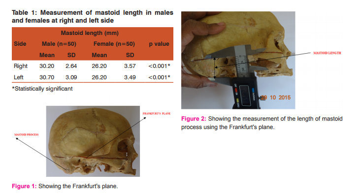
MATERIALS AND METHODS This cross sectional study included 100 dried skulls of known sex, (50 male and 50 female skulls) taken from KLE University’s department of Anatomy and Forensic Medicine. Written permission was taken from the respective departments. Adult normal skulls without any damage were taken for the study. Deformed skulls were excluded from the study. All were native of North Karnataka Region. Frankfurt’s plane was chosen and marked on the skull.
This line starts from the infraorbital margin, and upper border of the external acoustic meatus. (Figure 1). From this line on the mastoid process a vertical length was measured up to the tip of the mastoid process.10 With the help of digital verneir caliper length of the mastoid process was taken from the upper border of the external auditory canal at the level of the Frankfurt’s plane to the tip of the mastoid process on both sides. Figure 2 shows the measurement of the length of the mastoid process.
Statistical methods The mastoid process length of skulls was expressed as mean ± standard deviation (SD0 and median with range. The comparison of mean length was done using independent sample ‘t’ test. Probability value (p) of <0.050 was considered as statistically significant.
RESULTS AND DISCUSSION The results obtained from the observation showed that, the mean mastoid length in male was 30.2mm (Range- 25.2 to 36.65) with the standard deviation of 2.64 on right side. In female skulls the mean mastoid length was 26.2mm on the right side. (Range- 21.04 to 33.3).The standard deviation obtained was 3.57. (Table.No.1). Mastoid mean length is greater in males than in females which was significant (p value <.001). The male left side mastoid mean length showed 30.7mm (Range 24.02 to 36.7) with the standard deviation of 3.09. Whereas left side female mastoid mean length was 26.2 mm with the range of 21.03 to 33.30.
The standard deviation obtained was 3.49. (Table No. 1.).In left side also the sexual dimorphism was significant as the p value is <0.001. In males mean difference between Right and Left side is 0.49 with the standard deviation of 1.77. (Paired t=1.179). No significant mean difference found in both right and left side (p=0.053). In females mean difference between Right and Left side is0 .01with the standard deviation of 1.63 (Paired t=0.055). There was no significant mean difference found in Right and Left side (p=0.956). From the above study it showed that in male skulls the mastoid length was more as compared to females, p<0.001 which was significant. Applying Gausian distribution rule there was no significant mean length difference on right and left side mastoid length measurements. Hoshi H11. Studied the mastoid process length and divided the mastoid process into three categories viz, M, N and F types. Male, neutral and female categories. As the mastoid length is more in males as compared to females, skull lies on mastoid process in males and lies on occipital condyles in females when skulls are kept on flat surface.
Many studies have been conducted on mastoid length in western countries. In Caucasian population,12 the mean mastoid length was 28.06 mm in males and in females it was 25.10 mm. As compared to Negroes, mean length was less in Caucasians. In Nigroes male mastoid length was 30.32 mm whereas female mastoid length was 26.34mm. Our study is comparable to Negro population. Mastoid length measured by the use of points porion, mastoidale and asterion is the best method for the sexual dimorphism.13. Still mastoid length was used as the better predictor for the sexual dimorphism.14
Sumati and Puranik also computed that the sex determination by measuring the mastoid length in North Indian population.15 Same study also done in China by Song et al.16 and in India by Patil and Mody.17 In our study, it showed clearly that there was variation between male and female mastoid length. Statistically also it was proved that t-test and p values showed significant difference between male and female mastoid length i.e. p<.001. The present study supports the mastoid length as the tool for sex determination.
CONCLUSION Present study showed that mastoid length is less in females as compared to males which is also proved statically (p<.001). This shows that mastoid length is the reliable parameter for sexual dimorphism as our study supports other studies conducted worldwide and also in Indian population.
ACKNOWLEDGEMENT I am very thankful to Shri M. D. Mallapur Professor Department of Community Medicine who helped me in stastical analysis of this study. I also thankful to Mr. Harinarayan and Mr. Shalik who helped me in taking photographs in this study.
References:
1. Bilge Y, Kedici PS, Alacoc YD, Ulkuer KU, Likyaz YY. The identification of a dismembered human body; A multidisciplinary approach. Forensic Sci Int 2003; 137(2-3):141-6.
2. Bayers SN. Introduction to Forensic Anthropology. Boston: Pearson Education; 2008.
3. Phenice TW. A newly developed visual method of sexing the os pubis. Am J Phys Anthropol 1969;30:297-301.
4. Saini V, Srivastava R, Rai RK, Shamal SN, Singh TB, Tripathi SK. Sex estimation from the mastoid process. Rev Hosp Clin Fac Med S Palo 2012;58(1):15-20.
5. Bass WM. Human Osteology; A Labortary, Field manual of the Human Skeleton. Columbia: Missouri Archeological Society; 1971.
6. Spradley MK, Jantz RL. Sex estimation in forensic anthropology: skull versus postcranial elements. J Forensic Sci 2011;56(2):289-96.
7. Bachman DL, Pickring RB. The use of Forensic Anthropology. Boca Raton, FL: CRC Press; 1977.
8. Nagoka T, Shizushima A, Sawada J, Tomo S, Hoshino K, Sato H, et al. Sex determination using mastoid process measurements: Standards for Japanese human skeletons of the medieval and early modern periods. Anthropology Sic 2008;116: 105.3.
9. Paiva LA, Segre M. Sexing the human skulls through the mastoid process. Rev Hosp Clin Fac Med 2003;58/1:15-20.
10. Passey J, Mishra SR, Singh R, Sushobhana K, Singh S, Sinha P. Sex determination using mastoid process. Asian J Med Sci 2015;6(6):93-5.
11. Hoshi H. Sex difference in the shape of the mastoid process in norma occipitalis and its importance to the sex determination of the human skull. Okajma’s Folia Ana Japonica 1962;38:309-17.
12. Giles F, Elliot U. Sex determination by discriminant function analysis of crania. Am J Phys Anthropol 1963;21:53-68.
13. Nidugala H, Avadhani R, Bhaskar B. Mastoid process- A tool for sex determination, an anatomical study in South Indian skulls. International J Biomed Res 2013;04(02): 106-10.
14. Dasgupta A, Banerjee A, Kumar A, Rao SR, Jose J. Discriminant Function Analysis of mastoid Measurements in sex determination. J Life Sci 2012;4(1):1-5.
15. Patnaik SVVG. Determination of sex from mastoid process by discriminant function analysis. J Anat So India 2010;59(2):222- 8.
16. Song HW, Lin ZQ, Jia JT. Sex diagnosis of Chinese skulls using multiple stepwise discrinant function analysis. Forensic Sci Int 1992;54(2);135-40.
17. Patil KR, Mody RN. Determination of sex by discriminant function analysis ans stature by regression analysis a lateral cephalometric study. Forensic Sci In 2005;147(2-3):175-80.
|
 IJCRR
IJCRR 


 This work is licensed under a Creative Commons Attribution-NonCommercial 4.0 International License


