IJCRR - 13(2), January, 2021
Pages: 55-61
Date of Publication: 16-Jan-2021
Print Article
Download XML Download PDF
Comparative Study to Evaluate the Apical Sealing Ability of MTA Plus and Biodentin Using a Bacterial Leakage Model: In Vitro Study
Author: Revathi Bashyam, Ramesh Krishnan, Kruthika Murali, Nandhini B. Selvarajan, Suresh Kumar Vasaviah, Vinola Duraisamy
Category: Healthcare
Abstract:Introduction: The apical vessels may also be severed or damaged enough to interfere with the normal reparative process. Radicular lesions develop when microorganisms of sufficient pathogenicity and number gain access to periradicular tissues. When microorganisms are competent to colonize in an extraarticular biofilm, they may be principally resistant to abolition by host defence mechanisms and antimicrobial agents. Objective: To compare the apical sealing ability of two materials MTA plus andBiodentin as well as to evaluate bacterial microleakage using a bacterial leakage model for 28 days. Methods: Sixtysingle rooted extracted permanent teeth were selected. All the teeth should have straight pulp canals were included in the study while the tooth with root caries, multiple canals, lateral radicular canals Calcifications, periradicular resorptive changes excessive curvatures, developmental defects, root fractures, with internal resorption, previously end odontically treated cracks or root defects were excluded from the study. Samples used in this in vitro study had been extracted for orthodontic or periodontal reasons. Results: Biodentineand controls on day 1. There was no leakage observed for MTA plus and Biodentine. Only one sample of positive control leaked. The comparison was done using the Kruskal-Wallis test and the p-value was found to be 0.392 which was statistically not significant. Biodentineand control groups onday5.Onday5,13.33%ofMineral trioxide aggregate (MTA)plus group leaked(2 out of 15 samples) against 40% of positive controls leaked (6 out of 15 samples). Conclusion: The resent study concludes that MTA plus and Biodentinehave good apical sealing ability against E.faecalisat 28days. Biodentinewas better in performance than MTA plus in terms of apical sealing for accurately measuring the microleakage and quantify it further in-vitro models can be pursued.
Keywords: Pulpal Hyperaemia, Periradicular Tissues, Pulpotomy, Apexogenesis, Microorganisms
Full Text:
INTRODUCTION
Trauma to a tooth is invariably followed by pulpal hyperaemia, the extent of which cannot be always determined. Congestion and alteration in the blood flow in pulp initiate irreversible degenerative changes, which can result in pulpal necrosis. The apical vessels may also be severed or damaged enough to interfere with normal reparative process.1 Radicular lesion develop when microorganisms of sufficient pathogenicity and number gain access to periradicular tissues.2 Because of the complexity of the root canal system and the difficulty to completely clean it using the present techniques and instruments, root canals cannot always be adequately treated using a non-surgical orthograde approach.3 Periradicular surgery, when indicated should be considered an extension of non-surgical treatment as aetiology of the disease process and the objectives of the treatment are the same.
The fundamental goal of a root-end filling material is to give an apical seal that forestalls the development of microbes and the dispersion of bacterial items from the root waterway framework into the periapical tissues. It has been recommended that an ideal root-end filling material ought to cling to the planning dividers shaping a tight seal in the root trench framework. It ought to be anything but difficult to control, radiopaque, dimensionally steady, and non-absorbable. Also, an ideal root-end filling material should not be affected by the presence of moisture.5-7
MTA was first recommended as a root-end filling material when developed, but it has been used for pulp capping procedures, pulpotomy, apexogenesis, apical barrier formation in teeth with open apexes, repair of root perforations, and as a root canal filling material. The advantages of ProRootMTA (Mineral trioxide aggregate)as a root-end filling material, concerning the other mentioned alternatives, include greater sealing ability andbettermarginal.6-8 But it has certain clinical disadvantages such asit is tedious to handle and have a long setting time which could be overcome by MTA plus. But the material MTA plus is not much explored.9
Biodentine (SeptodontUSA) was recently introduced to the dental market. This new bioactive cement has dentin - like mechanical properties and can be used as a root-end filling material, as well as a repair material for root perforations and resorptions. Biodentinecan is used in both the root and crown.10-12 So the present study aims to compare the apical sealing ability of two materials MTA plus andBiodentin as well as to evaluate bacterial microleakage using a bacterial leakage model for 28 days.
MATERIALS AND METHODS
The present study was conducted in the department of Pedodontics at Vinayaka Mission’s Sankarachariyar Dental College, Vinayaka Mission’s Research Foundation (Deemed to be University), Salem, Tamilnadu, India. Sixtysingle rooted extracted permanent teeth were selected. All the teeth should have straight pulp canals were included in the study while the tooth with root caries, multiple canals, lateral radicular canals Calcifications, periradicular resorptive changes excessive curvatures, developmental defects, root fractures, with internal resorption, previously end odontically treated cracks or root defects were excluded from the study. Samples used in this in-vitro study had been extracted for orthodontic or periodontal reasons. The study was approved by the institutional research committee and institutional ethical committee. The sample size was determined scientifically. Considering Alpha: 0.05 Power of the study: 0.8 Effect size: 0.4. Therefore, the estimated sample size for the study was 15 for each group. This was calculated using the software G Power 3.1. 15 samples were randomly categorized to each of the experimental group and control group.
Group-1 : MTA PLUS group (n=15)
MTA plus was manipulated as per the manufacturer’s instructions and incrementally placed into the root end cavity and condensed well along the cavity walls and against a flattened file which was placed in the root canal. Initial set was allowed, after 48 hrs the k-file was removed. Each tooth was placed in sterile gauze piece soaked in saline for 48 hrs for the initial hard setting.
Group-2 : Biodentine group (n=15)
Biodentinewas mixed as per the manufacturer’s instructions in an encapsulator and incrementally placed into the root end cavity and condensed well along the cavity walls and against a flattened file which was placed in the root canal. The initial set was allowed, after 24 hrs the k-file was removed. Each tooth was placed in sterile gauze piece soaked in saline for 48 hrs for the initial hard setting.
Group-3 : Positive control (n=15)
ThermoplasticizedGP was used without sealer to fill the root end cavity and condensed against the flattened K - file. The file was removed after 48 hrs and placed in moist sterile gauze piece.
Group-4 : Negative control (n=15)
Root end preparations were filled with sticky wax and condensed against the k-file. After 48 hrs, the file was removed and placed in moist sterile gauze piece.
Materials
Materials used For control and experimental group were MTA plus (PrevestDenproLtd, Jammu, India), Biodentine (Septodont, Saint MaurdesFosses, France), Thermo plasticized Guttapercha(Bee fill 2 in 1 obturation device, Germany), Stickywax (Hiflex, UK). Materials used For canal preparation were ultrasonic diamond tips (Kistips),5.25% sodium hypochlorite (Hyposol, PrevestDenpro),17%EDTA(DoloEndogelTM,PrevestDenpro). Materials used For bacterial leakage model were scintillation vials, orthodontic resin, cyanoacrylate, phenollactosered broth (SigmaAldrich), E. Faecalisin TSB agar to1×109CFU/ml. Types of equipment used in the study were ethylenedioxide sterilization chamber
Statistical analysis
This was done using Kruskal-Wallis test and post-hoc-Tuckey test to compare the intergroup difference and statistical significance was set at the level of P=0.05.
RESULTS
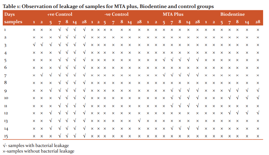
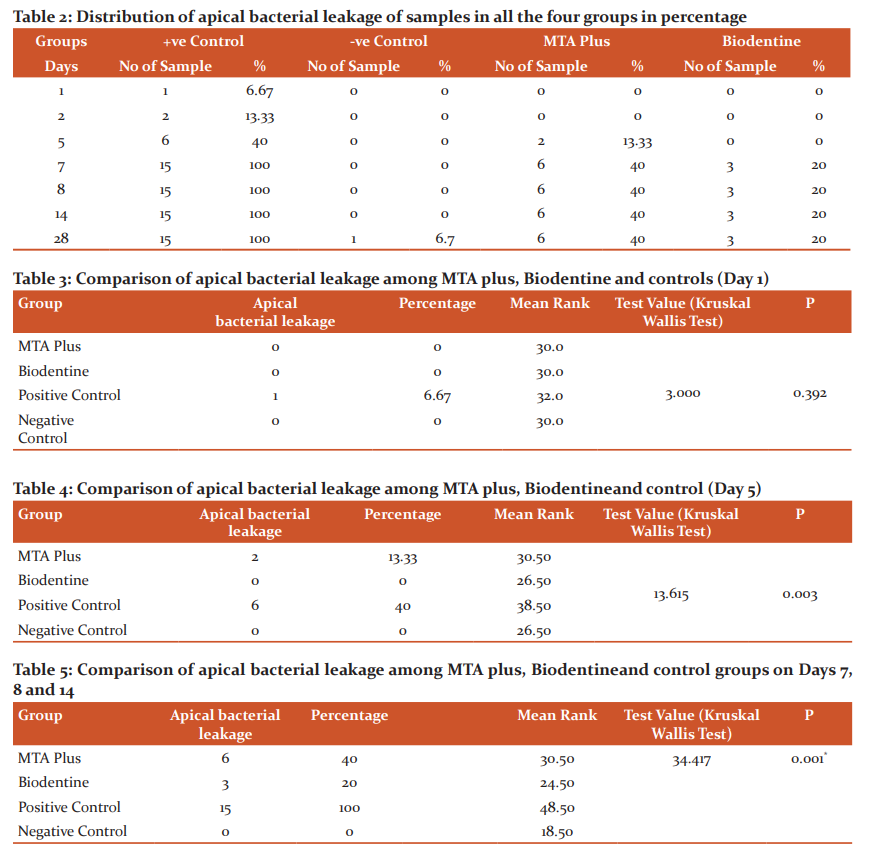
Leakage of samples for MTA plus, Biodentine and control groups and its percentage of failure has been shown in Table 1 and Table 2. Comparison of leakage of samples between MTA plus, Biodentine and controls on day 1. There was no leakage observed for MTA plus and Biodentine. Only one sample of positive control leaked. The comparison was done using the Kruskal Wallis test and the p-value was found to be 0.392 which was statistically not significant (Table-3)
On day 5, 13.33% of MTA plus group leaked (2 out of 15 samples) against 40% of positive controls leaked (6out of 15 samples). Statistical analysis was done using the Kruskal Wallis test and the p-value was found to be 0.003 which was statistically significant (Table-4)
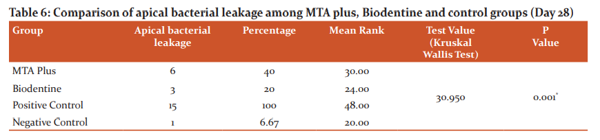
Table 5 illustrates the comparison of leakage of samples among MTA plus, Biodentine and control groups on day 7, 8 and 14. All the samples showed similar leakage on 7 th, 8th and 14th day. The percentage of samples leaked for, MTA plus was 40 % (6 out of 15 samples), whereas 20% of Biodentine showed leakage (3 out of 15 samples). When observed in control groups, 100% of positive controls leaked (15out of 15 samples); and negative control did not leak. Numerically, MTA plus samples leaked more than Biodentine group. Statistical analysis was done using the Kruskal Wallis test and the p-value was found to be 0.001 which was statistically significant.
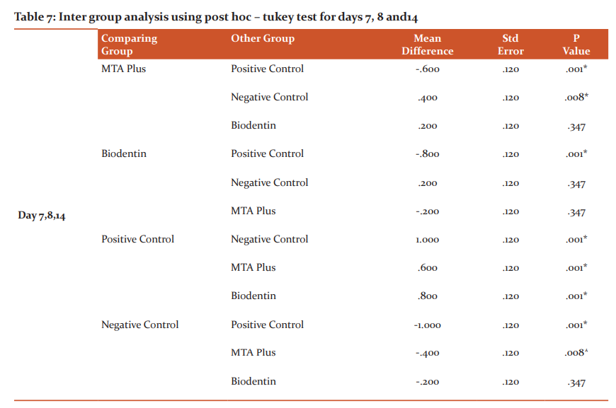
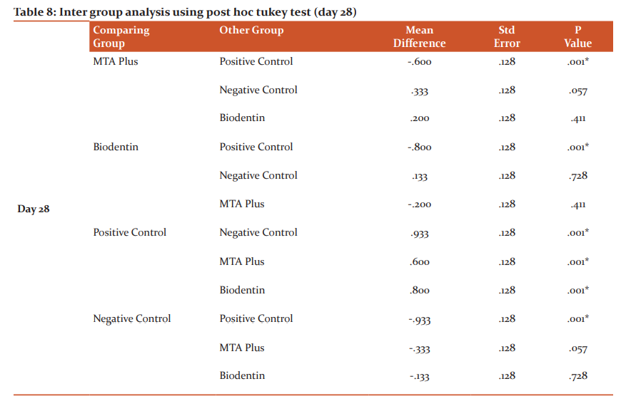
When the percentage of apical leakage was assessed at 28th day MTA plus samples showed 40%, Biodentine showed 20%, positive control showed100% and negative control showed 6.67% of leakage, in which MTA plus showed more leakage than Biodentine. Statistical analysis showed a significant p-value of 0.001 (Table-6). There was a statistically significant difference between the positive control and the MTA plus group and biodentine group and also negative control. MTA plus group showed a significant difference with both the control groups. Biodentine group showed significant difference with positive control. Intergroup comparison between MTA plus and Biodentine group had a mean difference of 0.200 and with a standard error of 0.120, the p-value was 0.347, which was statistically insignificant (Table 7). There was a statistically significant difference between the positive control and the MTA plus and the biodentine group and also negative control. Intergroup comparison between MTA plus and Biodentine group had a mean difference of 0.200 and with a standard error of 0.128, the p-value was 0.411, which was statistically insignificant (Table-8).
 Discussion
In the present study, these two tricalcium silicate cement are compared in terms of apical sealing ability. Several methods have been employed to evaluate apical microleakage. These include air pressure, neutron activation, radioisotope, electrochemical, fluid filtration, bacteria, and the use of the dyes. Numerous techniques such as scanning electron microscopy, transmission electron microscopy, and electron probe microscope analysis have been used to image and quantify leakage. There is no standardized leakage test to evaluate the sealing ability of endodontic materials.9 Bacterial leakage model can be used to study the bacterial penetration across the material. A bacterial leakage model was chosen for the present study because it is most relevant in clinical perspective.10 In vitro study with bacterial leakage test was conducted for 28 days. The thickness of the apical plugin this study is 3 mm as supported by Mehmet bani et al. 2015.11 The amount of apical microleakage was significantly lower for 3 and 4mm apical plugs than 1 and 2 mm subgroups of BiodentineandMTA in his study. In the present study, the rood end cavity was prepared with ultrasonic diamond tips. Khandelwal et al. 201512compared different retro preparations with MTA and Biodentine. Biodentinegroup prepared using ultrasonics for showed the best sealing than all the other tested groups. Irrespective of preparation techniques used, Biodentinestill showed better sealing than MTA. Preparation of the root end using ultrasonics showed less microleakage than but prepared teeth for both filling materials. In this study, machine trituration has done for biodentine as Gupta et al. 201513 reported more microleakage when Biodentine was manually manipulated. The setting time is one of the most clinically relevant factors to be considered. Hence in this study, all the samples were kept in moist gauze piece for a period of 48 hrs to allow an initial setting time. Long-setting period may induce clinical problems because of the failure of cement to maintain shape and support stresses during this period. Accelerated setting reduces the risk of dislodgement and contamination of MTA?like cement when used as root-end filling material, which is very well satisfied in Biodentine by addition of accelerators (CaCl2).14-16
In the present study, MTA plus showed more apical bacterial leakage than Biodentine. In the negative control group only one sample leaked at the end of the study. The leakage in negative control can be attributed to nail varnish failure. Among the positive control group, there was a 100% apical bacterial leakage indicating the need for an ideal apical sealing material for the retrograde fillings. Under the experimental conditions of this study, biodentine showed less leakage than MTA plus which was statistically insignificant. Biodentineand MTA plus showed significant difference than positive control which signifies that both the materials have the good apical sealing ability. The formation of CSH gel also reduces the porosity with time. The crystallization of the biodentine continues up to 4 weeks, therefore, improving the strength as well as other mechanical properties (sealing ability). The high mechanical strength of Biodentinemay is ascribed to the abolition of aluminates that lead to weakening and fragility of the set material as testified by the manufacturer. The thickness of the Ca?and Si-rich layers increased over time, and the thickness of the Ca?and Si-rich layer was significantly larger in Biodentine compared to MTA after 30 and 90 days, concluding that the dentine element uptake was greater for Biodentinethan forMTA.14,17,18
The sealing ability of a material is estimated by various phenomena such as porosity, marginal adaptation, and hydrophilicity. On mixing calcium silicate cement with water, many porosities and microchannels are produced and play a vital role in the hydration reaction, but may also influence the early sealing ability of the cement. Kokate and Pawar15 conducted a study that compared the microleakage of glass ionomer cement, MTA, and Biodentin when used as a retrograde filling material and suggested that Biodentinhas the least microleakage in comparison to other materials used which supports the current study.16,19 Sulthan17carried out a study to evaluate the pH and calcium ion release of MTA and Biodentin when used as root-end fillings. He concluded that Biodentine presented alkaline pH and the ability to release calcium ions similar to that of MTA. Blood contamination affected the push-out bond strength of MTA Plus irrespective of the setting time18.Formosa etal19. Found that the anti-washout gel changed the rheology and properties of the material. In particular, it was noted that while MTA mixed with water had a sandy consistency, MTA mixed with anti-washout gel had a far more vicious and rubbery consistency and are almost dough-like. This increased viscosity may explain from a purely physical standpoint, why MTA-AW developed the threshold strength of 3.92 MPa sooner than MTA. The anti washout gel added to MTA did not affect the radiopacity of resultant material the observed an increase in compressive strength of MTA-AW compared to MTA-W.
The present study has to be still explored with detail assessment of the leakage as it has certain experimental limitations invitro. It has certain limitations such as fewer sample size, limitation of in vitro model, quantifiable evaluation etc. The study can be further directed invitro by extending the longevity of the study and quantifying the microleakage. The above statements, however, should be addressed in future experiments before any conclusive recommendations can be made.
CONCLUSION
MTA plus and Biodentinehave good apical sealing ability against E.faecalisat 28 days. Biodentinewas better in performance than MTA plus in terms of apical sealing. For more accurate measurement of the microleakage further in vitro models can be pursued.
Acknowledgment: Authors acknowledge the enormous help received from the authors whose articles are cited and included in references to this manuscript. The authors are also grateful to authors/editors/publishers of all those articles, journals and books from where the literature for this article has been reviewed and discussed.
Conflict of Interest: Nil
Source of Funding: Nil
References:
-
McDonald RE, Avery DR, Dean JA. Management of trauma to the teeth and supporting tissues. 9th ed. St. Louis, Missouri: MOSBY. Dentis Child Adolesc 2011;404.
-
Siqueira JF, Lopes HP.Bacteria on the apical root surfaces of untreated teeth with periradicular lesions: a scanning electron microscopy study.Int Endodon J 2001;34:216.
-
TorabinejadM, Watson TF, Pitt Ford TR. Sealing ability of a mineral trioxide aggregate when used as a root-end filling material. J Endod1993;19:591–595.
-
Fogel HM, Peikoff MD. Microleakage of root-end filling materials. J Endod 2001;27(7):456 -458.
-
Ingle JI, Bakland LK. Endodontics. 5th ed. Baltimore: BC DeckerInc. (2002).
-
Torabinejad M, Watson TF, Pitt Ford TR. Sealing ability of a mineral trioxide aggregate when used as a root-end filling material. J Endod 1993;19:591–595.
-
Lee SJ, MonsefM, TorabinejadM. Sealing ability of a mineral trioxide aggregate for repair of lateral root perforations. J Endod1993;19(11):541-544.
-
TorabinejadM, Hong CU, Pitt Ford TR, Kettering JD.Antibacterial effects of some root-end filling materials. J Endod 1995;21(8):403–406.
-
Iwami Y, Shimizu A, Hayashi M, TakeshigeF, Ebisu. Three-dimensional evaluation of gap formation of cervical restorations. J Dent 2005;33:325-333.
-
Hirschberg CS, Patel NS, Patel LM, Kadouri DE, Hartwell GR. Comparison of sealing ability of MTA and EndoSequenceBioceramicRoot Repair Material: A bacterial leakage study. Quintessence Int 2013;44:e157–e162.
-
Bani M, Sungurtekin-Ekçi E, Odaba? ME. Efficacy of Biodentineas an Apical Plugin Nonvital Permanent Teeth with Open Apices: An In Vitro Study. BioMed Res Int 2015;3(5):231-335.
-
Khandelwal A, Karthik J, Nadig RR, Jain A.Sealing ability of mineral trioxide aggregate and Biodentineas root-end filling material, using two different retro preparation techniques-An in vitro study. Int J Contemp Dent Med Rev 2015;3:321-326.
-
Gupta PK, Garg, KalitaC, Saikia, SrinivasaTS, Satish G. Evaluation of Sealing Ability of Biodentineas Retrograde Filling Material by Using two Different Manipulation Methods: An In Vitro Study. J Int Oral Health 2015;7(7):111-114.
-
Khetarpal A, Chaudhary S, Talwar S, Verma M. Endodontic management of open apex using Biodentineas a novel apical matrix. Indian J Dent Res 2014;25:513-6
-
Kokate SR, Pawar AM. An in vitro comparative stereomicroscopic evaluation of marginal seal between MTA, glass ionomer cement &biodentineas root-end filling materials using 1 % methylene blue as the tracer. End odontol 2012;24(2):36-42.
-
Bolhari B, Ashofteh YK, Sharifi F, PirmoazenS. Comparative Scanning Electron Microscopic Study of the Marginal Adaptation of Four Root-EndFilling. J Dent (Tehran) 2015;12(3):226–234.
-
SulthanIR, RamchandranA, DeepalakshmiA, KumarapanSK. Evaluation of pH and calcium ion release of mineral trioxide aggregate and new root-end filling material. E J Dent 2012;2:166-169.
-
Aggarwal V, Singla M, MiglaniS, Kohli S. Comparative evaluation of push-out bond strengthofProRoot MTA, Biodentine, and MTA Plus in furcation perforation repair. J Conserv Dent 2013;16:462 -465.
-
Formosa LM, MalliaB, CamilleriJ. A quantitative method for determining the antiwash out characteristics of cement-based dental materials including mineral trioxide aggregate. Int Endod J 2013;46:179-186.
|
 IJCRR
IJCRR 


 This work is licensed under a Creative Commons Attribution-NonCommercial 4.0 International License


