IJCRR - 8(8), April, 2016
Pages: 37-43
Print Article
Download XML Download PDF
MAGNITUDE OF PULMONARY DISEASES - INCIDENTALLY DIAGNOSED ON AUTOPSY - AT LARGEST HOSPITAL AND MEDICAL COLLEGE OF RAJASTHAN
Author: Kalpana Mangal, Praveen Dhakar, Ajay Yadav, Karuna Gupta, Sapna Gandhi
Category: Healthcare
Abstract:Objective: A wide histopathological spectrum of preventable diseases can affect lungs. Many times radiological and clinical findings in pulmonary diseases are non specific hence pathology investigations can play important role in prompt diagnosis. In our 4 years cross-sectional study period (January2012- December2015), we have examined lungs of 1715 medico-legal autopsy cases. Objectives: 1. To know the histopathological spectrum of Lung diseases. 2. To know the frequency of pulmonary lesions in respect to age and sex. Materials and Methods: Representative sections from pulmonary lesions were taken and studied with the help of H and E and special stains wherever necessary. Results: The various pulmonary lesions were edema and congestion in 1325 (77.2%) cases, non tubercular pneumonia 137 (7.99%) cases, bronchopneumonia 15 (0.87%) cases, interstitial pneumonia 6 (0.35%) cases. Cases of tubercular pneumonia were 70 (4.08%) and pulmonary malignancies were 7 (0.41%) cases out of total pulmonary lesions. Pneumonias were occupying a large number, next to edema and congestion. Conclusion: The present study reveals the load and spectrum of lung disease in the largest Hospital and Medical College of Rajasthan, where infectious lesions (non-tubercular and tubercular pneumonias) are still the most common preventable causes of morbidity and mortality; hence autopsy study can be of great value in improving the vision and diagnostic setups for better clinical assessment, timely diagnosis and therapy.
Keywords: Edema and Congestion, Pneumonia, Autopsy, ARDS, Emphysema
Full Text:
INTRODUCTION The lungs are ingeniously constructed to carry out their cardinal function: the exchange of gases between inspired air and blood.1 During their functional course, lungs can become vulnerable for a wide range of inflammatory, neoplastic and other lesions, and almost always involved secondarily by terminal events of cardiovascular causes.2 Millions of people all over the world suffer by various chronic respiratory diseases, which can be prevented, if diagnosed timely.3 Most of the time clinical and radiological findings in pulmonary diseases are non specific hence prompt pathology investigations and diagnosis are essential to improve patient’s survival and to reduce further morbidity and mortality.4 Autopsy is an important and most useful way to find out the condition of internal organs, for this a systematic examination of organs was undertaken according to standardised research protocol, to evaluate diseases or injury that may be present and to determine cause and manner of death.5In present study, status of lungs was examined grossly and microscopically. On gross examination we observed various findings like consolidation, nodule, scarring, fibrosis, collapse, or hyper inflated lungs as well as congestion. Pulmonary diseases are the significant cause of death worldwide affecting mostly 20-59 years age group. We performed this retrospective study to describe spectrum of various histopathologically proven pulmonary lesions and their incidences in respect to age and sex over a 4 year period that is January 2012 to December 2015.
MATERIAL AND METHODS Study was conducted in the department of pathology, SMS medical college Jaipur on 1715 specimens of lung from deceased of 0 - >70 years of age. We examined all lungs grossly as well as microscopically. The medical and clinical histories were traced. Gross examination of lungs included colour, volume (collapsed or inflated), and consistency, presence of any consolidation, nodule, bullae, scarring, fibrosis, infarction and congestion. All specimens were adequately fixed in 10% formalin, weighed, and dimensions measured. Sections from representative areas were taken and paraffin blocks were made following standard protocol. Four micron sections were cut and stained with haematoxylin and eosin according to standard procedures.6 Special stains were used wherever required. All the cases were analysed by descriptive statistics.
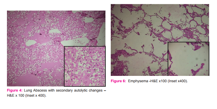
RESULTS In this study, we have observed that 1305 (76.09%) cases were from male deceased while 410 (23.91%) were of female, out of total 1715 cases. The male to female ratio was 3.18:1. Distribution of various pulmonary lesions out of 1715 autopsy cases was done (Table-1). After histopathological examinations, various pathological lesions were noted in 1549 (90.32%) cases while 166 (09.68%) cases were within normal limits. Out of these 1549 cases edema and congestion were the most common findings seen in 1325 (77.26%) cases, 1009 (76.15%) were males and 316 (23.85%) were females with male to female ratio (3.19:1). Most common age group affected was 20-59 years i.e.1106 cases (83.47%) and predominant age group in congestion and edema was 20-39 years with 621 cases (46.87%). Non tubercular pneumonia was the second most common finding noted in 137 cases (7.99%), 106 (77.37%) were males, females were 31 (22.63%) and male to female ratio 3.42:1. Most common age group affected was 20-59 years i.e. 108 (78.83%) cases. Tubercular pneumonia was the next common finding i.e. 70 cases (4.08%), 59 (84.29%) were males while 11 (15.11%) were female with male to female ratio 5.36:1. Most common age group affected was 20-59 years i.e. 59 (84.29%) cases. Next in order were malignant pulmonary lesions (including primary and secondary) 7 (0.41%) cases. All the cases found were males, most common age group affected was 40-59 years i.e. 4 (57.14%) cases, lung abscess and acute respiratory distress syndrome were 3 (0.17%) each with male to female ratio 1:2 in both the cases, as well as most common age group affected was 20-39 years in both the cases, as shown in table -2. Other findings were aspirational lung diseases 2 (0.12%), emphysema 2 (0.12%). Causes of death were taken out (Table-3). These were, due to unknown reason 812 (47.34%), sudden death 399 (23.27%), alcoholism 209 (12.17%), long illness 139 (8.11%), heart attack 122 (7.11%), poisoning 18 (1.08%), hanging 11 (0.63%) and drowning 5 (0.29%).
DISCUSSION The role and value of autopsy remains a vital component for the study and evaluation of the disease process, inspite of advances in diagnostic technology. There are large no. of cases of preventable respiratory diseases, still leads to morbidity and mortality.7, 8 and 9 In the present study age and sex wise distribution of pulmonary cases shows that the incidences were higher in 20-59 years age group. It is comparable to study done by V. Selvam et al where they found maximum incidences of pulmonary diseases in 3rd to 4th decades of life, men were more prone to death by diseases as compare to women 410 (23.91%)10 , while in study conducted by Chauhan et al, most common age group was 50->60 years.7 The reason may be that as men usually were bread earners and women usually indulged in household works. This makes the men more vulnerable for exposure of risk factors on their respective occupations as well as more addiction for smoking and alcoholism makes them prone for various diseases. Other studies were also showing comparable results with higher disease incidences in males as compare to females .11 In present study, we observed congestion and edema in 1325 (76.26%) cases while Chauhan et al found 182 (54.32%) and V. Selvam observed 32 (29.6%) cases. Non tubercular pneumonias were 137 (7.99%) in present study, while Chauhan et al and V. Selvam found.
Magnitude of Pulmonary diseases 14.62% and 10.2% respectively. Tubercular pneumonias in our study were 70 (4.08%) while these were 6.26% and 2.80% respectively in Chauhan et al and V. Selvam et al studies. Cases of emphysema were quite high in study conducted by V. Selvam et al i.e. 50% and Chauhan et al 7.06% while in present study they were only 0.12%. we found 0.17% each in lung abscess and ARDS cases, 0.12% cases of aspirational lung diseases. These cases were not found in studies conducted by Chauhan et al and V. Selvam et al. Malignant pulmonary lesions were found 0.41% in our study while it was 2.08% in study by Chauhan et al. No specific pathology was observed in 166 (9.68%) cases. [Table-4 ] The reason of difference amongst studies may be due to difference in sample size and pattern of studies.12 ,13 and 14
CONCLUSION The present study provides a comprehensive data about spectrum and frequency of Lung lesions, and up to our knowledge no such study has been done in our region. Autopsy studies can be of great value in improving the vision and diagnostic setup for better clinical assessment and timely diagnosis, to reduce further morbidity and mortality.
ACKNOWLEDGMENT
Authors wish to express and convey their sincere thanks and gratitude to all those who helped for completion of this research article. Authors acknowledge the immense help received from the scholars whose articles are cited and included in references of this manuscript. The authors are also grateful to author’s /editors/ publishers of all those articles, journals and books from where the literature for this article has been reviewed and discussed.
Authors acknowledge that we did not get any grants for this study; required funds were generated by ourselves. The Authors declare and acknowledges that we have no conflict of interest.


References:
1. Kumar Abbas, Aster, Robbins, Cotran. Pathologic basis of disease. South Asia Ed. Vol 2, Elsevier:8e
2. Manjit S Bal, P S Sethi, Anil K Suri, Vijay K Bodal, G Kaur. Histopathological pattern in lung autopsies, Journal of Punjab Academy of Forensic Medicine and Toxicology 2008; 8(2):29- 31.
3. John E Hall. Guyton and Hall Textbook of Medical Physiology, 13th Edition, Elsevier: Saunders, 2015.
4. Kasper, Fauci, Hauser, Longo, Jameson, Loscalzo. Harrison’s principles of internal medicine, 19th ed. Vol 2, Mc Graw Hill; Indian edition:2015.
5. KS Narayan Reddy, OP Murty. The essentials of Forensic Medicine and Toxicology, 33rd edition. JayPee Brothers:2014.
6. D. John Bancroft Manual of Histological techniques and their diagnostic application – Harry Charles 1994.
7. Chauhan, Madhuri Agrawal, Nirali Thakkar: Histopathological lesions in lung autopsy Journal of research in medical and dental science/vol.3/ issue 2/april-june.
8. Manjit et al: Journal of Punjab Academy of Forensic Medicine and Toxicology 2008.
9. Schwartz DA et al Herman CJ. Editorial response: the importance of autopsy in emerging and re emerging infectious diseases. Clin infect Dis 1996;23:248-54.
10. V. Selvam , R. Thamil Selvi, P.M. Subramaniam,Vijayanath: Journal of Pharmaceutical and Biomedical Sciences 2015.
11. Gupta BD et al: Journal of Forensic Medicine and Toxicology 2003.
12. Mangal K. et al : Incidence of Liver diseases – A retrospective Study of 1348 Autopsy cases at tertiary care centre Jaipur – Original Article, International Journal of Current Research – Vol.7,issue ,12,pp.23725-23729, Dec.2015.
13. Smita et al: study of liver pathology in autopsy cases-original article. International Journal of Current Research vol.6 issue 03 pp.5795-5797, March 2014.
14. Sotoudehamanesh R et al. silent liver diseases in autopsies from forensic medicine of Tehran. Archives of Iranian medicine 2006 oct.9(4):324-28.
|
 IJCRR
IJCRR 


 This work is licensed under a Creative Commons Attribution-NonCommercial 4.0 International License


