IJCRR - 11(4), February, 2019
Pages: 01-08
Date of Publication: 21-Feb-2019
Print Article
Download XML Download PDF
Ultrasonographic Biometry: Biparietal Diameter Measurement for Gestational Age Estimation in Singleton Pregnant Women of Karnataka
Author: Pranita R. Viveki, V. S. Shirol
Category: Healthcare
Abstract:Background: Appropriate intrauterine fetal growth and development are fundamental for newborn health and lifelong welfare. Bi-parietal diameter provides the closest correlation with gestational age in second trimester.
Objectives: 1. To establish the reference tables for bi-parietal diameter in normal singleton pregnant women from 20 to 38 weeks of gestation from Belagavi District, Karnataka. 2. To find out the predictive accuracy of gestational age determined by bi-parietal diameter measurements with gestational age by menstrual history.
Materials and Methods: The data was collected by using predesigned pretested questionnaire from September 2016 to January 2018. Total 768 singleton pregnant women with minimum 30 cases for each gestational week from 20 to 38 weeks of gestation were studied.
Statistical Analysis: Data was analysed by Percentages, mean, standard deviation, range, standard error, percentiles and regression equation etc.
Results: The regression equation derived for bi-parietal diameter (BPD) measurements was GA = (1.275 + (3.905 X BPD in cm) where \"R2\"- the proportion of variation in dependent variable (GA) was 0.944 and \"r\"- the correlation coefficient was 0.9. By using the common regression equations for GA from 20 to 38 weeks, there was a difference of 1.44 to 2.22 weeks in actual and predicted GA at 36 to 38 weeks of pregnancy. However, the same difference was less between 20 to 35 weeks of gestation.
Conclusion: The present study findings confirmed that the fetal BPD measurements significantly vary among different population groups. So generation of population specific reference tables by a large scale study is required for more precise reporting by ultrasonography.
Keywords: Biparietal Diameter, Gestational age, Ultrasonographic measurements
Full Text:
Introduction:
The correct clinical diagnosis of fetal growth disturbances has important implications for proper prenatal care and for determination of the delivery time. Many curves and reference tables for fetal biometry have been published in the literature, using mean values of the bi-parietal diameter (BPD), head circumference (HC), abdominal circumference (AC), and femur length FL, which allow estimation of the fetal weight. Fetal biometry by ultrasonography is the most widespread method used to establish gestational age, estimate fetal size and monitor its growth1,2. Researchers have been focusing in recent years on population specific fetal biometric parameter charts for various ethnic groups and the inter population variability in foetal growth patterns. Campbell S. et al3 and Waldenstrom U et al4 observed that bi-parietal diameter was more accurate predictive of expected date of delivery (EDD) than that calculated from the first day of last menstrual period (LMP).
Role of ethnicity on fetal biometry is a well known fact.5,6 Fetal nomograms need to be revised regularly.7 Acharya P. et al8 and various other studies9-11 have observed the smaller fetal measurements than the Caucasian fetal measurements and thereby they concluded that if western parameters are applied to all, the risk of over-diagnosis of intrauterine growth retardation (IUGR), and over or under estimation of gestational age (GA) and EDD in Indian population would be more.
Therefore, the present study was planned to measure the bi-parietal diameter by ultrasonography for different groups of GA from 20 weeks to 38 weeks in the normal singleton pregnant women from local population of Belagavi District, Karnataka and to find out the predictive accuracy of GA determined by BPD measurements with GA by menstrual history.
Objectives:
1. To establish the reference tables for bi-parietal diameter in normal singleton pregnant women from 20 to 38 weeks of gestation from Belagavi District, Karnataka.
2. To find out the predictive accuracy of gestational age determined by bi-parietal diameter measurements with gestational age by menstrual history.
Materials and Methods:
A random case series study was done from September 2016 to January 2018. 768 pregnant women with minimum 30 cases for each gestational week from 20 to 38 weeks of pregnancy referred to the Department of Radiology, Belagavi Institute of Medical Sciences (BIMS), Belagavi by antenatal clinic of Department of Obstetrics and Gynaecology (OBG) for routine antenatal scanning were studied after clearance from Institutional Ethics Committee. Antenatal cases with knowledge about exact date of LMP with regular menstrual cycles of 26-33 days12 for at least 3 cycles before conception, with delivery of a live baby with birth weight more than or equal to 2500 grams, fundal height corresponding to duration of pregnancy as per obstetricians finding, who delivered within one week of the expected date of delivery (EDD) and who delivered a newborn baby without any congenital abnormality were included in the study for analysis. Exclusion criteria were - pregnant women with age below 18 and above 35 years, with height below 140 cm, history of drug abuse, tobacco / gutkha use before and during pregnancy, oral contraceptive pills for 3 months prior to conception, and previous baby with low birth weight. Pregnant women with diabetes and hypertension detected during examination or developing later during pregnancy, women with multiple gestations, oligohydromnios, polyhydromnios, intrauterine growth retardation, or intrauterine death, women with uterine abnormalities like fibroids, bi-cornuate uterus, etc.
Method of collection of data:
A predesigned, pretested, structured proforma was used for each subject separately. The ultrasonographic examination of each pregnant woman fulfilling inclusion criteria, was done after submission of completely filled ‘Form F’ in compliance to PCPNDT (Pre-conceptional and Pre-Natal Dignostic Techniques) Act, duly signed by the women undergoing ultrasonography and the radiologist conducting ultrasonography. Using standard methodology, fetal BPD was measured from the leading edge of the echo from proximal skull surface to the leading edge of the echo from distal skull surface – outer to inner diameter. The reading of only first examination of each patient was included for the study purposes, although some patients underwent multiple ultrasonographic examinations during their pregnancy period.
The patients or close relatives were contacted for information about delivery like date of delivery, onset of labor (spontaneous or induced), mode of delivery (vaginal or caesarean section or assisted one), place of delivery, birth weight of the baby, any congenital anomaly detected in newborn baby, etc. The ultrasound examination was done by a single radiologist on one ultrasound machine - iU22 Philips make real-time machine with 3.5 MHz electronic curvilinear transducer.
Statistical Analysis:
The data was analyzed using MS Excel and Statistical Package for Social Sciences (SPSS) version 20. The basic categorical variables were reported as frequencies and percentages. The correlation of abdominal circumference with gestational age was plotted using scatter plots. The descriptive statistics (mean, standard deviation and range, standard error, percentiles and regression equation) were performed for abdominal circumference for each gestational week.
Results:
The present study included total 768 cases between 20 to 38 weeks of gestation for analysis ranging from 34 to 51 cases per gestational week. The average age of the study subjects was 23.59 + 3.28 years ranging from 18 to 35 years. The mean height observed was 151.13 + 3.43 cm. Majority of the subjects (53.65%) were educated up to secondary school, followed by higher secondary school (20.05%) with average education status of 9.14 + 3.14 standard. 42 subjects (5.47%) were illiterates. Almost all (99.61%) were housewives/home makers and around 2/3rd cases were from rural area. 42.97% cases were primigravidae and 427 (55.60%) were from below poverty line family. Majority (79.30%) of the cases delivered in a government health institutes and 89.19% cases delivered normally. 47.79% newborns were females and 62.89% newborns were weighing between 2500 to 2700 gms with average birth weight of 2712.22 + 181.66 gms.
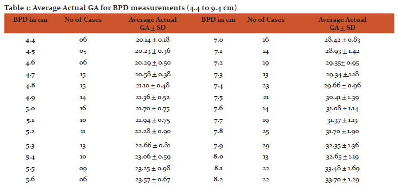
As seen in Table 1, the average GA observed in the present study with reference to BPD measurements from 4.4 to 9.4 cm along with number of cases for BPD measurements. The BPD measurements went on increasing with advancing GA. The average GA was found to be 21.70 + 0.75 weeks for the BPD measurements of 5.0 cm in 16 cases, while it was 32.65 + 1.19 weeks (13 cases) and 37.06 + 1.3 weeks ( 14 cases) for BPD measurements of 8 and 9 cm respectively
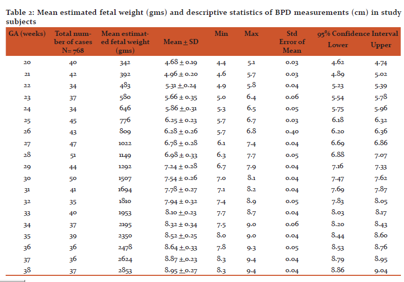
Table 2 shows the mean fetal weight in each GA, descriptive statistics like mean + SD, Minimum and maximum values, standard error of mean, 95% confidence interval for BPD measurements for each GA from 20 to 38 weeks. The mean fetal weight observed was 342 grams, 1507 grams and 2853 grams at 20 weeks, 30 weeks and 38 weeks of GA respectively. For 20 weeks of GA, the mean BPD was found to be 4.68 + 0.19 cm ranging from 4.4 to 5.1 cm with standard error of 0.03 cm and 95% confidence interval (4.6 cm – 4.74cm), while it was 7.54 + 0.26 cm ranging from 7.0 to 8.1 cm with standard error 0.04 with 95% confidence interval (7.47 cm - 7.62 cm) and 8.95 + 0.27 cm, ranging from 8.3 to 9.4 cm with standard error 0.04 with 95% confidence interval (8.86 cm - 9.0 cm) for GA of 30 weeks and 38 weeks respectively.
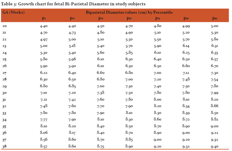
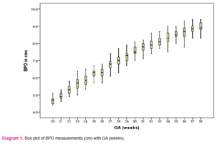
Diagram 1 shows the box plot describing BPD measurements about its median, first quartile, third quartile, minimum and maximum observations. Table 3 shows the growth chart for fetal BPD measurements for each GA. For 20 weeks of GA, the 5th, 50th and 95th percentile values of BPD measurements were 4.40 cm, 4.70 cm and 5.00 cm respectively, while it was 8.57 cm, 8.90 cm and 9.40 cm for 5th, 50th and 95th percentiles at 38 weeks of GA. At 20 weeks of GA, 50% of the subjects were having BPD value below 4.7 cm, while 50% of them were having below 8.9 cm at 38 weeks of GA.
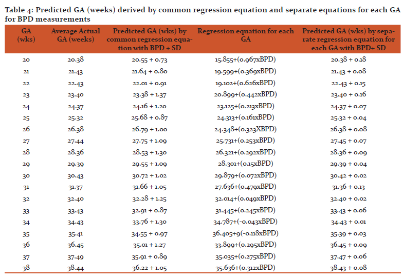
The common regression equation considering all gestational weeks derived for GA estimation by BPD measurements was 1.275 + (3.905 X BPD in cm, where “R2”- the proportion of variation in dependent variable (GA) was 0.944 and “r”- the correlation coefficient was 0.9
Table 4 shows predicted GA (weeks) derived by common regression equation and separate equations for each GA for BPD measurements. By using the common regression equations for GA from 20 to 38 weeks, there was a difference of 1.44 to 2.22 weeks in average actual and predicted GA at 36 to 38 weeks of pregnancy. However, the same difference was less between 20 to 35 weeks of gestation. The GA estimated by separate regression equations for each GA was more accurate as compared to that with common regression equation.
Discussion:
The literature is replete with articles that focus on predicting menstrual age using ultrasound measurements of the fetus. A common theme among these articles is that the variability in predicting menstrual age increases as pregnancy advances for all fetal parameters and the increase in variability is undoubtedly due to actual differences in fetal size, because it has been demonstrated in populations with optimal menstrual histories, with known dates of conception and in whom age was established early in pregnancy by use of crown-rump length measurements.13
Considering the above facts, present study was conducted to measure BPD by ultrasonography for different GA groups from 20 to 38 weeks of gestation in a normal singleton pregnant women from Belagavi District, Karnataka and to find out the predictive accuracy of GA determined by USG parameters with actual GA by LMP.
The BPD has received the greatest attention in literature as the means of establishing GA.14-17 Virtually many studies have demonstrated a progressive increase in variability in from 20 weeks to a term, but the extent to which the variability increases in the late third trimester of gestation has been a subject of some disagreement in the available literature.15-17 In various studies on antenatal cases with known LMP or in whom GA was confirmed by in early pregnancy by CRL, the variability of the late third trimester BPD age predictions has been repeatedly demonstrated to be approximately + 3.5 weeks.16
Benson CB and Doubilet PM18 confirmed the large variability associated with BPD measurements in third trimester among the cases whose menstrual histories had been established early in pregnancy by CRL. However, in this study no attempt was made to eliminate multiple gestations or cases with potential growth disturbances. This study concluded that the variability in predicting menstrual age using BPD reached a peak of approximately 4.1 weeks (2 SD) in the late trimester of pregnancy.
Many studies have demonstrated a progressive increase in variability in BPD measurements from 20 weeks to term, but the degree to which the variability increases in the late third trimester of pregnancy has been a subject of some disagreement in the literature. In the studies of patients with optimal menstrual histories, the variability of late third trimester BPD age predictions has been consistently demonstrated to be approximately + 3.5 weeks.13
Fetal BPD measurement is the most reliable in the second trimester and using both A and B scans. Fetal maturity can be predicted when used between 20-30 weeks and the values become more scattered around the mean in later weeks. In high risk pregnancies like diabetes and toxaemia, statistically significant difference in the fetal BPD is seen.19 Studies on BPD in certain populations showed linear correlation between BPD and GA and fetal weight in normal fetuses.20,21
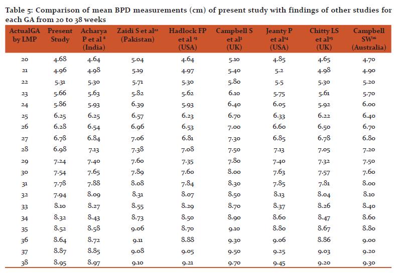
Table 5 shows mean BPD values obtained in the present study against the published standard values of the other studies by Acharya P et al8, Zaisi S et al22, Hadlock FP et al23, Campbell S et al3, Jeanty P et al24, Chitty LS25and Campbell SW26. The average GA was found to be 21.70 + 0.75 weeks for the BPD measurements of 5.0 cm in 16 cases, while it was 32.65 + 1.19 weeks (13 cases) and 37.06 + 1.3 weeks ( 14 cases) for BPD measurements of 8 and 9 cm respectively. The study by Hadlock FP et al13 reported almost same values of average GA for BPD measurements of 5 cm (21.2 weeks), 8 cm (32.5 weeks) and 9 cm (37 weeks). The mean values of BPD almost comparable till 30 weeks of GA, but thereafter the mean values in the present study were found to be lower than the other study values.
Many studies have demonstrated a progressive increase in variability in BPD measurements from 20 weeks to term, but the degree to which the variability increases in the late third trimester of pregnancy has been a subject of some disagreement in the literature. In the studies of patients with optimal menstrual histories, the variability of late third trimester BPD age predictions has been consistently demonstrated to be approximately + 3.5 weeks.
The earliest measurement of GA taken in pregnancy should usually be accepted as the definitive assessment, subsequent examinations reflecting only fetal growth in the intervening period. The ultrasound assessment of GA confirms the menstrual dates, if the measurements taken after the first trimester are within one week of GA taken from menstrual dating. Reduced accuracy of prediction of GA after 20 weeks must be appreciated.26
The observed GA was found to be almost same in a study by Hadlock FP et al 13 like that in the present study for BPD values from 20 to 38 weeks of GA. The use of reference tables from other populations may lead to an inappropriate and incorrect assessment of pregnancy.This study has provided the reference charts of BPD for normal singleton pregnant women from 20 to 38 weeks of gestation which will have relevant clinical impact. It will help to improve the accuracy of diagnosis of GA and fetal growth disturbances like IUGR, macrosomy, etc. However, the measurements of other parameters like head and abdominal circumferences, femur length would have increased the accuracy. The influence of fetal sex, maternal height, age, parity, weight, etc on fetal growth27, were not taken into account in selection of study subjects. A larger multicentered study is required to be undertaken for more accurate and valid assessment of fetal growth and GA in local population.
Conclusion:
There was a discrepancy in measurements of bi-parietal diameter in present study and in western and other different population groups, which was more especially in later part of third trimester. Use of charts derived from a different population especially in late third trimester may lead to errors in estimation of GA, fetal growth and development and deciding expected date of delivery. For this, use of reference tables prepared from local population will enhance the clinical accuracy. The normalcy of fetal parameters should be judged against local population standards.
Ethical considerations:
Ethical clearance was obtained from institutional ethics committee of KLE Academy of Higher Education and Research Belagavi. A permission letter was obtained from Belagavi Institute of Medical Sciences, Belagavi. The voluntary nature of participation and the right to withdraw at any time were emphasized. Written informed consent was obtained from every participant. Confidentiality was maintained throughout the study.
Source of Funding : Nil
Conflict of Interest : Nil
Authors’ Contribution:
All the authors participated in the designing, data collection, analysis, and writing of the study. The authors have read and approved the final paper.
Acknowledgments:
Authors acknowledge the immense help received from the scholars whose articles are cited and included in references of this manuscript. The authors are also grateful to authors / editors / publishers of all those articles, journals and books from where the literature for this article has been reviewed and discussed.

References:
-
Snijders RJ, Nicolaides KH. Fetal biometry at 14–40 weeks gestation. Ultrasound in Obstetrics and Gynecology 1994; 4:34–48.
-
Salomon LJ, Duyme M, Crequat J, et al. French fetal biometry: reference equations and comparison with other charts. Ultrasound in Obstetrics and Gynecology 2006; 28:193–198.
-
Campbell S, Warsof SL, Little D, et al. Routine ultrasound screening for the prediction of gestational age. Obstetrics and Gynecology 1985;65:613-620.
-
Waldenstorm U, Axellson U, Nilsson S. A comparison of the ability of a sonographically measured biparietal diameter and the last menstrual period to predict the spontaneous onset of labor. Obstetrics and Gynecology 1990;76:33-36.
-
Yeo GS, Chan WB, Lun KC, et al. Racial differences in fetal morphometry in Singapore. Annals of the Academy of Medicine, Singapore 1994;23:371–376.
-
Jacquemyn Y, Sys SU, Verdonk P. Fetal biometry in different ethnic groups. Early Human Development 2000;57:1–13.
-
Giorlandino M, Padula F, Cignini P. Reference interval for fetal biometry in Italian population. Journal of Prenatal Medicine 2009;3(4):62-68.
-
Acharya P, Acharya A. Evaluation of Applicability of Standard Growth Curves to Indian Women by Fetal Biometry. South Asian Federation of Obstetrics and Gynecology 2009;1(3):55-61.
-
Kinare AS, Chinchwadkar MC, Natekar AS, et al. Patterns of Fetal Growth in a Rural Indian Cohort and Comparison with a Western European Population. Journal of Ultrasound in Medicine 2010; 29(2):215-223.
-
Garg A, Pathak N, Gorea RK, et al. Ultrasonographical age estimation from biparietal diameter. Journal of Indian Academy of Forensic Medicine 2010;32(4):308-33Robin B, Kailash, Chervenak F. Sonogaphic determination of gestational age. TMJ Science Journal 2009;59(2):202-208.
-
Gorea RK, Mohan P, Garg A, et al. Fetal age determination from length of femur and humerus by ultrasonography. Medico-legal Update 2009;9(1):23-26.
-
Hadlock FP, Deter RL. Estimating fetal age: Effect of head shape on biparietal diameter. American Journal of Roentgenology 1981;137:83-85.
-
Galan HL, Pandipati S and Roy A. Ultrasound evaluation of fetal biometry and normal and abnormal fetal growth. Text book of Ultrasonography in Obstetrics and Gynecology by Peter W. Callen, 5th edition, Elsevier, New Delhi, 2011:225-247.
-
Hadlock FP, Harrist RB, Martinez-Poyer J. How accurate is second trimester fetal dating? Journal of Ultrasound Medicine 1992;10:557-560.
-
Kurtz AB, Wapner RJ, Kurtz RJ, et al. Analysis of biparietal diameter as an accurate indicator of gestational age. Journal of Clinical Ultrasound 1980;8:319.
-
De Crespigny LC, Speirs AL. A new look at biparietal diameter. Aust New Zealand Journal of Obstetrics and Gynecology 1989;29:26-29.
-
Hadlock FP, Deter RL, Harrist RB, et al. Fetal biparietal diameter: Rational choice of plane of section for sonographic measurement. American Journal of Roentgenology 1982;138:871-875.
-
Benson CB, Doubilet PM. Sonographic prediction of gestational age: Accuracy of second and third trimester fetal measurements. American Journal of Roentgenology 1991;157;1275-1278.
-
Campbell S. The prediction of fetal maturity by ultrasonic measurement of the biparietal diameter. BJOG: An International Journal of Obstetrics and Gynecology 1969;76(7):603-609.
-
Mador ES, Ekwempu J, Mutihir T, et al. Ultrasonographic biometry: Biparietal diameter of Nigerian foetuses. Nigerian Medical Journal 2011;52(1):41-44.
-
Okupe RF, Coker OO, Gbajumo SA. Assessment of fetal biparietal diameter during normal pregnancy by ultrasound in Nigerian women. British Journal British Journal of Obstetrics and Gynecology 1984;91(7):629-632.
-
Zaidi S, Shehzad K, Omair A. Sonographic foetal measurements in a cohort of population of Karachi, Pakistan. Journal of Pakistan Medical Association 2009;59(4):246-249.
-
Hadlock FP, Deter RL, Harris RB, et al. Estimating fetal age: Computer assisted analysis of multiple fetal growth parameters. Radiology 1984;152:497-501.
-
Jeanty P, Rodesch F, Delbeke D, et al. Estimation of gestational age from measurements of fetal long bones. Journal of Ultrasound in Medicine 1984;3:75–79.
-
Chitty LS, Altman DG, Henderson A, et al. Charts of fetal size: 2. Head measurements. British Journal of Obstetrics and Gynaecology1994;101(2):35-43.
-
Australian Society for Ultrasound in Medicine. Promoting excellence in ultrasound: Policies and Statements D7- Revised in 2001. Available on link https://www.signosticsmedical.com/pdf/P03622_SignosRT_User_Manual_US-CAN.pdf
(last accessed on 22/3/2018)
-
Kiserud T, Piaggio G, Carroli G, et al. The World Health Organization fetal growth charts: A multinational longitudinal study of ultrasound biometric measurements and estimated fetal weight. PLOS Medicine 2017;14(1):1-36.
|


 This work is licensed under a Creative Commons Attribution-NonCommercial 4.0 International License