IJCRR - 11(1), January, 2019
Pages: 01-05
Date of Publication: 10-Jan-2019
Print Article
Download XML Download PDF
A Three and a Half Years Histopathological Study of Fibroepithelial Breast Lesions in a Tertiary Care Hospital
Author: Rohi Wani, Sheema Sheikh, Abdul Maajed Jehangeer, Salma Bhat, Isma Niyaz, Bilques Khursheed
Category: Healthcare
Abstract:Breast specimens are one of the most frequent entries in our department of pathology. Out of these surgical specimens, fibroepithelial lesions are received almost every day.
Aim: To study and provide an overview of fibroepithelial lesions of breast. Further to stratify and classify various fibroepithelial lesions into fibroadenomas and phyllodes tumor.
Methodology: All the 386 cases of fibroepithelial lesions received over a period of 3 1/2 years from Jan 2015-June 2018 were retrieved and studied in detail. All the associated histological changes as also the clinical details were noted down.
Results: Out of the total 386 fibroepithelial lesions, there were 372 fibroadenomas and 14 phyllodes tumors. Among all phyllodes tumors there was a single case each of borderline phyllodes tumor and malignant phyllodes tumor.
Conclusion: Fibroepithelial lesions show a wide range of morphology. A strict histopathological assessment with classification leads to proper diagnosis and thus proper treatment in such cases.
Keywords: Fibroepithelial lesions, Fibroadenoma, Phyllodes tumor
Full Text:
Introduction
Fibroepithelial lesions are the common lesions of breast. They are morphologically and behaviourally heterogeneous tumor with different clinical behaviours and treatment protocols. These constitute the commoner fibroadenomas and the rarer phyllodes tumor1. Fibroadenomas are the commonest benign biphasic fibroepithelial neoplasms occurring in 2nd and 3rd decade of life2. Phyllodes tumor occurs in an older age group usually around 45-49 years of age3. Almost all of the phyllodes tumor are reported as fibroadenomas on Fine needle aspiration cytology (FNAC) since the clinical presentation, as also the cytology findings of these overlap. Although higher grade phyllodes tumors are rare neoplasms but need urgent attention. Accurate diagnosis and grading of phyllodes tumors are important for patient management and prognosis, as grade broadly correlates with increasing local recurrence risk, and metastasis occurs only in malignant tumors4.
Materials and Methods
This study is a descriptive, retrospective type of study done on the breast specimens received in our Histopathology department over a period of 3½ years (from Jan 2015- June 2018). All the breast cases which included lumpectomy, excision biopsies, tru cut biopsies, mastectomy specimens and blocks for review were included in the study irrespective of age and sex. Specimens were received in 10% formalin and were subjected to routine haematoxylin and eosin stains. Fibroepithelial lesions were segregated and studied in detail. Clinical data of patients was retrieved from medical records.
All the sections were studied in detail, clinical history was noted down and morphological diagnosis was made. Cases were clustered as fibroadenomas and phyllodes tumors. Necessary details and variations in histopathology were noted down. All these lesions were classified according to the WHO classification of tumors5 of breast and tabulated as such.
Results
A total of 759 specimens of breast were received over this period of three and a half years. Out of these fibroepithelial lesions, fibroadenomas constituted the major portion constituting almost half of the total number of breast specimens. Thus the most common breast lesion diagnosed was a fibroadenoma accounting for 372 cases whereas phyllodes tumor was diagnosed in 14 constituting of one malignant phyllodes, one borderline phyllodes and 12 benign phyllodes tumors. A total of 386 cases of fibroepithelial lesions were encountered in this period.
Discussion
Fibroepithelial lesions of breast are neoplastic proliferations of specialised stroma which secondarily distorts lobules and ducts, incorporating these within a well defined mass. Although the resulting lesions do contain epithelial components, but it is only the stromal component which is neoplastic. These lesions encompass a variety of benign and malignant lesions ranging from fibroadenoma to malignant phyllodes tumor6. They encompass a heterogeneous group of lesions that range from benign to malignant, each exhibiting differing degrees of stromal proliferation in relation to the epithelial compartment 7.
These lesions comprise a morphologically and biologically spectrum of biphasic tumors with epithelial and stromal components that demonstrate widely variable clinical behaviour. Fibroadenomas are common benign tumors with a number of histologic variants, most of which pose no diagnostic challenge. Cellular and juvenile fibroadenomas can have overlapping features with phyllodes tumors and should be recognized. Phyllodes tumors constitute a spectrum of lesions with varying clinical behaviour and are graded as benign, borderline or malignant based on a set of histologic features according to recommendations by the World Health Organization (WHO)4.
Fibroadenomas are benign tumor masses arising from both the epithelium and stroma of breast consecutively. They constitute the most common breast lesions especially in young age groups. It arises from terminal duct lobular unit from the intralobular stromal mesenchymal cells (Fig 1), along with the hyperplasia of intralobular ductal and acinar epithelium2.
All the cases of fibroepithelial lesions in our study were fibroadenomas except the 14 phyllodes tumor cases. There was a wide age range of 13 to 50 years in our cases. Size varied from as small as less than 1cm to as big as 10 cm. There were three cases of fibroadenomatosis in which more than two fibroadenomas were resected from a single patient at one point of time from the same breast or contralateral breasts.
Grossly all the fibroadenomas were round, nodular, discrete firm swellings with cut surface showing homogenous firm greyish white surface with slit like spaces. Bilateral breast adenoma was seen in 10 (2.6%) of cases.
A wide variety of proliferative changes can be seen in the epithelial components of fibroadenoma2,8. A lot of the common microscopic variations of fibroadenoma were encountered in our study also. Out of these simple fibroadenoma, which had no other pathological association was seen in 76% of cases. The most common pathological association was fibrocystic change seen in about 16% followed by adenosis which was seen in 13 of fibroadenomas (Table 1). The most frequent association of fibroadenoma with fibrocystic disease was similar to studies as that of Geethamala K et al2.
Epithelial hyperplasia was present in a total of 7 cases with mild hyperplasia in 4 and moderate hyperplasia in 3 cases. This is much less (47/372) than was seen in a study by Geethamala K, et al2 and Kuijper et al (43.9%)8. There were 2 cases each of complex fibroadenoma, fibroadenoma with apocrine metaplasia and Juvenile fibroadenoma. A single case of myxoid fibroadenoma, giant fibroadenoma and fibroadenoma with stromal hyalinisation was also seen
All these histological variants usually do not pose any problem with the diagnosis, however cellular and Juvenile fibroadenoma (Fig 2) have some overlapping features with benign phyllodes tumor and thus should be recognised since the course of disease and treatment is quite different from one another4,6,9. Although there are no clear cut boundaries but strict histologic assessment of features with classification ultimately reveal the correct diagnosis and thus provide useful clinical information10.
Phyllodes tumor is a rare fibroepithelial lesion as compared to fibroadenoma, with wide spectrum of morphology. It has risk of local recurrence and uncommon metastasis. Although microscopic distinction between fibroadenoma and phyllodes tumor especially benign phyllodes tumor is difficult, strict histologic assessment of a combination of histologic features with classification help to achieve the correct diagnosis and provide useful clinical information10.
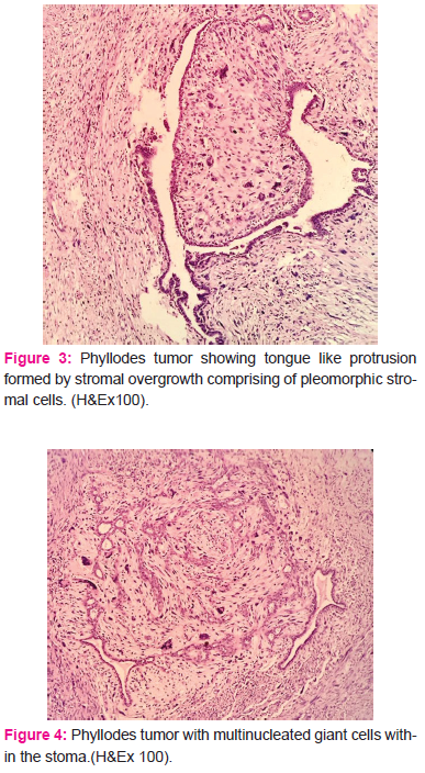
Phyllodes tumors were classified according to three tier grading system of WHO classification into benign, borderline and malignant4 (Table2). Out of the total cases of 14 phyllodes tumor, 1 (7%) was borderline, 1 (7%) was malignant and the rest (12=86%) were all benign (Fig 3 &4). There was another case of breast sarcoma but it was a primary case of Fibrosarcoma without any evidence of it arising from or any association with phyllodes tumor.
The value of FNAC in the diagnosis of phyllodes tumor has always remained controversial3. In all our cases of phyllodes tumor FNAC was done as a first line investigation and it was reported as fibroadenoma in 11 out of the 12 benign phyllodes tumors. Only in a single case of benign phyllodes tumor, it was reported as phyllodes tumor on cytology. There is a significant overlap of phyllodes tumor with fibroadenoma in cytological diagnosis and it’s always difficult to diagnose phyllodes tumor on FNAC alone11. The sensitivity for diagnosing phyllodes tumors by FNAC is only 40%, although increased sensitivity can be achieved by combining cytohistological and radiological results12. Clinical presentation of a patient giving a diagnostic clue towards phyllodes tumors rather than fibroadenoma should always be investigated through radiology and core needle biopsy which has high sensitivity since the surgical management is the mainstay and moreover local recurrence has been associated with inadequate local excision3.
In a study done by Karim et al, the clinical and pathological features of a large single institutional series of ethnically diverse patients with phyllodes tumours was done to determine which characteristics were predictive of outcome. Sixty five phyllodes tumors were analysed; 34 were benign, 23 borderline and eight malignant (34 low grade and 31 high grade PTs on a two tiered grading system). Out of these Nine patients (15%) had local recurrences. A greater percentage of higher grade tumours recurred and women of Asian origin had a higher recurrence rate than those of non-Asian origin. The 5 year disease-free survival was 81% as also the time to recurrence was significantly lower in the high grade group. There were no metastases or deaths from disease. The mean age at diagnosis and the mean tumour volume, both significantly increased with grade. Thus tumour grade was the only parameter related significantly to outcome of these patients13.
In the benign phyllodes tumor, the size varied from smallest 4cm to largest measuring 15cms. The age varied from 17 to 45 years. One of the benign phyllodes tumors showed areas of tubular adenosis with foci of hyalinisation and fibrocystic changes in the adjacent areas.
Another phyllodes tumor was seen in a 30 years old female who presented with a huge swelling in her left breast. She had noticed it four months back as small swelling, with a history of recent rapid increase in size and had reached the present diameter of 10x10 cm. In the meanwhile patient had seeked medical help in which FNAC of the swelling was done and reported as diagnostic possibilities of either a ductal carcinoma with osteoclast type giant cells or ductal carcinoma with increased stromal cellularity. Ultrasonography was reported as Fibroadenoma- BIRADS II. Trucut biopsy was done and reported descriptively as Malignancy with fibromyxoid stroma. Radical mastectomy was performed and we received a huge mastectomy specimen measuring 20x16x7 cms with axillary tail measuring 14x7x4 cms. On cut section a huge growth measuring 10x10x6 cms was occupying all quadrants sparing just the portion of upper inner quadrant. Cut surface of growth was variegated having friable, necrotic and haemorrhagic areas. On microscopic examination it came out to be stromal cell tumor with pleomorphic cells showing moderate to marked atypia (Fig 3&4). The mitotic rate of >5/10hpf was counted, although such high mitotic count was present in focal areas only. All the lymphnodes were free from tumor. Benign phyllodes tumor was identified in 70-80 %of the serial sections. So the final diagnosis of borderline phyllodes tumor was given.
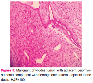
In our study a single case of malignant phyllodes tumor (Fig5) was seen. A young 25 years old female presented with swelling right breast. The duration of swelling was a few days only and tru cut biopsy was done straightway on the basis of clinical features and radiological suspicion of malignancy. It was reported as malignant phyllodes on biopsy and surgery (partial mastectomy was performed within days of diagnosis). Grossly we received a partial mastectomy specimen with a globular , greyish white, well circumscribed firm area measuring 7x4x3 cc. On cut section it was variegated with areas of hemmorhage. Microscopy revealed a pleomorphic cell population with mitotic rate of >15/hpf. There was also an area of malignant smooth muscle differentiation (Fig 5). The diagnosis of Malignant phyllodes tumor with Leiomyosarcomatous element was rendered. IHC was advised for confirmation and it came out positive for desmin and smooth muscle actin.
Conclusion
Fibroepithelial lesions of breast are one of the most common lesions especially in young females. Out of these fibroadenomas are quite common and the rarer phyllodes tumor can cause a lot of clinical concern. Moreover the need to differentiate fibroadenomas from phyllodes tumor due to the different surgical procedures required for these tumors and the tendency of malignancy in phyllodes needs to be considered seriously.
Acknowledgement
Authors acknowledge the immense help received from the scholars whose articles are cited and included in references of this manuscript. The authors are also grateful to authors / editors / publishers of all those articles, journals and books from where the literature for this article has been reviewed and discussed.
Source of Funding: Nil
Conflict of interest: Nil


References:
1.Daramola AO, Oguntunde OA, Awolola NA. Audit of fibroepithelial tumors of the breast in a Nigerian tertiary institution. Niger. J Clin Pract 2016; 19:645-8
2.Geethamala K, Vani BR, Murthy VS, Rhada M. Fibroadenoma: A harbour for various histopathological changes. Clin Cancer Investig J 2015; 4:183-7
3.Mishra SP, Tiwary SK, Mishra M, Khanna AK. Phyllodes Tumor of Breast: A Review Article. Hindawi Publishing Corporation. ISRN Surgery. Volume 2013, Article ID 361469, 10 pageshttp://dx.doi.org/10.1155/2013/361469
4.Krings G, Bean GR, Chen YY. Fibroepithelial lesions; The WHO spectrum. Semin Diagn Pathol 2017 Sep; 34(5):438-452
5.Lakhani S, Ellis I, Schnitt S, et al.: WHO Classification of Tumours of the Breast, 4th ed. Lyon, IARC, Press, 2012
6.Giri D. Fibroepithelial Lesions; Arch Pathol Lab Med. 2009; 133:713–721
7. Tan BY, Tan PH. A Diagnostic Approach to Fibroepithelial Breast Lesions. Surg Pathol Clin. 2018 Mar; 11(1):17-42
8. Kuijper A, Mommers ECM, Wall E, van Diest PJ. Histopathology of fibroadenoma of the breast. Am J Clin Pathol 2001; 115:736-42
9. Yasir S, Nassar A, Jimenez RE, Jenkins SM, Hartmann LC, Degnim AC, Frost M, Visscher DW. Cellular fibroepithelial lesions of the breast: A long term follow up study. Ann Diagn Pathol. 2018 Aug; 35:85-91
10. Zhang Y, Kleer CG. Phyllodes Tumor of the Breast. Histopathologic Features, Differential Diagnosis, and Molecular/Genetic Updates. Arch Pathol Lab Med. 2016; 140:665–671
11. Tse GM, Ma TK, Pang LM, Cheung H. Fine needle aspiration cytologic features of mammary phyllodes tumors. Acta Cytol. 2002 Sep-Oct; 46(5):855-63
12. Ward ST, Jewkes AJ, Jones BG, Chaudhri S, Hejmadi RK, Ismail T. The sensitivity of needle core biopsy in combination with other investigations for the diagnosis of phyllodes tumours of the breast. Int J Surg. 2012; 10(9):527-31.
13. Karim RZ, Gerega SK, Yang YH, Spillane A, Carmalt H, Scolyer RA, Lee CS. Phyllodes tumours of the breast: A clinicopathological analysis of 65 cases from a single institution. Breast. 2009 Jun; 18(3):165-70.
|


 This work is licensed under a Creative Commons Attribution-NonCommercial 4.0 International License