IJCRR - 10(16), August, 2018
Pages: 08-10
Date of Publication: 27-Aug-2018
Print Article
Download XML Download PDF
Rare Case of Capillary Hemangioma of Bilateral Lower Limb: A Case Report
Author: Vipan Kumar, Anu Yarky, Aakash Parashar, Devinder Kumar
Category: Healthcare
Abstract:Aim: The aim of this case report is to bring to light an unusual case of soft tissue tumor in a young female.
Case Report: Hemangiomas are commonly occuring soft tissue tumors, but it rarely involves the feet. We are reporting a twenty two year old female with capillary hemangioma of her left ankle and right foot which was painful. Her excision biopsy was done and microscopic investigation revealed capillary hemangioma. The patient has had no recurrence since the surgery.
Discussion: Few similar cases have been reported in the past in children, adults and elderly patients and none of them experienced any recurrence after treatment by excision or sclerotherapy or embolisation.
Conclusion: Insidious onset and gradually progressive painful swellings can often be misdiagnosed as vascular malignant neoplasms. Here we have reported an unusual case of capillary hemangioma of bilateral lower limb in a young female.
Keywords: Soft tissue tumors, Capillary hemangioma, Excision biopsy
Full Text:
Introduction :
Hemangiomas are benign soft tissue tumors. They can occur anywhere in the body be it skin, muscle, bone or our organs. Capillary hemangioma is the most commonly occurring hemangioma. Often surgeons, clinically misdiagnose patients with gradually progressing painful swellings as malignant. Thus the next step in diagnosis is radiological intervention and histopathological diagnosis. Here we are reporting an unusual case of a young female who was diagnosed to have capillary hemangioma in bilateral lower limbs.
Case scenario:
Our patient a young 22 year old female came to the out patient department with complaints of Swelling in the dorsum of right foot and left ankle since one and half year. She was apparently alright when she noticed swelling over her right foot, dorsal aspect and similar swelling over medial aspect of left ankle. Swelling was initially the size of a peanut but gradually progressed to the size of lemon within one and half year. It was painful and bluish brown in colour. There was no history of fever, discharge from the swelling, or pedal oedema. She complained of difficulty in walking due to pain, difficulty in bearing weight over a single limb and restriction of movement. There was no past history of Diabetes, hypertension, tuberculosis and no history of similar swellings in past. Family history was also inconclusive.
On examination, our patient had a bluish brown coloured swelling over her left ankle which was approximately of size 3*4 cm and another swelling over dorsum aspect of right foot which was approximately of the size 3.5*4 cm. It was tender, compressible, non reducible. There was no discharge or sinus present. There was no pedal oedema associated with the swelling. Peripheral pulses were normally palpable.
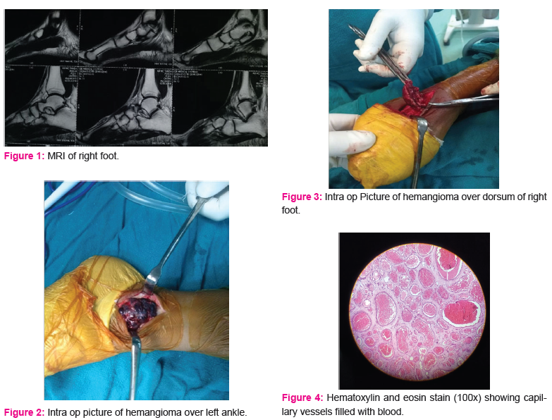
MRI- Magnetic resonance Imaging of right foot was done. (Figure a) It was suggestive of a well defined, lobulated, T1 Hypo mixed STIR hyperintensity lesion on dorsum of foot in subcutaneous plane on post contrast enhancement. It was of size 4*1*3 cm and was separated from the underlying extensor tendons. Underlying metatarsals were normal. Bones comprising the ankle joint showed normal configuration. Joint space was normal. Talocalcaneal and talo navicular joints were normal, Achilles tendon was normal. Plantar soft tissues were also normal. A possibility of Slow flow vascular malformation was kept.
Patient underwent Excision biopsy (Figure b and Figure c). and the tissue was sent for histopathological analysis which was suggestive of capillary hemangioma (Figure d). Post op patient was asymptomatic and no similar swellings were present till one year of follow up period.
Discussion :
A hemangioma is a benign tumor of blood vessels. They can occur anywhere in the body, including in skin, muscle, bone, and internal organs. Mostly they occur on the surface of the skin or just beneath it. Hemangiomas are rarely cancerous and do not require any medical treatment but some hemangiomas can be cosmetically disfiguring and may require treatment. There are some cases of hemangioma in which surgery may be necessary like when they are deep in muscle or bone, or for tumors on the skin that cause problems with vision, breathing, or eating.
Capillary hemangioma is the most common type of hemangioma.(1) It is made up of small capillaries that are normal in size but high in number. They form a tightly packed group held together by thin connective tissue. Because of their proximity to the skin, capillary hemangiomas are typically brighter red in color. They can be small or large; flat or raised; or may protrude out as a nodule. Diffuse hemangioma or angiomatosis appears as a spongy mass that covers an entire extremity. Other types of hemangiomas are compound hemangioma, cavernous hemangioma and lobular capillary hemagioma.
Types of hemangiomas according to their presentation can be infantile and congenital also. Hemangiomas in Muscle, Bone, and Internal Organs are not as common as hemangiomas of the skin but cases like ours have been reported in the past. Intramuscular hemangioma can develop at any age, but most often occur in young adults. Capillary hemangiomas are more common in muscle than cavernous and compound hemangiomas. These tumors are often painful and require treatment. Bone hemangiomas occur in the skull or spine typically and are most common in people who are 50 to 70 years of age.
A great deal of research is being done to learn more about hemangiomas. The development of medication directed at halting the development of blood vessels (anti-angiogenic drugs) is an exciting area of research for many types of tumors.
Yetkin H et al in 2001 reported a case of multiple hemangiomas of foot. They reported a case of a forty-seven-year-old female with multiple painless hemangiomas on her left foot. During surgery there were seven well defined masses that were totally excised after ligation of penetrating vessels. The microscopic investigation revealed mixed-type (capillary and cavernous) hemangioma. They did not experience any recurrence after two years follow up period.(2) Capillary hemangiomas that occur in adults and on the lower extremities are uncommon.
Cione et al in 2002 reported a similar uncommon case of capillary hemangioma involving surgical treatment of the lesion on an adult foot .(3)
Uslu M et al in 2014 presented a report of 2 cases of intramuscular hemangioma in children, 1 treated by excision and 1 by percutaneous sclerosis. They stated that Surgical excision, ultrasound or fluoroscopic-guided percutaneous sclerotherapy, and angiographic embolization are all worthy treatment options. Surgical excision is the most prevalent form of therapy, although this can be difficult when it is in the hands and feet. For this reason, ultrasound- and fluoroscopic-guided percutaneous sclerotherapy is a useful treatment option for pedal intramuscular hemangioma.(4) In matter of recurrence, our patient has not had any recurrence of similar swellings till date and has been asymptomatic for the past one year of follow up.
In 1986 Nappi et al reported a clinico-pathologic study of two cases of disseminated lobular capillary hemangioma which were misdiagnosed clinically as having malignant vascular neoplasms.(5) Reported cases had recurrence of multiple satellite lesions after surgical removal of a solitary capillary hemangioma. One patient developed skin nodules in several sites, including the mouth, knee, thumb, and foot, after surgical removal of a solitary oculocutaneous neoplasm. The second patient had more than 700 skin lesions, distributed over the entire body.
Conclusion
Several classification systems have been proposed for classifying peripheral vascular malformations.
Example 1: Classification of vascular malformations according to flow dynamics:
Low Flow : Venous, Lymphatic and mixed malformations
High Flow : Arteriovenous malformations and fistulas.
Example 2: International Study of Vascular Anomalies classification of vascular anomalies into tumours and simple malformations.
Vascular tumor : Hemangioma and others;
Simple vascular malformations : Capillary, venous, lymphatic.(6)
Haemangiomas are benign vascular tumours. They are characterised by cellular proliferation, rapid early proliferative stage and a later stage of involution. Low-flow lesions are some of the most common types of vascular malformations, with prevalence of up to 1% in the general population. The extremities encompass approximately 40% of the sites involved, with low-flow venous Peripheral vascular malformations being the most common types encountered. Other locations include the head and neck (40% of cases) and the trunk (20%).(6) Our patient presented to us with subcutaneous swellings in right foot and left ankle, which was diagnosed as a low flow vascular malformation on Magnetic Resonance Imaging and as capillary hemangioma on histopathology report of excised biopsy tissue. The purpose of our study is to present such rare case of capillary hemangioma over bilateral lower limbs in a young female.
Acknowledgement : Authors acknowledge the immense help received from the scholars whose articles are cited and included in references of this manuscript. The authors are also grateful to authors / editors / publishers of all those articles, journals and books from where the literature for this article has been reviewed and discussed.
Conflict of Interest: There is no conflict of interest.
References:
Hemangioma - OrthoInfo - AAOS [Internet]. [cited 2018 Aug 1]. Available from: https://orthoinfo.aaos.org/en/diseases--conditions/hemangioma
2. Yetkin H, Kanatli U, Guzel VB, Poyraz A. Multiple hemangiomas of the foot: A case report. Foot Ankle Int. 2001;
3. Cione JA, Cozzarelli J. Capillary hemangioma of the foot. J Am Podiatr Med Assoc [Internet]. 2002 Mar [cited 2018 Aug 1];92(3):155–7. Available from: http://www.ncbi.nlm.nih.gov/pubmed/11904329
4. Uslu M, Be?ir H, Turan H, Bozkaya H, Erdem H. Two different treatment options for intramuscular plantar hemangioma: Surgery versus percutaneous sclerotherapy. J Foot Ankle Surg. 2014;
5. Nappi O, Wick MR. Disseminated lobular capillary hemangioma (pyogenic granuloma). A clinicopathologic study of two cases. Am J Dermatopathol. 1986;
6. Madani H, Farrant J, Chhaya N, Anwar I, Marmery H, Platts A, et al. Peripheral limb vascular malformations: an update of appropriate imaging and treatment options of a challenging condition. Br J Radiol [Internet]. 2015 Mar [cited 2018 Aug 1];88(1047):20140406. Available from: http://www.ncbi.nlm.nih.gov/pubmed/25525685
|
 IJCRR
IJCRR 


 This work is licensed under a Creative Commons Attribution-NonCommercial 4.0 International License


