IJCRR - 10(14), July, 2018
Pages: 01-03
Date of Publication: 18-Jul-2018
Print Article
Download XML Download PDF
Entrapment of the Plantaris Tendon - A Rare Anomaly
Author: Krishna Kanta Biswas, Pradipta Ray Choudhury, Bijon Chandra Dutta
Category: Healthcare
Abstract: Introduction: The plantaris muscle originates from the lower part of the lateral supracondylar ridge of the femur, passes between the gastrocnemius and soleus muscles, and inserts by a long slender tendon into the calcaneus. It is a vestigial muscle in humans and may vary in its origin, insertion, number of muscle belly and course of the plantaris tendon. These variations may influence the surgical outcome of the operations around the knee joint and the posterior compartment of leg.
Aim: The aim of the present study is to report a case of plantaris muscle, where the tendon of the muscle was entrapped between the tibial nerve and the nerve to the soleus.
Case Report: During routine dissection of the popliteal fossa and the posterior compartment of leg for the first year MBBS students, a plantaris muscle was reported with abnormal course of the plantaris tendon. The dissection was performed following Cunningham's manual of practical anatomy. The tendon of the plantaris muscle passed between the tibial nerve and the nerve to the soleus on the right side of a 60 year old male cadaver.
Discussion: The present variation has been reviewed and discussed with the previous studies.
Conclusion: The pull of the plantaris tendon may press upon the nerve to the soleus thereby causing compression neuropathy..
Keywords: Tibial nerve, Nerve to the soleus, Compression neuropathy
Full Text:
INTRODUCTION
The plantaris muscle has a small fusiform belly, which originates from the lower part of the lateral supracondylar ridge and adjacent popliteal surface of the femur. It ends in a long slender tendon that passes between the gastrocnemius and soleus muscles and inserts into the calcaneus, medial to the calcaneal tendon. It is a vestigial muscle in humans1.
During its course, the plantaris tendon passes posterior to the tibial nerve and popliteal vessels. Distal to the popliteal fossa, the tibial nerve gives off a branch to the soleus, which supplies the muscle through its superficial and deep surfaces2.
The plantaris muscle is subjected to many variations. It may vary in its origin, insertion, number of muscle belly and course of the plantaris tendon. The plantaris muscle may originate from the lower part of the linea aspera; the lateral condyle of the femur above the origin of the lateral head of the gastrocnemius; the posterior ligament of the knee joint; the fascial covering of the popliteus; the oblique line of the tibia, under cover of the soleus and the fibula, between the flexor hallucis longus and peroneus longus3-5.
The plantaris tendon may also insert into the soft tissues between the muscle bellies of the gastrocnemius and soleus; the flexor retinaculum; the fascia of the leg; the inner border of the calcaneal tendon; the bursa between the calcaneal tendon and the calcaneus; the dorsomedial surface of the calcaneal tendon at its insertion; the fibrous and fatty tissues situated in front of the calcaneal tendon and the plantar aponeurosis1,3-5. The plantaris muscle may be double or absent6,3. The plantaris tendon may also pass between the tibial nerve and nerve to the soleus, thereby causing entrapment of the tendon7-9.
The aim of the present study is to report a rare variation of the entrapment of the plantaris tendon between the tibial nerve and the nerve to the soleus.
CASE REPORT
During routine dissection of the popliteal fossa and the posterior compartment of leg for the first year MBBS students, a plantaris muscle was reported with abnormal course of the plantaris tendon. The dissection was performed following Cunningham’s manual of practical anatomy and the course of the plantaris tendon was observed, and photographed10. The ethical clearance for the study has been obtained from the Institutional Ethical Committee.
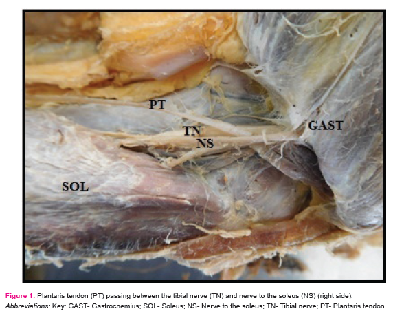
The plantaris muscle took origin from the lateral supracondylar ridge and the adjacent popliteal surface of the femur, in the right side of a 60 year old male cadaver. The muscle then formed a long slender tendon, which passed between the gastrocnemius and soleus muscles and inserted into the calcaneus, medial to the calcaneal tendon. The plantaris tendon passed between the tibial nerve and the nerve to the soleus, thereby causing entrapment of the tendon.
DISCUSSION
In humans, the plantaris acts along with gastrocnemius to help in plantar flexion, due to common insertion into the calcaneus. But, in many mammals, it inserts directly or indirectly into the plantar aponeurosis1. This is due to the evolutionary changes towards the erect posture of man, where the insertion of the plantaris muscle gradually shifted to the calcaneus3.
The different variations of the plantaris muscle regarding origin, insertion, number of muscle belly and course of the plantaris tendon may help the surgeons during surgeries of the popliteal fossa and the posterior compartment of leg. The knowledge of such variations may also help the clinicians during diagnosis of a posterior knee injury and/or tennis leg7.
Das et al8 reported a case in which the plantaris tendon took origin from the lateral supracondylar line and the oblique popliteal ligament, and its tendon passed between the gastrocnemius and soleus muscles, and inserted into the medial border of the calcaneal tendon. Here, the plantaris tendon passed between the tibial nerve and the nerve to the soleus.
Nayak et al7 reported a case in which they found an additional tendon of the plantaris muscle arising from the fascia covering the popliteus, which then joined the original tendon of the plantaris to form a single tendon and inserted into the calcaneal tendon. During the course, the plantaris muscle was entrapped between the tibial nerve and its branch to the soleus.
Saha et al9 reported a case, in which they observed a plantaris muscle with double bellies, of which the inner belly originated from the fascia covering the politeus and the outer belly originated from the lower part of the lateral extension of the linea aspera, above the origin of the lateral head of the gastrocnemius. Both the bellies then fused to form a common tendon and inserted into the calcaneal tendon. During their passage, the muscle bellies were entrapped between the tibial nerve and the nerve to the soleus.
In the present case, similar to Das et al8, Nayak et al7 and Saha et al9, the plantaris tendon passed between the tibial nerve and the nerve to the soleus, thereby causing its entrapment. No other associated anomaly in relation to its origin, insertion and number of muscle belly were observed in the present case. Such an anomaly of entrapment of the plantaris tendon is relatively rare, which, if misdiagnosed may lead to severe complications.
CONCLUSION
The pull of the plantaris tendon may press upon the nerve to the soleus, thereby causing compression neuropathy. Also, such an entrapment may complicate the surgical exploration of the structures of the posterior compartment of leg and interpretation of MRI scans and ultrasounds during evaluation of muscle tears surrounding the knee joint.
ACKNOWLEDGEMENT
Authors acknowledge the immense help received from the scholars whose articles are cited and included in references of this manuscript. The authors are also grateful to authors/ editors/ publishers of all those articles, journals and books from where the literature for this article has been reviewed and discussed.
Conflict of interest: None.
Source of funding: None.
References:
-
Standring S. Leg. In: Tubbs RS, editor. Gray’s Anatomy The anatomical basis of clinical practice. 41st ed. Elsevier; 2016. p.1410.
-
Rosse C, Gaddem-Rosse P. Limbs. In: Hollinshead Textbook of Anatomy. 5th ed. Philadelphia: Lipincott-Raven; 1997. p.372-4.
-
Daseler EH, Anson BJ. The plantaris muscle: An Anatomical Study of 750 Specimens. J Bone Joint Surg Am. 1943;25:822–7.
-
Henle J. Muskellehre. In: Handbuch der Systematischen Anatomie des Menschen. Braunschweig: Friedrich Vieweg und Sohn; 1871 [cited 2018 July 22]. Available from:
https://archive.org/details/handbuchdersyste58henl
-
Le Double AF. Tome II. In: Traite des variations du systeme musculaire de l’homme et de leur signification au point de vue de l’anthropologie zoologique. Paris: Schleicher Freres; 1897 [cited 2018 July 21]. Available from:
https://ia800302.us.archive.org/15/items/traitdesvariat01ledo/traitdesvariat01ledo.pdf
-
Rana K, Das S, Verma R. Double plantaris muscle: a cadaveric study with clinical importance. Int J Morphol. 2006;24(3):495-8.
-
Nayak SR, Krishnamurthy A, Prabhu LV, Madhyastha S. Additional tendinous origin and entrapment of the plantaris muscle. Clinics (Sao Paulo). 2009;64(1):67-8.
-
Das S, Vasudeva N. Entrapment of plantaris tendon between the tibial nerve and its branch: a case report. Eur J Anat. 2006;(10):53–5.
-
Saha S, Mahajan A. An unilateral rare variant of plantaris muscle belly and its entrapment: a clinic-anatomical study. Int J Health Sci Res. 2015;5(10):343-6.
-
Romanes GJ. The leg and the foot. In: Cunningham’s manual of practical anatomy.15th ed. Oxford university prsss; 2011. p.160-5.
|
 IJCRR
IJCRR 


 This work is licensed under a Creative Commons Attribution-NonCommercial 4.0 International License


