IJCRR - 10(11), June, 2018
Pages: 20-25
Date of Publication: 11-Jun-2018
Print Article
Download XML Download PDF
Anencephaly: Incidence, Risk Factors and Biochemical Analysis of Mother
Author: Shilpa K., Priya Ranganath, Sumathi S.
Category: Healthcare
Abstract:Introduction: Anencephaly is a common & lethal neural tube defect (NTD) which occurs due to the defective closure of rostral pore of neural tube. The study aims to identify the risk factors associated with anencephaly in our population. Attempt was made to correlate the incidence of anencephaly with associated systemic and congenital anomalies, maternal factors, biochemical analysis of mother for ACE, alpha fetoprotein, folic acid & random blood sugar.
Material & Method: The present study included 60 anencephalic fetuses of 20-30 weeks & mothers of the fetuses from Victoria & Vani Vilas hospital attached to Bangalore Medical College & Research Institute. Study was conducted over a time period of 3 years from August 2014 to June 2017.
Results: The incidence of anencephaly in Victoria and Vanivilas hospital was 1.04 in 1000 births. 23 (38.4%) were males & 37(61.6%) were females. The alpha fetoprotein is high in 100% of cases & ACE level was normal in 85 % cases, in 1.6% it was high & in 13.4% cases it was low. In 15%, folic acid was low & 15% cases were hyperglycemic.
Conclusion: Better knowledge of unexpected fetal loss is the promise for better parental counseling & for prevention of recurrences. Understanding & identifying the risk factors associated with anencephaly in our population, allows approaches to avoid them & there by lower the incidence of anencephaly in our population.
Full Text:
Introduction:
Anencephaly is a common & lethal neural tube defect (NTD) which occurs due to the defective closure of rostral pore of neural tube. It is also known by other name like acrania (absence of skull), acephaly (absence of head), merocrania & meroanencephaly. Anencephaly can diagnosed by ultrasound examination (USG) & by elevated maternal alpha feto protein1 & Amniocentesis2. Studies show that a woman who had one child of NTD such as anencephaly will also have 3% risk of having another child with a NTD3. Women taking certain medications for epilepsy & women with insulin-dependent- diabetes mellitus have a higher risk4. The study aims to identify the risk factors associated with anencephaly in our population. Attempt was made to correlate the incidence with associated systemic anomalies, maternal factors, biochemical analysis of mother for ACE, Alpha fetoprotien, Folic acid & RBS, correlation of biochemical analysis with associated anomalies.
Material & Methods:
The present study included 60 anencephalic fetuses (23 males & 37 females) of 20-30 weeks & mothers of the fetuses from Victoria & Vani vilas hospital attached to Bangalore Medical College & Research Institute, Bangalore. Study was conducted over a time period of 3 years from August 2014 to June 2017. The total number of deliveries during this period was 57429. All the procedures were approved by research board & ethical committee of BMCRI, Bangalore. Consent for autopsy was requested compassionately, respectfully & fully informed. Complete history regarding the age of mother, febrile illness during pregnancy, medications taken during pregnancy, maternal diabetes, family history for NTDs & Folic acid supplementation were taken. The fetuses were dissected & the dissecting instruments required for fetal autopsy are small scissors & forceps & scalpels. Measurements of crown heel, crown rump, head circumference, foot length, & weight are taken for comparison with standard chart. Dissection was performed by positioning of the body. The body was kept in supine position with a wooden block under the shoulder to keep the neck in extended position.
A curved incision was made bilaterally from the acromion process through the medial border of shoulder joint to mid-axillary line laterally; this continued to the iliac crest over the inguinal ligament to meet pubic symphysis. The skin with the superficial tissue flap was reflected up the root of the neck, then to the inferior margin of mandible bilaterally, taking care not to injure the neck structures and rectus sheath. This way, whole of the front of the neck, chest and abdomen was exposed5.
Opening the abdominal cavity: A paramedian incision was made on rectus near pubic symphysis up to xiphoid process.
Opening thoracic cavity: Sternum was removed by cutting at costochondral junction and then separating sternoclavicular joint. All the major organs (lungs, heart, spleen, kidney, adrenal gland) were weighed & data recorded with expected values. Photographs were taken. The samples were fixed with 10% buffered formaldehyde approximately to 24 hours. The associated abnormalities were grouped according to main organ system to which they belonged.
Maternal analysis: 2 ml of blood with EDTA & 2 ml of whole blood was withdrawn from the mother before delivery; sent for analysis of alpha fetoprotein, folic acid, acetyl cholinesterase and blood glucose (random blood sugar).
Anencephaly & associated anomalies were compared with the biochemical analysis of mother. The observed data were subjected to Fisher's exact tests and the significance was determined with P< 0.05 for statistical significance. The statistical tests were performed using software SPSS 15 (Statistical Package for Social Sciences).
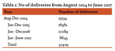
Results: The number of deliveries conducted between August 2014 to June 2017 at Victoria Hospital, Bangalore is shown in Table 1. The total number of deliveries during this period was 57429. Of these deliveries 60 fetuses (males 23 &37 females) with anencephaly, of age 20 to 30 weeks were collected from Department of OBG, Victoria & Vani Vilas hospital attached to Bangalore Medical College & Research Institute, Bangalore.
Classification of mothers according to age showed that maternal age in 1.6% were < 20year, 83.4% were 21-35 years, 13.4% were 36-40 years and 1.6% was >40 years. The mean of maternal age is 24.4 years. When mothers were classified according to their level of education, it was noticed that 91.6% were found to be high school were 3.4%. The study showed that 76.7% of mothers were unemployed (housewives) & 46% of the respondents were found to have regular visits to antenatal care centers. When respondents were inquired about experiencing any form of febrile illness during pregnancy (specifically the first trimester), 86.6% of mothers had no febrile illness during pregnancy; only 14.4% of them reported so. Season of birth were 40% were born in winter, 16.6% were in spring, summer & 26.7% in autumn. The study showed that 8.3% of mothers had multiple abortions, 1.6% had abortion with asthma, diabetes with obesity & hypertension, 5% were diabetic & 81.6 % were normal. 10% of mothers reported taking medications during pregnancy, 1.6% mentioned antiemetic, 5% NSAID (paracetamol & aspirin), 1.6% mentioned herbal medication. None of the respondents was found to have been exposed to any form of radiation during pregnancy. 15% mothers got consanguineous marriage, all were 1st cousins & remaining 51 (85%) cases were unrelated.
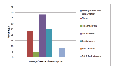
The study showed that 76.7% of mothers were taking folic acid during pregnancy. Regarding the timing of folic acid consumption during pregnancy, only 5% took folic acid preconception (Chart 1).
The comparison of control group (normal fetuses) & case group (anencephalic fetuses) indicates that there is no difference seen. The weight of fetus with anencephaly is more than the control group, and weight of liver was less than control group, and these were statistically significant.
Associated anomalies were present in 42 (70%) fetuses (Table 2). Out of 42 fetuses, those who had associated anomalies were 17 (40.4%) males and 25 (59.6%) were females. All the fetuses had acrania (100%) & 19 (45.3%) fetuses had spina bifida; there were no anomalies found in reproductive system.
The 6 cases (14.3%) had facial defects (4.7% were males & 9.5% were females). The most common defect was cleft palate & cleft lip (9.5% cases), it was found to be statistically significant. In 19 cases (45.3%), spina bifida was very common, in 1 (2.3%) along with spina bifida, cleft lip & palate was also found. The spina bifida upto sacral region was seen in 4.7% of cases & it was found to be statistically significant.
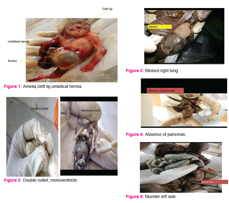
Anomalies in limbs: 1 case (2.3%) had Amelia of right lower limb, congenital dislocation of right elbow joint, malrotation of gut towards left, which was found to be statistically significant (Figure 1).
CVS: in 1 male fetus (2.3%), there was a single ventricle (mono ventricle) on the left side, with two outlets (aorta & pulmonary trunk), which was found to be statistically significant (Figure 2 a&b).
Lungs: in 1 case (2.3%), left lung had 2 fissures & 3 lobes, in 1 (2.3%) right lung had 3 fissures with 4 lobes. In 2 cases (4.7%) both lungs had single oblique fissure with 2 lobes (Fig 3). In 1 case (2.3%), there was malrotation of gut towards left side & also absence of pancreas (Fig 4).
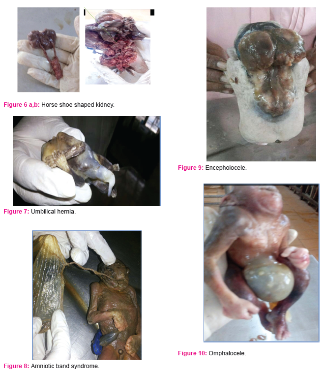
Urinary system: 1 case (2.3%) had unilateral biureter on left side (Fig 5) & 1 case (2.3%) had horse shoe shaped kidney; both are statistically significant (Figure 6a& b).
There were no anomalies found in reproductive system. In 2 male cases (4.7%), there was umbilical hernia (Fig 7). Diaphragmatic hernia was not seen. Amniotic band syndrome (fig 8), encephalocele (fig 9) & omphalocele (fig 10) were seen in 2.3% of cases.
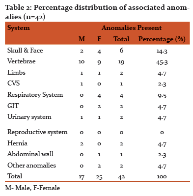
Male fetuses had 40.4% of overall associated anomalies & female fetuses 59.6% of associated anomalies. The female anencephalic fetuses were found to have more number of associated anomalies.
Biochemical Analysis of mother: In 100% of mothers AFP were high, this was a confirmatory test for anencephaly (NTDs).
Normal Values
AFP - upto 10 IU/ml
Folic acid -3.1-17.5ng/ml
Acetylcholine esterase-1700-5778U/L
Blood glucose (RBS) –upto 140 mg/dl
ACE- The 85% of cases ACE level were normal, 13.3% low & 1.7% of cases it was high. Folic acid level in 68.3% cases were normal, 28.3% of cases were low & 3.4% cases were high. RBS were high in 15% of cases all mothers were diabetic, 3.4% cases low & 81.6% were normal.
The Mean, SD & P values are given in (Table 3).
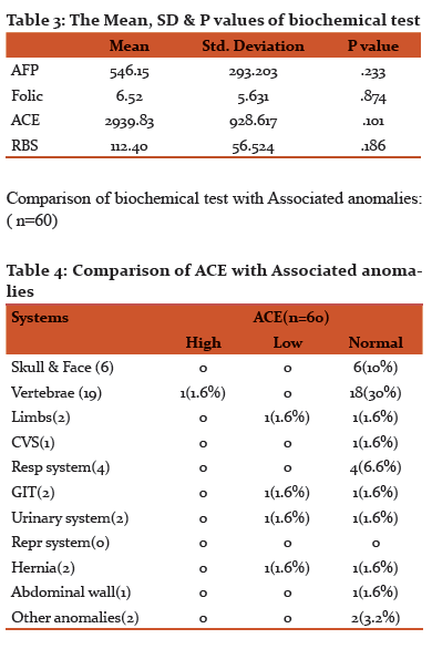
ACE level were high in 1.6% of cases of spina bifida & it was low in anomalies of limb , GIT, Urinary system & umbilical hernia (1.6%cases).
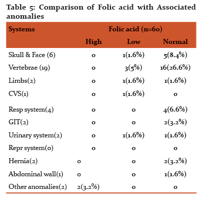
Folic acid was low in 1.6% of cases with anomalies of skull & face, limb, CVS & urinary system & high in 5% of cases with spina bifida.
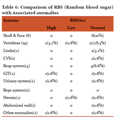
RBS was high in 1.6% of cases with anomalies of GIT, Urinary system and other anomalies & 3.2% with spina bifida. It was low in 1.6% of cases with spina bifida & umbilical hernia.
Discussion: Incidence of anencephaly is reported to be 1:1000 to 1:20000. In the present study the incidence of anencephaly in Victoria and Vanivilas hospital was 1.04 in 1000 births.
Literature showed that anencephaly is more common in females6,7. In the present study, 23(38.4%) were males & 37(61.6%) were females. The birth was of 1st order in 40% of cases , 35% cases were 2nd order, 21.6% were 3rd order & 3.4% cases were 4th order.
Age of mother in 1.6% cases were <20, 83.4% were 21-35, 13.4% cases 36-40 & in 1.6%, >40.
Season of birth: 16.7% cases were born in spring, 26.7% in autumn, 40% in winter & 16.6%were summer which was similar in some studies8.
Maternal occupation: 76.7% cases mothers were house wives, 10% cases farmers, 1.6% were tailor & teacher & 3.4 % were group D.
Family history of both the mother & father was taken in which 1.6% cases had Down’s syndrome for 1st cousin & in 1.6% father of the fetus had hyperthyroidism.
15% mothers got consanguinity, all were first cousins & 85% were unrelated which was similar in few studies9,10,11
The rate of anencephaly could be higher in underdeveloped countries due to possible misdiagnosis, most probably due to poor diet; improper maternal health care, inappropriate treatment & environmental factors also contribute to it9. The ratio of anencephaly is higher in Iranian population than compared to European population. It has been calculated that in Iranian population 13.1/10,000 newborns had anencephaly whose mothers aged >35 years & consanguineous marriage contributes 36 % to anencephaly which was found to be similar in present study12.
Congenital anomalies vary in different studies. The study of malformation is greatly helpful in genetic counseling & prenatal diagnosis in successive pregnancies13. Anomalies were found in 42 cases (70%) in the present study & it is compared with the other studies.
In present study, out of 42 cases, 25 females (41.7%) & 17 males (28.3%) showed associated anomalies; majority of anomalies were in female anencephalic fetuses which was similar to some studies. Cleft lip & palate were the most common defect (14.3%) & 19 (45.3%) had spina bifida. There were no reproductive system anomalies in the present study.
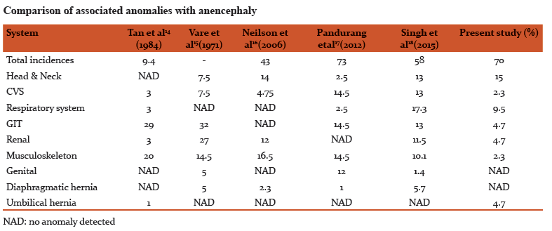
Most of the organs in the present study were anatomically normal when it was compared with the control group & some studies19.
Literature showed that anencephalic fetuses were successful donors of hearts & kidneys for transplantation7.
The intake of 4mg per day of folic acid intake is recommended in mothers with history of neural tube defect7,20.
The alpha fetoprotein is high in 100% of cases which was similar in a study21 & ACE level was normal in 85 % cases, in 1.6% it was high & in 13.4% cases it was low (22,23,24,25,26
In 15%, folic acid was low & 15% cases were hyperglycemic20
The intake of 4mg per day of folic acid intake is recommended in mothers with history of neural tube defect7,20.
In present study ACE level were high in 1.6% of cases of spina bifida & it was low in anomalies of (1.6%cases) Limb, GIT, Urinary system & umbilical hernia.
Folic acid was low in 1.6% of cases with anomalies of skull & face, limb, CVS & Urinary system &high in 5% of cases with spina bifida.
RBS was high in 1.6% of cases with anomalies of GIT, Urinary system and other anomalies & 3.2% with spina bifida. It was low in 1.6% of cases with Spina bifida & umbilical hernia.
Conclusion:
Better knowledge of unexpected fetal loss is the promise for better parental counseling & for prevention of recurrences. Understanding & identifying the risk factors associated with anencephaly in our population, allows approaches to avoid them & thereby lower the incidence of anencephaly in our population. To prevent the NTD, dietary supplements should be provided to low socioeconomic pregnant females. Peri-conceptional & 1st trimester folic acid supplementation is of prime importance. All pregnant mothers have to go in for triple marker tests; that is, beta HCG, alpha fetoprotein and estradiol.
Amniocentesis could be made compulsory for mothers with a history of an anencephalic child. The mother has to be counseled regarding the risks of another such fetus. The family has to be told about pedigree charts, incidence and occurrence of anencephaly in the population.
References:
-
Moore LK, Persaud TV. Editors. The Developing Human Philadelpia: Saunders; 1998, p.478-9
-
Clara G, Jenny N, Mona S. Testing & pregnancy. In: Genetics in Family medicine. The Australian Handbook for General Practitioners. Australia: Dr Kristine BS; 2007:18
-
Cowcock S, Ainbender E, Prescott G. The recurrence risk for neural tube defects in united states : A collaborative study. American Journal of Medical Genetics 1980: 5(3): 309-14.
-
“Anencephaly ” Genetics Home Reference. National Institute of Health. 22 August 2011.
-
Patowary AJ: the fourth incision –few modifications in autopsy incisions , J Indian acad forensic med 32(3) pg 234-238 ISSN 0971-0973
-
Kheir AEM, Eisa WMH, Neural tube defects; clinical patterns, Associated risk factors & maternal awareness in Khortoum state Sudan, Journal of medicine & medical research 2015,3(1)1-6.
-
Gupta P, Nain P & Singh J. Anencephaly:A Neural tube defects –A review, American journal of pharmtech research, 2016; 2(3).
-
Ranganath P, Rajangam S, Perinatology - Journal of perinatal and neonatal care vol 9, 2007 (1):43-47
-
Hassan M, Muhammed N, Gul F, Ahmad S, Frequency of Anencephaly in Hazara Division Of Pakistan, American Journal of Drug Delivery & therapeutics 2015,2(1)039-043
-
Kaur N, Kaur A, Gupta S. Ultra sound evaluation of fetal CNS anomalies & its correlation with postnatal outcome IJCRR,2017; 5(3).
-
Reddy J, Ramanappa MV Anencephaly and its associated anomalies in antenatal scans,J.Evid Based Med .Healthc,pISSN-2349-2562,eISSN-2349-2016;Vol.3(23),1033-1035.
-
Golalipour MJ, Najafi L, Keshtkar AA, Prevalence of Anencephaly in Gorgan , northern Iran , Arc Iran Med 2010;13:34-7
-
Naik S, Venkataramanababu P, Study of various congenital anomalies in fetal & neonatal autopsies, International Journal of Research in medical sciences May 2015,3(5)1114-1117
-
Tan KB, Tan SH, Yeo GS, Anencephaly in Singapore: a ten year series 1993-2002. Singapore Med J , 2007;48:12-5
-
Vare AM, Bansal PC. Anencephaly: An Anatomical study of 41 anencephalies Indian journal Pediatric 1971; 38: 301-305
-
Neilson LAG, Maroun LL, Graem N. Neural tube defects & associated anomalies in a fetal &perinatal autopsy series, Acta patho Microbial Immunol scand 2006;114:239-246
-
Panduranga C, Kangle R, Suranagi V, Pilli GS, Anencephaly: A pathological study of 41 cases J sci soc 2012 :39:81-84
-
Singh J, Kapoor K, Kochhar S. Incidence of Anencephaly in a tertiary care hospital in North west India International journal of scientific & research publications 2015,5(9)2230-315
-
Potters pediatric pathology volume 1 & volume 2, second edition Elsevier.
-
Lisa Moore, Anencephaly, Journal of Diagnostic Medical Sonography 2010, 26 (6)286-289
-
Brock DHK, Alpha fetoprotien & neural tube defects, J. Clin.Path, 29, 10; 157-164
-
Engl N, J Med. 322:669-674 March 8, 1990 DOI: 10:10056/NEJM 199003083221006.
-
Incore V, Ninios AP, Pavlik J, Hsu CD, Prenatal diagnosis of acrania associated with Amniotic band syndrome: Obstet Gynecol 2003
-
Macri JN, Weiss RR. Prenatal serum alpha feto protein screening for neural tube defects. obstet gynecol 1982;59:633-9
-
Amniotic-fluid alpha feto protein measurement in antenatal diagnosis of anencephaly and open spina bifida in early pregnancy: second report of U.K. collaborative study on alpha feto protein in relation to neural tube defects. Lancet1979;2:651-62.
-
Report of collaborative Acetylcholine esterase study .amniotic fluid acetylcholineesterase electrophoresis as a secondary test in the diagnosis of anencephaly and open spina bifida in early pregnancy.lancet1981; 2:321-4
|


 This work is licensed under a Creative Commons Attribution-NonCommercial 4.0 International License