IJCRR - 10(2), January, 2018
Pages: 22-24
Date of Publication: 19-Jan-2018
Print Article
Download XML Download PDF
A Rare Case Report of Hydrometrocolpos in a Female Newborn
Author: Khadija Kanso, Bassem Abou Merhi, Marwan Zeidan, Soukaina Ibrahim, Fatima Ghandour, Fadi Iskandarani, Eliane El-Houwayek, Fatima Yassine, Imad Chokr
Category: Healthcare
Abstract:Introduction: Congenital hydrocolpos and the hydrometrocolpos is a rare disorder caused by accumulation of cervico-vaginal secretions that present with cystic pelvic mass in neonates.Prenatal diagnosis is important to prevent complications like sepsis and renal failure and to affect prognosis. We will report a case of a newborn female admitted to our NICU with abdominal mass and imperforated anus diagnosed to have hydrometrocolpos, septated vagina with bicornate uterus and urogenital sinustreated surgically.
Case Report: This report will help us to develop a high index of suspicion of hydrometocolpos in female newborn with abdominal
mass.
Keywords: Hydrocolpos, Hydrometrocolpos, Neonatal Intensive Care Unit (NICU).
DOI: 10.7324/IJCRR.2018.1025
Full Text:
Introduction:
Hydrocolpos and hydrometrocolpos is uncommon disorder caused by vaginal distention and fluid accumulation (1). Congenital hydrocolpos can be associated with other genitourinary anomalies or it can be a part of a syndrome. Prenatal diagnosis either by ultrasound or magnetic resonance imaging is essential to prevent complications (1, 2).
Case Report:
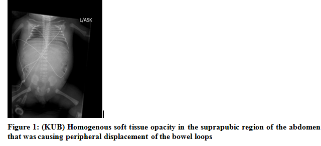
We report a case of 35weeks and 5 days preterm girl with history of normal vaginal delivery to a 19 years old mother, G1P1A0 admitted to our neonatal intensive care unit (NICU) with abdominal distension, lower limbs swelling and imperforated anus. The pregnancy though uncomplicated was poorly followed and no antenatal ultra-sonograms (US) were available. On physical examination, the baby had a palpable mass in the lower mid abdomen. The superior margins of the mass were well felt and appreciated. Both lower limbs found to have pitting pedal edema. She had low imperforated anus. The child was referred to Department of Radio- diagnosis for evaluation of the abdominal mass. A plain radiograph of the abdomen (KUB) was initially obtained and showed homogenous soft tissue opacity in the suprapubic region of the abdomen that was causing peripheral displacement of the bowel loops(Figure 1).
Ultrasound of the abdomen was done at the same time and showed that there was a huge fluid filled structure occupying almost all the abdomen and showing a midline septations. This mass was causing displacement of the liver superiorly. Bowel loops were being displaced peripherally. There was also bilateral hydronephrosis. Rest of the intra- abdominal organs appeared to be normal (Figure 2).

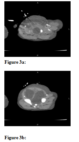
Following ultrasound, a preliminary diagnosis of neonatal hydrometrocolpos was made. A computed tomography scan (CT Scan) of the abdomen and pelvis was done immediately after KUB and US to reveal a cystic mass 8.5x8.2x6 cm occupying the abdomen and pelvic cavity with median septations and double thin walls extending down to the rectum with severe bilateral hydronephosis. The uterus was not well dilneated (Figures 3a, 3b, and 3c).

The child was transferred to the operating room (OR) where imperforated hymen,septated vagina with bicornate uterus, hydrometocolpos and urogenital sinus (vagina was connected to the urethra) were identified. Drainage of milky intravaginal collection and closure of the communication between urethra and vagina was performed. Foley catheter was inserted through the cloaqual canal into the vagina for continuous drainage and lastly colostomy was performed. After stabilization in the NICU the patient was discharged from our institute to be followed up later for more exploration of the ano-genital area firstly for anatomical definition and secondly for surgical anastomosis and closure of the colostomy after 6 months.
Discussion and Review of Literature:
Hydrocolpos is an uncommon disorder that can be associated with genitourinary anomalies ranging from persistent urogenital sinus to cloacal dysgenesis (1). It can be defined as vaginal distention with fluid accumulation and it can be due to either a combination of increased activity of secretory cervical glands and vaginal obstruction or the presence of urogenital sinus with urine collection (1, 2). Hydometrocolpos is characterized by uterine distention that might result if the accumulated fluids stretchthe cervical canal as well as the body of the uterus (2).
The causes of accumulation of cervico-vaginal secretions are diverse and include imperforate hymen which is defined as a membrane occluding the lower third of vagina, transverse vaginal septum, vaginal atresia and malformations of cloaca including urogenital sinus (2,3). Cloacal malformations form a group of non-hereditary ano-rectal malformations. The cloaca is defined as a single common chamber of the caudal intestinal, urinary, and genital tract; it is normally present in the 4th to 5th week embryogenesis. Later in the fetal life the cloaca will be divided into the urogenital sinus anteriorly and ano-rectum posteriorly. The urogenital sinus then becomes the urinary bladder and urethra with a portion transformed into the vagina and hymen. The type of cloacal anomalies will results from the timing of developmental arrest (4). Hydrometrocolposis rare with an incidence of 0.006% approximately (2, 3). Bischoff et al. reported a high incidence of hydrometrocolpos in patients with cloacal malformations (28%) (5). Child with this disorder presents clinically with increasing abdominal distension (3). Prenatal diagnosis of hydrometrocolpos by radio-imaging may be difficult but is essential in that it can substantially affect prenatal and immediate neonatal treatment and it can help improving prognosis (4).Ultrasonography and in particular magnetic resonance imaging can be helpful in the antenatal diagnosis of congenital hydrometrocolpos. Magnetic resonance imaging (MRI) provides informationin cases in which the diagnosis is unclear or additional anomalies cannot be adequately evaluated by ultrasound. MRIproved to be better when ultrasonography is limited because of late presentation or by the presence of obesity or oligohydramnios (3, 4). Antenatal imaging will be helpful for preoperative planning (4) and for appropriateprenatalcounseling to parents. It may improve the outcomes especially related to obstructive uropathies(1). Association of an imperforate hymen with other genitourinary anomalies is well known such as imperforate anus, cloacal anomalies, renal agenesis, uterine anomalies or persistent urogenital sinus. It may be a part of a syndrome like McKusick-Kaufman syndrome (autosomal recessive disorder which includes vaginal atresia and secondary hydrometrocolpos, polydactyly, congenital cardiac anomalies and hydrops fetalis), Ellis Van Creveld syndrome, Bardet- Biedl syndrome, VACTERL association (Vertebral, anal, cardiovascular, tracheooesophageal, renal and limb anomalies), and MURCS association (Mullerian duct aplasia/hypoplasia, renal agenesis/ectopia and cervico-thoracic somite dysgenesis such as Klippel Feil abnormality, anomalous ribs or Sprengel deformity) (1, 2, and 3).The most frequent complication of congenital hydrometrocolpos includesurinary tract obstruction, leading to hydronephrosis and renal failure. Other complications include repeated urinary tract infections, sepsis, pyocolpos, rupture and peritonitis (1, 2, and 3). Drainage of the collection which can be performed under ultrasound guidance was associated with decreased risk of urinary obstruction and associated complications (3, 4). The recognition of hydrocolpos prenatally can facilitate rapid treatment at birth (4). Infants with prenatal diagnosis of pelvic mass should be categorized as high risk deliveries and should be promptly followed by multidisciplinary team soon after birth (1).
Conclusion:
Hydrocolpos or hydrometrocolpos is uncommon disorder of neonates that can present with a midline abdomino-pelvic mass. Ultrasound and magnetic resonance imaging can help in the prenatal diagnosis and early surgical treatment for better outcome and prognosis. This report will aid us to the development of high index of suspicion of congenital hydrocolpos in neonates with pelvic mass for early and even antenatal diagnosis that lead to further prevention of secondary complications such as hydronephrosis and gastrointestinal obstruction.
Acknowledgement:
Authors acknowledge the immense help received from the scholars whose articles are cited and included in references of this manuscript. The authors are also grateful to authors / editors / publishers of all those articles, journals and books from where the literature for this article has been reviewed and discussed.
References:
- Murthy V, Costalez J, Weiner J, Voos K. Two Neonates with Congenital Hydrocolpos. Case Reports in Pediatrics, vol. 2013, Article ID 692504, 3 pages, 2013.
- Shetty D, Varma R. Neonatal Hydrocolpos: A Case Report and Review of Literature. SSRG International Journal of Medical Science. 2017;4.
- B R, Basavalingu D, Paramesh VM, NagendraPDK. Radiological Diagnosis of Neonatal Hydrometrocolpos- A Case Report. J Clin Diagn Res. 2016; 10(3): TD18–TD19.
- Winkler N,Kennedy A, Woodward PJ. Cloacal Malformation. J Ultrasound Med. 2012; 31:1843–1855.
- Bischoff A, Levitt MA, Breech L, Louden E, Pe˜na A. Hydrocolpos in cloacal malformations. Journal of Paediatric Surgery. 2010;45:1241–45.
|
 IJCRR
IJCRR 


 This work is licensed under a Creative Commons Attribution-NonCommercial 4.0 International License


