IJCRR - 8(13), July, 2016
Pages: 50-57
Date of Publication: 12-Jul-2016
Print Article
Download XML Download PDF
IMMUNOHISTOCHEMISTRY STUDY FOR HER-2/NEU EXPRESSION IN LESIONS OF UTERINE CERVIX
Author: Pramod Sarwade, Sunita Patil, Rajan Bindu
Category: Healthcare
Abstract:Objectives:
1. To study expression of HER-2/neu in lesions of uterine cervix by immunohistochemistry.
2. To correlate expression of HER-2/neu with histological type, grade and stage of cervical malignancy.
Methods: Total 50 cases were included during the period from October 2013 to October 2015. Immunohistochemical studies were done on formalin-fixed paraffin embedded tissue blocks with HER-2/neu antibody.
Results: Out of fifty cases, 10 cases (20%) of adenocarcinoma (ADC), 1 case (2%) of adenosquamous carcinoma, 4 cases (8%) of CIN, 3 cases (6%) of chronic cervicitis, 2 cases (4%) of chronic cervicitis with squamous metaplasia, 30 cases (60%) of squamous cell carcinoma of cervix (SCC). And It Amongst 9 cases of poorly differentiated SCC cases, 2 cases showed 1+ positivity, 2 cases showed 2+ positivity, 1 case showed 3+ positivity. Out of 16 cases of moderately differentiated SCC, 4 cases showed 1+ positivity, 4 cases showed 2+ positivity, 2 cases showed 3+ positivity. Among 5 cases of well differentiated SCC, 2 cases showed 1+ positivity. Out of 10 cases of adenocarcinoma of cervix, 2 cases of well differentiated adenocarcinoma showed 1+ positivity. Each 2 cases of moderately differentiated adenocarcinoma showed 1+ and 2+ positivity. Cervical carcinoma cases with involvement of lymph node and parametrium showed stronger HER-2/neu staining.
Conclusion: Higher HER-2/neu expression was noted in malignant lesion as compared to benign lesions. Stronger HER-2/neu expression was noted among higher stage tumors and those with parametrial and lymph node involvement.
Keywords: HER-2/neu expression, Immunohistochemistry, Cervical lesions
Full Text:
INTRODUCTION
According to the global cancer statistics for 2012, cervical cancer is the fourth most common cancer affecting women worldwide and it is also the fourth most common cause of cancer death in women worldwide. Almost 70% of the global burden falls in areas with lower levels of development and more than one fifth of all new cases are diagnosed in India.[1] Carcinoma cervix is one of the common malignancy in women in India with an incidence of 9 to 44 per 100,000 women.[2] Current treatment is failing to cure locally advanced disease. Therefore next step in treatment is testing of molecular targeted therapies to improve outcome of cervical cancer patients. [3] The c-erbB-2 proto-oncogene, also called HER-2/neu, is located on Chromosome 17q21 which encodes 185kDa transmembrane glycoprotein with tyrosine kinase activity. Overexpression leads to constitutive activation of tyrosine kinase residues.[4] The epidermal growth factor receptor (EGFR/HER) family of receptors has been associated with aggressive biological behavior and metastatic potential.[5] The expression of HER family members in gynaecological cancers and their relationship with disease stage, grade and response to treatment controversial.[6] HER2/neu expression is not limited to cervical cancers but is also seen in other tumors like breast, stomach, ovary, uterine serous endometrial carcinomas, colon, bladder, lung, head and neck and esophagus.[7]
MATERIALS AND METHODS
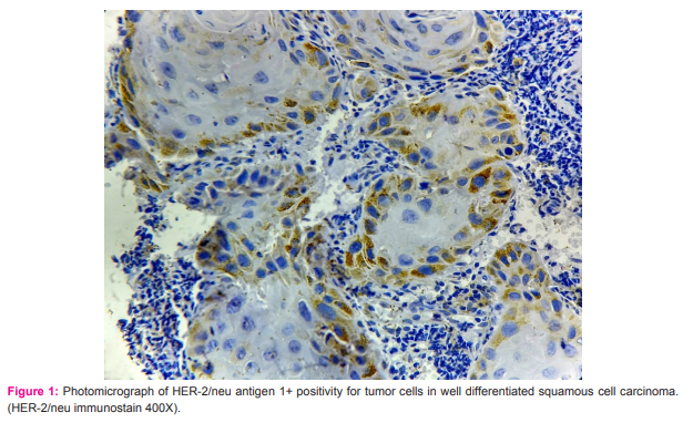
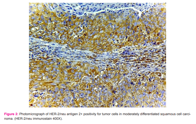
A total of 50 cases of cervical tissue were obtained from October 2013 to October 2015. The study consists 10 cases (20%) of adenocarcinoma, 1 case (2%) of adenosquamous carcinoma, 4 cases (8%) of cervical intraepithelial neoplasia (CIN), 3 cases (6%) of chronic cervicitis, 2 cases (4%) of chronic cervicitis with squamous metaplasia, and 30 cases (60%) of squamous cell carcinoma of cervix. Routine Hematoxylin and eosin stained slides were screened to obtain the best section for immunohistochemistry with HER-2/ neu antibody. 3-5 micron sections were cut on poly L-lysine coated slides. Antigen retrieval was done by heating the sections in citrate buffer at pH 6.0 using microwave oven. 100 µl prediluted primary antibody HER2/neu (Monoclonal, Immunogen: A synthetic peptide corresponding to residues near the C-terminus of human HER2, Clone: EP1045Y, Species: Rabbit, Ig class: IgG, Protein Conc.: 50mg/ml, Catalog no.: AN471-5ME, BioGenex, USA) were applied so as to cover the tissue sections. Standard Streptovidin-biotin peroxidase method is used. A golden brown membrane and cytoplasmic staining was taken as a positive reaction. Intensity of HER-2/neu expression was graded according to the 2014 ASCO/CAP guidelines[8] as complete intense circumferential membrane staining within >10% of tumor cells (3+); circumferential staining that is incomplete and/ or weak/moderate within >10% of tumor cells or complete intense circumferential membrane staining within 10% of tumor cells (1+) and no staining is observed or shows membrane staining that is incomplete and is faint/barely perceptible and within <10% of tumor cells (0). Statistical analysis of data was performed using Fischer’s Exact Test. P value <0.05 was taken as statistically significant.
RESULTS
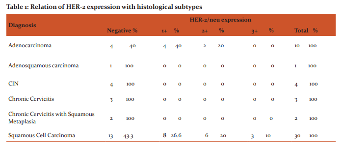
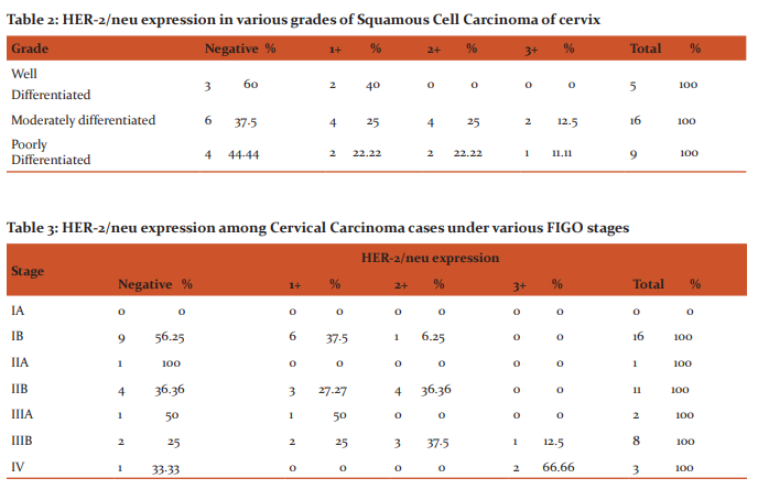
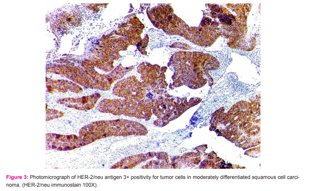
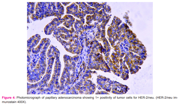
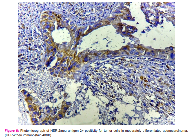
Out of 50 cases, 27(54%) were negative, 12(24%) were having 1+ positivity HER-2/neu expression, 8(16%) were showing 2+ positivity HER-2/neu expression and 3(6%) were showing 3+ HER-2/neu expression. All chronic cervicitis cases, chronic cervicitis with squamous metaplasia cases, CIN cases and adenosquamous carcinoma case were negative HER-2/neu immunostain [Table 1]. Out of 30 cases of Squamous Cell Carcinoma (SCC), 5 cases (16.66%) were well differentiated, 16 cases (53.33%) were moderately differentiated and 9 cases (30%) were poorly differentiated. Among 5 cases of well differentiated SCC, 2 cases showed 1+ positivity [Figure 1] while remaining 3 cases were negative. Out of 16 cases of moderately differentiated SCC, 4 cases showed 1+ positivity, another 4 cases showed 2+ positivity [Figure 2], 2 cases showed 3+ positivity [Figure 3] and remaining 6 cases were negative. Out of 9 cases of poorly differentiated SCC cases, 2 cases showed 1+ positivity, 2 cases showed 2+ positivity, 1 case showed 3+ positivity and remaining 4 cases were negative. Overall it was found that among 6 cases of SCC with 2+ positivity, 4 c a s e s were (66.66%) moderately differentiated and 2 cas es were (33.33%) poorly differentiated. Also out of 3 cases of SCC with 3+ positivity, 2 cases were (66.66%) moderately differentiated and 1 case was (33.33%) poorly differentiated. However a statistically correlation was not established between grade of SCC and intensity of HER-2/neu staining (p=0.935) [Table 2]. Out of 10 cases of adenocarcinoma of cervix (ADC), 2 cases of well differentiated ADC showed 1+ positivity [Figure 4] and 3 cases were negative. Each 2 cases of moderately differentiated ADC showed 1+ and 2+ positivity [Figure 5] and remaining 1 case was negative. However significant correlation was not established between grade of adenocarcinoma and intensity of HER-2/neu staining (p=0.46)
Out of 41 cervical carcinoma cases, 16 cases (39.02%) were under FIGO stage I, 12 cases (29.26%) were under FIGO stage II, 10 cases (24.39%) were under FIGO stage III and 3 cases (7.31%) were under FIGO stage IV. Among the 16 cases under stage IB, 6 cases showed 1+ positivity, 1 case showed 2+ positivity. 1 case under stage IIA was negative for HER-2/neu staining. Of the 11 cases under stage IIB, 3 cases showed 1+ positivity, 4 cases showed 2+ positivity. 1 case under stage IIIA showed 1+ positivity. Out of 8 cases under stage IIIB, 2 cases, 3 cases and 1 case showed 1+, 2+ and 3+ positivity respectively. Out of 3 cases, 2 cases under stage IV showed 3+ positivity. However, significant correlation between stage of cervical carcinoma and intensity of HER-2/neu staining was not established (p=0.073) [Table 3]. Among 41 cervical carcinoma cases, it was found that 17 cases (41.46%) had no parametrial or lymph node involvement, 11 cases (26.82%) had only parametrial involvement and 13 cases (31.70%) had involvement of both lymph node and parametrium. On analyzing variation in intensity of HER-2 staining with lymph node and parametrial involvement, it was found that among 8 cases with 2+ positivity, 1 case had no lymph node or parametrial involvement, 4 cases had only parametrial involvement and 3 cases showed both involvement. Out of 3 cases with 3+ positivity, all 3 cases had involvement of both. Even though cervical carcinoma cases with involvement of lymph node and parametrium showed stronger HER-2/neu staining, a significant correlation between these variables was not established (p=0.107) [Table 4].
DISCUSSION
Three cases of chronic cervicitis and two cases of chronic cervicitis with squamous metaplasia were studied in the present study. HER-2/neu staining was negative in all cases. Gupta et al [9] noticed out of 20 cases, most of the cases exhibited no or faint (1+) staining, except one case of chronic cervicitis exhibited moderate (2+) intensity of staining. Cases of squamous metaplasia were negative for HER-2/neu expression.
Four cases of CIN were encountered in the present study. HER-2/neu positivity was not noted in all CIN cases. In contrast to present study HER-2/neu expression was noted in 37.5% of CIN cases by Li et al [10] while 60% of CIN cases showed HER-2/neu positivity in a study by Gupta et al. [9] Protrka et al [11] reported HER-2/neu expression in 66% of CIN cases. Joseph et al [12] stated HER-2/neu positivity in 70% of CIN cases. The reason for discordance between studies could be due to the fact that less number of cases was analyzed in the present study. HER-2/neu expression was noticed in 23 cases (56%) of cervical carcinoma in present study. According to various studies expression of HER-2/neu in cervical carcinoma ranged from 12.1% to 100%.[9-19] This variability may be possibly due to the difference in number of cases analyzed, variation in stage of tumor and in experimental procedure. Costa et al [13] stated HER-2/neu expression in 77% of cervical carcinomas while Lakshmi et al [14] reported HER-2/neu expression in 96.8% of squamous cell carcinomas of cervix. Ndubisi et al [15] observed HER-2/neu expression in 34 cases out of 150 (22%). In the present study, 10 cases of adenocarcinoma were analyzed. 2 cases of well differentiated adenocarcinoma showed 1+ positivity while each 2 cases of moderately differentiated adenocarcinoma showed 1 + a n d 2+ positivity. However a significant correlation was not established between HER-2/neu expression and grade of adenocarcinoma (p=0.46). Similar to this study, Kihana et al, [16] Mandai et al [17] and Joseph et al [12] also did not find any relation between HER-2/neu expression and grade of adenocarcinoma. Squamous Cell Carcinoma belonging to moderately and poorly differentiated carcinomas showed 2+ and 3+ positivity where as well differentiated carcinoma shows only 1+ positivity. Also among 3 cases with 3+ positivity 2 cases (66.66%) belonged to moderately differentiated carcinoma while remaining 1 case (33.33%) belonged to poorly differentiated carcinoma. But no correlation was established between HER-2/neu expression and grade of SCC (p=0.935). Similar to the present study, Mandai et al, [17] Ray et al [18] and Joseph et al [12] also did not find any correlation between HER-2/neu expression and grade of SCC. It was found that higher intensity of HER-2/neu expression among cases under stage III and IV. A statistically significant relationship was not established between HER-2/neu expression and stage of the tumor. Contrary to this Ndubisi et al [15] and Gupta et al [9] found a significant correlation between HER-2/neu expression and higher stage of cervical carcinoma. Ndubisi et al [15] had studied HER-2/neu expression in 150 cases of cervical carcinoma while Gupta et al [9] had analyzed 65 cases of cervical carcinoma. The confrontation between the present study and other studies could be due to the small number of cases analyzed in the present study. Similar to present study, Joseph et al [12] did not find any relation between HER-2/neu expression and higher stage of cervical carcinoma. Joseph et al [12] studied HER-2/ neu expression in 24 cases of cervical carcinoma. Mandai et al [17] and Yong et al [19] analyzed HER-2/neu expression in 39 and 74 cases of cervical adenocarcinoma respectively and reported that a significant correlation existed between HER-2/neu expression and lymph node metastasis. Yong et al [19] found the expression of c-erbB-2 in 34 cases. Gupta et al [9] analyzed HER-2 expression in 65 cases of cervical carcinomas and stated that intensity of staining correlated with presence of parametrial extension. The confrontation between the present study and the above mentioned studies could be due to the less number of cases analyzed. Similar to our present study, Joseph et al [12] did not find any relation between HER-2/neu expression with parametrium and lymph node involvement Lakshmi et al [14] and Gupta et al [9] reported gradual progression in HER-2/neu expression from premalignant to malignant. Overall squamous cell carcinomas showed a significantly stronger staining (3+) with HER-2/neu. Gupta et al [9] also reported a significantly higher expression of HER-2/neu in squamous carcinoma of cervix. As stronger HER-2/neu expression was noted among higher stage tumors and those with parametrial and lymph node involvement it can be assumed that HER-2/neu expression in cervical carcinomas is a poor prognostic factor. In the present study, HER-2/neu expression did not correlate significantly with grade of the tumor, stage at presentation or with presence of parametrial/ lymph node involvement.
CONCLUSION
Incidence of HER-2/neu expression in cervical cancer suggests the role of this gene in tumor genesis. A statistically significant correlation was not established between HER-2/ neu expression and various parameters that were analyzed namely stage of tumor, grade of tumor and presence of lymph node and parametrial extension. Detailed study of various factors associated with HER-2/neu over expression and its positivity rates in cervical tumors is required to enrich our knowledge concerning the role of Her-2/neu in path physiology of malignancy, response to treatment and promise of monoclonal antibody therapy as well as other approaches.
ACKNOWLEDGMENT
Authors wish to express and convey their sincere thanks and gratitude to all those who helped for completion of this research article. Authors acknowledge the immense help received from the scholars whose articles are cited and included in references of this manuscript. The authors are also grateful to author’s /editors/ publishers of all those articles, journals and books from where the literature for this article has been reviewed and discussed. Authors declare and acknowledge that we have no conflict of interest.
References:
1. Ferlay J, Soerjomataram I, Ervik M, Dikshit R, Eser S, Mathers C et al. GLOBOCAN 2012 v1.0, Cancer Incidence and Mortality Worldwide: IARC CancerBase No. 11 [Internet]. Lyon, France: International Agency for Research on Cancer; 2013. Available from: http://globocan.iarc.fr, accessed on 23rd July 2014.
2. Wadhwani R, Bamnia R, Meena M. Clinico-pathological analysis of cancer cervix in tertiary care centre. J Evol Med Den Sci. 2013;2(39):7381-85.
3. Dueñas-Gonzalez A, Cetina L, Mariscal I, de la Garza J. Modern management of locally advanced cervical carcinoma. Cancer Treat Rev. 2003;29:389-99.
4. Singleton TP, Niehans GA, Gu F, Litz CE, Hagen K, Qiu Q, et al. Detection of c-erbB-2 activation in paraffin-embedded tissue by immunohistochemistry. Hum Pathol 1992 Oct:23(10);1141-50.
5. Nevin J, Laing D, Kaye P, McCulloch T, Barnard R, Silcocks P, et al. The significance of Erb-b2 immunostaining in cervical cancer. Gynecol Oncol 1999;73:354-8
6. Reyes HD, Thiel KW, Carlson MJ, Meng X, Yang S, Stephan J, et al. Comprehensive profiling of EGFR/HER receptors for personalized treatment of gynecologic cancers. MolDiagnTher. 2014;18(2):137-51.
7. Iqbal N, Iqbal N. Human Epidermal Growth Factor Receptor 2 (HER2) in Cancers: Overexpression and Therapeutic Implications. Mol Biol Int [Internet]. 2014;2014:1-9. Available from: http://www.hindawi.com/journals/mbi/2014/852748/
8. Wolff AC, Hammond MEH, Hicks DG, Dowsett M, McShane LM, Allison KH, et al. Recommendations for Human Epidermal Growth Factor Receptor 2 Testing in Breast Cancer. American Society of Clinical Oncology/College of American Pathologists Clinical Practice Guideline Update. Arch Pathol Lab Med. 2014; 138: 241–256.
9. Gupta N, Singh S, Marwah N, Kumar S, Chabra S, Sen R. HER2/neu expression in lesions of uterine cervix: Is it reliable and consistent?. Indian J PatholMicrobiol. 2009;52:482-5.
10. Li G, Yu D, Zhong-Qiu W. Significance of EGFR, HER-2 and VEGF expressions in cervical intraepithelial neoplasia and squamous cell carcinoma. Cancer Res Clin. 2013;25(11):750-3.
11. Protrka Z, Mitrovi? S, Arsenijevi? N, Baskic D, Radosavljevic G, Stankovic M, et al.HER-2 expression in uterine cervix carcinogenesis. J BUON. 2007;12(1):91-7.
12. Joseph T and Raghuveer CV. HER-2/neu expression in cervical intraepithelial neoplasia and cervical carcinoma. International Journal of Biomedical and Advance Research 2015; 6(01):47- 52.
13. Costa MJ, Walls J, Trelford JD. c-erbB-2 oncoprotein overexpression in uterine cervix carcinoma with glandular differentiation. A frequent event but not an independent prognostic marker because it occurs late in the disease. Am J Clin Pathol. 1995;104:634-42.
14. Lakshmi S, Nair MB, Jayaprakash PG, Rajalekshmy TN, Nair MK, Pillai MR. c- erbB-2 oncoprotein and epidermal growth factor receptor in cervical lesions. Pathobiology. 1997;65:163-8.
15. Ndubisi B, Sanz S, Lu L, Podczaski E, Benrubi G, Masood S. The prognostic value of HER-2/neu oncogene in cervical cancer. Ann Clin Lab Sci. 1997;27(6):396-401.
16. Kihana T, Tsuda H, Teshima S,Nomoto K, Tsugane S, Sonoda T, et al. Prognostic significance of the overexpression of cerbB-2 protein in adenocarcinoma of the uterine cervix. Cancer. 1994;73:148-53.
17. Mandai M, Konishi I, Koshiyama M, Komatsu T, Yamamoto S, Nanbu K, et al. Altered expression of nm23-H1 and c-erb B-2 proteins have prognostic significance in adenocarcinoma but not in squamous cell carcinoma of the uterine cervix. Cancer. 1995;75:2523-9.
18. Ray A, Naik SL, Sharma BK. Distribution of prognostically unfavorable product of C-erb B-2 oncogene and EGFR in carcinomas of the breast and uterine cervix. Indian J PhysiolPharmacol. 2002;46(4):423-33.
19. Yong H, Shumo C, Shaoyin Y, Daren S. Expression of c-erbB-2 and PCNA in cervical adenocarcinoma and its signification. Chin J Canc Res. 1998;10(1):68-70.

|


 This work is licensed under a Creative Commons Attribution-NonCommercial 4.0 International License