IJCRR - 4(17), September, 2012
Pages: 103-109
Date of Publication: 14-Sep-2012
Print Article
Download XML Download PDF
INTERESTING FACTS ON THYMUS GLAND CHANGES-A REVIEW
Author: Praful S. Jevoor, MathadaV. Ravishankar, Sandhya Dharwadkar, Amit Magadum, Somashekhar Biradar
Category: Healthcare
Abstract:In most of the cadavers procured for the dissections in the department of anatomy belongs to male adult cadavers, which were devoid of active thymus gland and often the gross features of this gland was ignored because of its remnants were found with variable degree of shape and sizes. During dissections in most of the cadavers we have often noticed the morphology of most thymus remnant from simple flat mass to a well defined solid mass of thymic tissue. To test our curiosity on microscopic studies we have noticed features like, abundant number of iscrete lymphocytes along with Hassall?s corpuscles were appreciated under hematoxylin and eosin staining. The thymus being a central lymphoid organ develops from endoderm of third pharyngeal pouch along with the parathyroid gland. It remains active till the age of 14 years and later shows gradual inclination towards its involution as conventionally it is believed to be an involuting organ with advancing age. Thymus being an important immune competent organ which has created a curiosity regarding its changes from advancing age to number of invasive and non invasive experimental studies, such interesting facts were considered to drag the attention of involuting organ.
Keywords: Acetylcholine, Adipocytes, Hassall?s corpuscles, Immunosenescence
Full Text:
INTRODUCTION
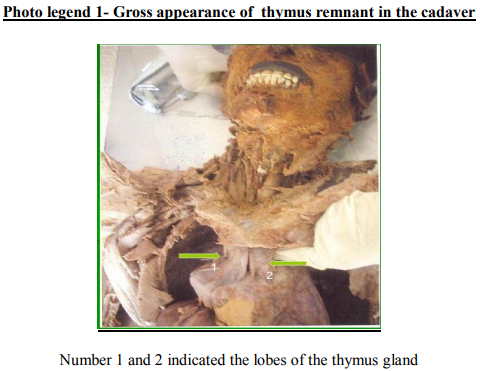
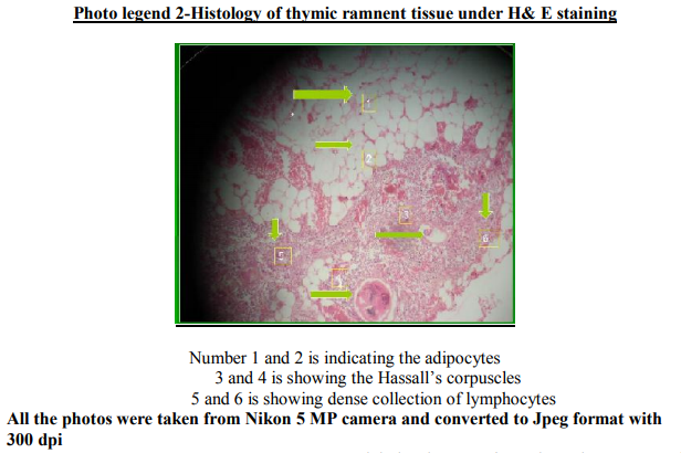
Thymus is a central lymphoid organ located in the superior mediastinum; many times it was ignored during the dissections of adult cadavers practical point of view its morphology was less stressed as it is replaced by the fatty tissue in most of the adult cadavers. Immunologically it is considered to be an important gland which programs the stem cells which are originating from bone marrow for its specific antigen activity. Regarding the involution of the thymus gland and its significance is sited in the texts of histology, this organ created curiosity in connection with its versatile changes in different age, sex and disease conditions, such as immune deficient ailments varying from opportunistic susceptibility of body to simple viral infections to life threatening condition like acquired immunodeficiency syndrome(AIDS). The thymus arises from the third pharyngeal pouch of thymic primordia at the end of the fourth week in the form of endodermal proliferations, these endodermal proliferation forms a hallow tubes that invade the underlying mesoderm which later transforms into solid branching cords, these cords are the primordia of the polyhedral thymic lobules. The whorl like Hassall?s corpuscles within the medulla apparently arises from the ectodermal cells of the third pharyngeal cleft shortly after the formation of thymus is subsequently infiltrated by lymphocytes. As the thymus is developed along with many structures in the primitive pharyngeal floor during the fetal life, often it tend to take a different pathway and destination with or without influencing its accompanied structures 1,2 . Such rare incidence of embedded thymic follicles within the section of thyroid gland was accidentally found during the histopathological investigations showing its diverse destination3 . The congenital anomaly like digeorge syndrome is often associated with maldevelopment of thymus gland. The thymus is highly active during the perinatal period and continues to grow throughout childhood reaching its maximum size at puberty and later on the gland involutes rapidly which is replaced subsequently with fatty tissue4 . The importance of the thymus often reviewed in adults with the suspected clinical locomotors functions, most of such signs and symptoms are often proved with persistent activity of thymus. In case of adults the thymus often start producing its antigens against own body components which can lead to condition like myasthenia gravis this condition can be handled effectively by thymectomy 5 . During routine dissections in most of the cadaveric gross thymic remnant was noticed as a mass resembling the original gland as shown in the photo legend (1) and its microscopic appearance has shown in the photo legend (2).
DISCUSSION
If we observe the whole life span of an individual as the age advances some of the bodily organs are showing obvious changes from simple atrophy to involution, often these changes are so subtle that the functional derangements are correlated with advanced structural changes in number of organs. During the early period of post natal life thymus gland plays a significant role of programming the cells which are reaching the gland for their charged specific tasks, such cells are destined to be involved in immune mechanism, our body defense activities are influenced by factors like age, sex, environment, food, nutritional status, genetic factor, hormonal status etc. During the dissections in most of the male cadavers often we have noticed and collected a lobulated mass situated at the upper and anterior part of the pericardium in the region of superior mediastinum(Photo legend-1), where usual thymus gland is situated. Such mass was studied thoroughly from out side and was later subjected to histological tissue processing, embedding, microtome sectioning and it was stained with hematoxylin and eosin, later on microscopic analysis it has shown a classical involuntary changes in most of the areas which resembles the thymic tissue and the Hassall?s corpuscles (Photo legend-2) were sparsely spread among dense fatty tissue along with large number of scattered lymphocytes. These findings have confirmed that it is an involuted thymic remnant. Thymus gland shows a linear increase in its weight as the age advances till the 16 years of age, later it undergoes gradual loss in its gross weight 6 . Thymus is being a primary lymphoid organ plays an important role in the development of other secondary lymphoid organs. This fact can be realized by the non-development or immature development of other secondary lymphoid organs followed by thymectomy during the early postnatal life, such dependent activity makes one to become more susceptible to infections from simple to complex nature. The activity of thymus begins in the early fetal life itself as a source of production of diversified T cell production which is further boosted in the early stages of post natal life, but its function declines as the age advances. The neonatal thymectomy results in reduced number of T cell counts in short and long term complications which shows its indispensable functional significance through infection prone attitude in infants with advancing ageing process. 7 The developmental anomalies like digeorge syndrome often associated with the absence of thymus gland, during such situations clinical trials associated with the transplantation of the thymus gland in early childhood was found with formation of new T-cells after a span of 5 months suggesting immune reconstitution which was able to render an essential support for host to fight against the entry of foreign bodies 8,9.It is commonly seen that the advanced ageing process affects all aspects of immune system followed by thymectomy during infancy a particular type of “ T “cell activity in an individual is often characterized by lower CD4 + and CD8+ cell number and diversity. The removal of thymus in infancy results in premature onset of decline in immune competency, often such individuals are susceptible for infectious diseases, such clinical findings shows premature signs of immune aging and Immunosenescence which was experimentally correlated with the consequence of thymectomy in the early postnatal period 10. There are some experimental results correlating that the early thymectomy in case of the female rats are showing the precautious sexual maturity but the same procedure in case of the male rats showing no significant effects 11. A prospective randomized study treatment with growth hormone in HIV infected adults were showing changes in the body in the form of increased thymic mass along with increase T cell receptor rearrangement, such studies are indicating that the growth hormone is an important factor which can regulate the T cell development which can reverses the adverse influence on thymopoiesis in rodents. The thymus being a primary site of de novo cell production it will modulate the recovery in immune deficient cells, which supports the hypothesis of enhancement of thymus activity by growth hormone administration in immune deficient conditions. The possibility of the functional reinstate of the thymus was observed in a condition where the growth hormone treatment can revive the thymic function by enhancing the thymopoiesis 12. Such immune reconstitution studies can support the highly virulent HIV infections which are targeting the thymus where it shows a strong correlation associated with reversible changes in the remnants of thymus gland 13, 14 .The age factor of an individual and the capacity of thymus reversibility are seems to be interdependent one where the immune modification process shows difference in childhood and adulthood 15 . A relatively common clinical condition where the derangement in functional activity of the primary lymphoid organ due to autoimmune factors targets the normal neuromuscular mechanisms leading to condition like myasthenia gravis where the surgical evidence of thymectomy and its clinical impact on the prognosis of the myasthenia patients have shown that the thymus is profoundly involved in pathogenesis by synthesizing autoantibodies against acetylcholine (Ach) receptors. In more than 70% of all the myasthenics the thymus gland shows lymphofollicular hyperplasia, where the thymectomy appears to be one of the most rewarding therapeutic measure16 . Though the thymus is showing its involution process in either sex, indeed as the age advances the gland is obviously showing some discrimination. These changes could be due to different hormones involved in driving the advance age related changes in an individual. Stage like pregnancy has overall influence on the body of an individual which was studied in mammals has shown that the thymus loses its weight and cellularity with a marked involuntary changes in the cortical portion. The increase in the levels of hormones in pregnancy is generally believed to be one of the causative factors for its altered morphology and function. The thymus cortical part is the site of the cell trafficking in and out of the thymus, the small MER (Medulla termed epithelial medullary ring) seen in virgin mice with the advancing age, where the MER will enlarge and much more expansion is seen in its size during pregnancy. In the later stages of pregnancy the cortical epithelial cells appears too effete and most of the macrophages become heavily laden with apoptotic cells followed by drastic changes in the thymic microcirculation which could affect the secretion of cytokines and other products of the thymus 17 . The collection of thymus in diversified age group of people and when they were subjected to micro anatomical studies have found that during the biopsies obtained from the patients immediately prior to partial thyroidectomy for nontoxic nodular goiter, on its microscopic examination 50% of the glandular cell activity declination was recognized especially in late adolescent individuals and 10% in case of late middle aged individuals. In men the age involution is linear whereas in women it is biphasic. In humans during second and third decades the proportion of the gland parenchyma is relatively less in women when compared to men. But with advanced ageing process there is a little sex difference where the medulla shows a continuous linear involution which is similar in both the sex. But with respect to cortical part of the thymus in case of men which has shown accelerated atrophy in contrast with the changes in age matched women. Probably
the sex related changes in the thymus attributed to hormonal levels in the body which were experimentally correlated 18 . The number of invasive and investigative studies were showing some interesting structural changes in many organs with the help of improved scanning techniques where the drastic revolution in the field of radio diagnostics has helped us to understand and correlate the structural and functional changes of many more organs19,20 .
CONCLUSION
Thymic parenchyma atrophies with advancing age and it is replaced by fatty tissue it may be diffuse or focal. The thymic remnants are showing wide changes from infancy to adulthood, in different sex, stressful and disease status of the body. Unlike our conventional thoughts its capability of functional and structural reversibility in immune deficient adults certainly taps possibility of its functional revival, where the thymus gland is showing its versatile morphological and histological pattern in the body is associated with a wide range of immune mediated response. Indeed such peculiar experimental out come makes one to go further in the direction of the research related to immune modulation where the thymus stands as a peculiar and interesting organ.
COMPETING INTERESTS-
Here the authors declares that they don?t have any competing interests.
ACKNOWLEDGEMENT-
The author would like to acknowledge the support provided by Dept. of Anatomy, JN medical college, KLE University, Belgaum.
FUNDING-
This work has not utilized any funding or financial aid from any of the sources.
References:
1) Sadler T W. Langmans medical embryology,Chapter 16,Head and neck,11th edition, Lippincott?s Williams and Wilkins,2010: 271-277.
2) Drake R l, Vogal A W,Mitchell A M. Conceptional overview: Gray?s anatomy for students. 2nd Ed. Churchill living stone. Elsevier, Philadelphia,2009: 709.
3) Maradi K, Sharma J. Ectopic intra thyroidal thymus-rare finding in an adult. The internet journal of head and neck surgery. 2009; 3 (1). www.ispub.com/journal/the-internet-journal-ofhead-and-neck-surgery.(Date of access 14-12- 2012)
4) Edwin et al. Age related changes in the cellular composition of the thymus in children. The journal of allergy and clinical immunology. 2005; 115(4):834-840.
5) Papatestas A E et al. Effects of thymectomy in Mysthenia grevis. Annals of surgery.1987;206(1): 79-88.
6) Begum M, Paul U K, Alam M J. Age related changes in weight of the Thymus Gland of Bangladeshi people. Bangladesh j anat. 2010; 8(1): 10-12.
7) Afifi A, Raja S G, Pennington D J and Tsang V T. For neonates undergoing cardiac surgery does thymectomy as opposed to thymic preservation have any adverse immunological consequences .Interactive cardiovascular and thoracic surgery. 2010; 11(3): 287-291.
8) Markert ML,Devlin BH,Mccarthy EA. Thymus transplantation.Clinical immunology.2010; 135(2):236-46.
9) Markert ML, Devlin BH, Alexieff MJ, Li J, McCarthy EA, Gupton SE,Chinn IK, et al. Review of 54 patients with complete DiGeorge anomaly enrolled in protocols for thymus transplantation: outcome of 44 consecutive transplants. Blood. 2007; 109(10):4539-4547.
10) Sauce D,Larsen M,Fastenackels,Duperrier A,Keller M,Loebenstein BG, et al. Evidence of premature immune aging in patients thymectomised during early childhood. The journal of clinical immunology.2009; 119(10): 3070-3078.
11) Jr. Ahearn T F. "The Relation of the Thymus Gland to Sexual Maturity" http://ecommons.luc.edu/luc_theses/435 ( date of access 26-4-2012)
12) Napolitano L A et al. Growth hormones enhances the thymic function in HIV infected adult. J clin investigation, 2008; 18(3): 1085- 1098.
13) Hazra R, Mackall C. Thymic functions in HIV infections. Current HIV/ AIDS report. 2005;(2):24-28.
14) Savino W, Dardenne M, Marche C, Trophilme D, Dupuy J M, Pekovic D, et al. Thymic epithelium in AIDS. An immunohistological study. AJP1986 ;122(2):302-307.
15) Ping Ye, Denise E, Kirschner and Athena P, Kourtis, The thymus during HIV disease: Role in pathogenesis and in immune recovery. Current HIV research.2004;2(2) :177-183.
16) Melms A, B.C. Schalke B C,Kirchner T, Muller-Hermilink H K, Albert E and Wekerle H. Thymus in mysthenia grevis. Isolation of T lymphocytes in lines specific for the nicotinic acetyl choline receptor from thymuses of myasthenia gravis patients, journal of clinical investigation. 1988; 81 (3):902-908.
17) Kendall M D and Clarke A G. The thymus in the mouse changes its activity during pregnancy , a stage of the microenvironment , journal of anatomy 2000: 197(3); 393 -411.
18) Simpson J G, Grey E S and Beck J S. Age involution in the normal human adult thymus, Clinical experimental immunology 1975;19(2):261-265.
19) Moor A V et al. Age related changes in the thymus gland CT- Pathologic correlation. Am J Radiol. 1983; 141: 241-246.
20) Daga B V, Chamangokar V A, Dhamangaokar V B. Case report-CT diagnosis of thymic remnant cyst/ thymopharyngeal duct cyst. Ind J radiol imaging. 2009; 19: 293-295.
|
 IJCRR
IJCRR 


 This work is licensed under a Creative Commons Attribution-NonCommercial 4.0 International License


