IJCRR - 4(17), September, 2012
Pages: 85-94
Date of Publication: 14-Sep-2012
Print Article
Download XML Download PDF
BACTERIOLOGICAL APPLICATIONS OF QUANTUM DOTS
Author: Nithish.U.S, Sarah Sunitha
Category: General Sciences
Abstract:Quantum Dots are nanocrystals which are fluorescent in nature and their unique optical properties depend on their size. Quantum Dots have replaced conventional fluorophores which have disadvantages like photobleaching, narrow absorption spectra, stability and short period of fluorescence. Due to the possibility of conjugating the Quantum Dots to various types of bio molecules like streptavidin, antibodies, mannose etc there have been numerous applications in the detection, enumeration and differentiation of various bacteria. Quantum Dots can be applied to various matrices like food, water, tissue and blood samples. Quantum Dots have been used to detect pathogenic bacteria like E.coli, Staphylococcus aureus, Salmonella typhimurium, Bacillus anthracis, Listeria monocytogenes and Mycobacterium tuberculosis. Quantum dots can detect bacteria at the levels of 103- 10 CFU/ml in contaminated water samples. The minimum time for detection of bacteria using Quantum Dots ranges from 15 minutes to 2 hours. The major hindrance in using the Quantum Dots is its cost of production. This review summarises the properties, synthesis and applications of Quantum dots in the detection of bacteria which can have major implications in food and water safety evaluation, diagnosis of bacterial diseases and environmental enumeration of bacteria.
Keywords: Quantum Dots, Optical properties, Detection, Enumeration
Full Text:
INTRODUCTION
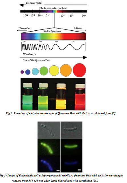
Quantum Dots (QD) are highly crystalline semiconductor nanocrystals. These are crystals whose dimension is less than that of the Exiton Bohr radius [1].Due to their nanoscale size; they have discrete electronic energy state giving rise to unique optical and electronic properties. They also possess the property of size dependent photo-emission which is due to the phenomenon of Quantum Confinement [2]. Their stability against photobleaching has attracted many researchers to develop bio-imaging and Quantum computing techniques. Due to their unique properties they have wide range of applications in electronics, computing and biology. QDs are made up of elements like Cadmium, Selenium, Indium, Tellurium, Phosphorous, Sulphur etc. These elements make up the core of a Quantum dot, which are then coated with a shell made of material of higher band energy. The outer shell also forms a protective layer on the Quantum dots. To make QDs biologically compatible one or more layer of organic polymers are added and are conjugated with organic ligands. The diameter of QDs varies from 1-20nm which consists of 100-100,000 atoms [2]. Due to their distinctive properties, QDs have improved the sensitivity of cellular analysis and molecular detection by few folds. The Quantum Dots were first studied by A.I. Ekimov and A.A. Onushchenko [3]. The size of microscopic CuCl crystals grown in transparent dielectric matrix varied from tens to hundreds of angstroms. Later high quality CdSe, CdS and CdTe semiconductor nanocrystals were synthesized based on pyrolysis of organometallic reagents [4]. The particles sizes were as small as 20 angstroms in diameter. Syntheisis of water soluble Quantum dots paved way for their conjugation to bio molecules and thereby opened up different applications in biology [5, 6]. In Quantum Dots, the energy levels of the valence electrons are discrete and have large differences. Thus they show the characteristics of an atom and hence they are also called artificial atoms [8]. The reduction in the size of the crystal brings the valence electrons closer thereby increasing their overall energy. This leads to splitting up of different electron levels giving it a discrete electron energy level system and also increasing their band gaps. This results in merging of valence band and conduction band making it a very good conductor of electricity. The splitting of energy level leads to excitement of the electron by absorption of energy and transition to higher energy levels. But these electrons return to their ground state by emitting a photon of energy either equal or lesser than that of the absorbed energy. The emitted photon has lesser energy when the electron rests in a metastable level for a brief period of time. This gives QDs their fluorescent property making them applicable in imaging and detection of molecules or cells. Thus as the size reduces, the gap between energy levels broadens up and the electron has to return from a much higher level which increases the emitted photon energy. Therefore as the size reduces, the emitted photon shifts from the red region of visible light spectra to the blue region. This gives rise to the unique fluorescent emission of the Quantum Dots.
PROPERTIES
The exceptional properties of the Quantum Dots like photo stability, broad range of absorption spectra, ease in change of emission spectra, sensitivity, brightness and ability of coupling with biological molecules, make them very useful in the field of biological imaging of different types of cells. Quantum Dots emitting different colours can be excited by monochromatic light. The time taken to fluoresce upon excitation is very less compared to conventional fluorophores and this property lasts longer for a given Quantum Dot. The Quantum Dots are very stable and resistant to photobleaching and overcome the disadvantages exhibited by fluorophores, which are important for imaging applications. SYNTHESIS QDs can be synthesized by colloidal synthesis, Efield method or by fabrication through etching or self-assembly.
Colloidal chemistry method
This method is also called Hot-Injection method [2].Here Quantum Dots are prepared as a batch by quickly adding appropriate amount of precursors into hot solvent and organic ligand system. First the precursors are decomposed to monomers by chemical or physical means like temperature. This results in nucleation process where the monomers starts binding to each other. This reaction is fast in the beginning and slows down in the end due to change in concentration.
E-field technique
In this method, Quantum Dots are synthesised between the interfaces of heterojunction of monomers [8]. On applying an electric field on the interface, 2D Electron Gas [2DEG] is created. Later these charges are confined to the interface by external devices. Thus the voltage and the 2DEG permit rearrangement of atoms, finally giving a Quantum Dot at the interface.
Fabrication
Etching or Lithographic method
In this method, first a Quantum Well or Double Barrier Heterostructure [DBH] are grown by the Epitaxial Method. Then pillars are etched out of it and they are further etched to give nanocrystals like Quantum Dots [8]. These pillars have the dimension of a Quantum Wire, which is then metalized on each terminal. Application of electric current on the terminals of Quantum Wire leads to the breakdown of the Quantum Wire and formation of Quantum Dots.
Self-assembly growth technique
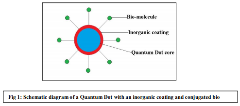
Here Quantum Dots are formed as an out growth in the Epitaxial technique [8]. The effect is similar to the condensation of water droplets on cold surface. Generally, the epitaxial growth proceeds by forming a layer of atoms at once. But lattice mismatch and difference in surface energy between the atoms induces the formation of islands, thus Quantum Dots are formed. It is seen that bare Quantum Dots are unsuitable for biological application. This is because; they are insoluble in water and toxic due to their heavy metal composition, small size and active surface [9]. To overcome these problems, the surface of Quantum Dots is treated with stabilizing and coating substances. Most of the coating substances are organic polymers [10] which are amphiphilic in nature, also making them water soluble and easy to apply for biological analysis by conjugating with proteins, antibodies and other molecules. Additionally, it is observed that coating provides better photo stability [11]. It is also seen that some bio molecules like proteins can be effective nucleating and stabilizing agents for Quantum Dots [12].
APPLICATIONS OF QUANTUM DOTS IN BACTERIOLOGY
The superior optical properties of Quantum Dots entail them for numerous applications in labelling of cells. They are highly suitable for labelling, as the intense emission of light helps in detection of even a small number of the cells. They find applications in enumeration of bacterial cells in various matrices, detection of pathogens, food contaminants, and in the study of micro flora in bio films.
Enumeration
Here the number of bacterial cells can be enumerated by relating the intensity of light emitted by the Quantum Dots to the cell count. For example, Alteromonas species inhabiting the surface of small marine animals like Copepods were enumerated using QDs [13]. The CdSe Quantum Dots were conjugated with streptavidin so that QD can specifically bind to the anti-antibody against Alteromonas species. It is also possible to detect wide range of bacteria from a sample such as sewage water using water soluble Quantum Dots [14]. Quantum Dots can also be used to detect a particular strain of a bacterial species as supported from the result of an experiment where mannose conjugated CdS Quantum Dots could detect a strain Escherichia coli producing FimHlectin which binds specifically to mannose [15]. In a study on fluorescence detection of count of Escherichia coli and Staphylococcus aureus using Quantum Dots, spectrofluorometer was used for roughly counting the cells [16]. The low detection limit for this method was in order of 102CFU/ml. In a different study the same detection limit was obtained when the Quantum Dots were labelled to bacteria covalently using glutaraldehyde [17]. It was also seen that there was a linear relationship between fluorescence intensity and total bacterial count. By using Quantum Dots it is possible to distinguish between wild type and auxotrophic strain of a bacteria belonging to same species. This technique involves conjugating Quantum Dots with the biomolecule needed for bacterial growth but which cannot be synthesized by the bacteria. This method is applied for detection of purine auxotrophs of Bacillus subtilis and Escherichia coli where Quantum Dots conjugated with AMP were taken up preferentially by purine-auxotrophs rather than the wild type [18]. The Quantum Dots can also be coupled to the antibodies used in ELISA which can give us an intense signal even in a minute concentration of antigen. This method was applied for detection of Escherichia coli in water samples where the target bacteria were separated by antibody coated magnetic beads and then detected by ELISA using Quantum Dots conjugated to secondary antibodies [19]. Thus Quantum Dot based enumeration of bacteria can be rapid and sensitive compared to the conventional methods.
Detection of pathogens
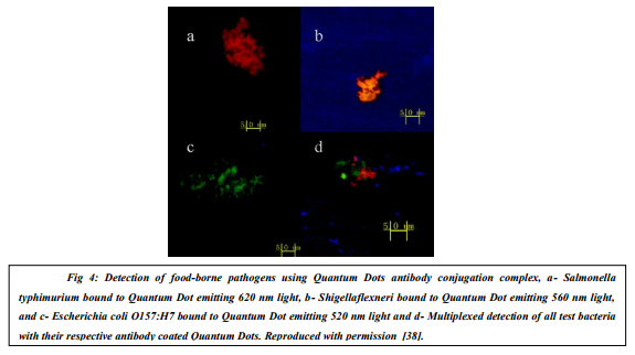
Quantum Dots have better optical properties than other dyes or fluorophores; it is widely applicable in detecting pathogens from various clinical samples and to detect a particular pathogen from a cluster of bacteria. There are number of studies where Escherichia coli and Salmonella typhimurium were detected simultaneously using antibodies against these two species and conjugating different antibodies with Quantum Dots emitting light of different wavelength [20]. Multiplexed detection was also applied for Escherichia coli and Salmonella enteritidis [21]. Denatured Bovine serum albumin stabilized Quantum Dots were conjugated to secondary antibodies for detection of Escherichia coli and Listeria monocytogenes [22]. This type of conjugation to the secondary antibodies gives us an advantage of universal usage of the antibody for the detection, ease in their production and circumventing the tedious process of labelling individual primary antibodies. The interactions between antibody and antigen are weak and these are further weakened by conjugation of bulk substance like Quantum Dot. This reduces the staining property of Quantum Dots conjugated to antibodies, hence, it is preferred to conjugate small molecules like biotin to antibody and then bind them to Quantum Dots conjugated to streptavidin. The above method was also adopted for detection of E.coli [23] and Listeria monocytogenes where the limit of detection was found to be 2-3 CFU/ml with a detection time of 1.5 hrs. [24]. In another study Mycobacterium bovis BCG and Mycobacterium tuberculosis was detected using genus specific antibody and biotinylated antiantibody which would bind to streptavidin coated Quantum Dots [25]. The result gave a limit of detection of 103 bacteria/ml. This method was also applied for detection of Escherichia coliO157:H7 strain with a limit of detection of 10 3 CFU/ml with a detection time less than 2hrs [26]. In one of the studies Escherichia coli was imaged using organic acid stabilized Quantum Dots [27]. Here the Quantum Dots were internalized by the bacteria giving rise to fluorescence signal. It was found that none of the other bacteria interfered with the test. Quantum Dot labelling of the cells also aids Flow cytometric separation and analysis of pathogenic bacteria from other non-pathogenic ones. This method is a fast and effective technique for separation. In a study pathogenic Escherichia coli strain O157:H7 were separated from non-pathogenic bacteria using Quantum Dots and Flow cytometer [28]. The limit of detection was 1% of the pathogenic strain in the mixture of cells.
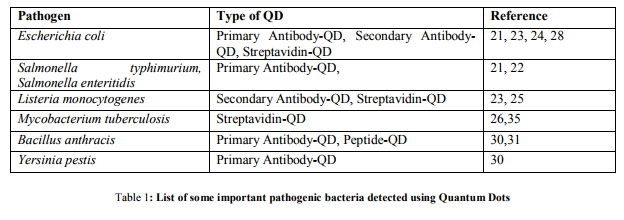
It is also possible for detection of strain and metabolism specific bacteria using Quantum Dots conjugated to a biomolecule which might bind to the receptor on the particular strain or may be used as a substrate for a metabolic pathway [29]. In another study different antibodies are conjugated to Quantum Dots of different emission peaks for multiplexed analysis of Bacillus anthracis spores and Yersinia pestis by Flow cytometry [30]. It also possible to detect one spores of Bacillus anthracis from other similar bacterial spores by conjugating Quantum Dots by BABA peptides [31] or a short peptide chain from gamma-phage lysine protein [32]. This analysis can be performed using methods like Flowcytometry, Confocal laser scanning microscopy and Spectrofluorometer with single cell resolution. An innovative yet specific method of labelling bacteria is using phage conjugated with Quantum Dots. This method can be used for recognising both slow growing and highly infectious bacteria. This method was studied for detection of Escherichia coli using biotinylated phage and streptavidin coated Quantum Dots [33]. Quantum Dots coated with zinc [II]-dipicolylamine could detect only mutant and pathogenic Escherichia coli that lacks O-antigen and could also facilitate in vivo optical imaging of the infection in the animal [34]. It is also possible to identify the bacterial species by detecting the species specific DNA sequence. In this method of detection the Quantum Dots are made to bind to a probe which is complementary to the species specific DNA sequence. In a study this method was used for detection of Mycobacterium tuberculosis and Mycobacterium avium subsp. paratuberculosis where the biotinylated probe hybridized with the DNA was detected by streptavidin coated Quantum Dots [35]. This method was 70 to 90% accurate compared to real time PCR and had a limit of detection of 12.5 ng of DNA in 20µl. This technique can be implied in diagnostic assay for rapid, specific and sensitive method of pathogen detection. In another study Quantum Dots based molecular beacons were used to detect the antibiotic resistant β-lactamase genes in pUC18 of Escherichia coli [36]. The mechanism of detection used in the study was a single step FISH, which gave an excellent signal on genes in the plasmid. There was an attempt to use ferrichrome conjugated to Quantum Dots for detection of bacteria with receptors for ferrous ions [37]. This method was successful in detecting Pseudomonas fluorescens having ferrous receptors.
Detection of bacterial food contaminants
Many bacterial infections are spread by contaminated food. Thus, detection of bacterial contaminants in food is important to monitor outbreaks of food borne infections. Three food-borne pathogens namely Salmonella typhimurium, Shigella flexneri and Escherichia coli were detected by antibodies against each bacteria which was conjugated to Quantum Dots of different emission wavelength [38]. The same method of antibody conjugated Quantum Dots were developed for specific detection of Staphylococcus aureus in food and environment [39]. The bacteria were detected under a fluorescent microscope and limit of detection was found to be 900 CFU/ml. Most of the contaminants are found in meats, thus Quantum Dots can be used to analyse contaminant in meat sample. In a recent study, Quantum Dots were used to detect pathogens like Escherichia coli and Salmonella in ground beef by conjugating Quantum Dots to specific antibodies against the pathogens [40]. In a study chicken carcass wash water was used as a sample for detecting Salmonella typhimurium contaminant in chicken meat [41]. Here contaminant was separated from sample with antibody conjugated magnetic beads and then reacted to biotin tagged secondary antibody which binds to streptavidin coated Quantum Dots. In this method the limit of detection was in order of 103 CFU/ml. Also there has been a report of use of indirect immunofluorescence coupled to Quantum Dots for labelling and detection specific bacterial serotype of pathogen Vibrio parahaemolyticus attached to small marine animals which are pathogen carriers [42]. This method can also be extended for detection of other Vibrio species like Vibrio cholerae. With the use of Quantum Dots in biosensors and microarrays, the size of the instrument have been reduced and also permitted for automation thus increasing rapidity and sensitivity of the test. These techniques have been used in assaying Salmonellae [43]. Nowadays array systems have been developed for rapid and sensitive detection of bacteria as indicated by a study where Escherichia coli O157:H7 was detected using Quantum Dots conjugated to antibody and the concept of sandwich ELISA [44]. The above method gave a limit of detection of below 10 CFU/ml. Escherichia coli was also detected using colistin-functionalised Quantum Dots which gave a limit of detecting as low as 28cells/ml [45] with a short analysis time of 15 min. excluding preparation and photoactivation time. Detection of bacteria in Oral bio films It is observed that many bacteria exhibit a symbiotic relationship between each other. These bacteria form biofilm which is a complex cluster of bacteria. These biofilms are found widely in our environment and also in our body like mouth etc. It is desirable to study symbiosis for evolutionary studies. Quantum Dots can be used for specific detection of bacteria in oral biofilms. This method has been used in a study where specific human oral bacteria namely Streptococcus gordonii DL1, Streptococcus mutans UA159 and Veillonella spp. strain R1 were detected in a biofilm in vivo and in vitro [46]. Here immunofluorescence method of imaging was used, and this method does not depend on detection of bacteria based on their morphology, which makes this rapid technique amenable to automation, enabling the detection from numerous samples. These studies also give an insight on the spatial relationship between bacteria and interspecies interaction in biofilm. Presence of specific gene in the biofilm can give a large insight on the species of the bacteria and also function of that gene in the biofilm. In a report Bacillus spoOA gene was analysed in a biofilm using DNA-Quantum Dots system [47]. The hybridization of target DNA to the probe was detected using flowcytometer and had a limit of detection of 0.02 nM. It has been observed that biofilms made of Streptococcus sp. and Veillonella sp. are formed in early stage of the plaque. These results were confirmed by using Quantum Dot based immunofluorescence on the enamel surface [48].
TOXICITY OF QUANTUM DOTS TO BACTERIA
We have seen the brighter side of the Quantum Dots but it has been demonstrated that these nanoparticles are toxic to living cells including bacteria. Formation of reactive oxygen species by the interaction of Quantum Dots with other bio molecules or release of the heavy metal ions which constitute the Quantum Dots have been the mechanism of inducing toxicity to the cells. The toxicity of Quantum Dots on four different strains of bacteria namely Pseudomonas aeruginosa, Staphylococcus aureus, Bacillus subtilis and Escherichia coli were studied [49]. The result indicated that Gram-positive strains were more resistant to the toxicity than the Gram-negative bacteria. This might be due to large amount of peptidoglycan on cell wall of Gram-positive bacteria. The study also indicated that Gram-positive bacteria previously exposed to Quantum Dot showed lack of reproducibility.
CONCLUSION
Today, the major hindrance for using Quantum Dots in bacteriological applications is its toxicity. It is suggested that by eliminating the heavy elements from Quantum Dots, the toxicity can be reduced. Thus new Heavy Metal-free Quantum Dots like carbon quantum Dots are being produced [50]. But their effective usage is still under research. The cost of production of Quantum Dots is also topic of concern when they have to be mass produced and used as diagnosis & analysing tools. Many methods for synthesizing biocompatible Quantum Dots using bacterial cells like Escherichia coli have been proposed [51]. The efficacy of these Quantum Dots in various applications will have to be proved. In conclusion, it can be said that Quantum Dot based techniques are rapid, sensitive and reliable tools for the enumeration, detection and differentiation of Bacteria.
References:
1. Poole CP, Owens FJ (2009) Introduction to nanotechnology. John Wiley & Sons INC, Delhi.
2. Guan J (2008) Synthesis and Structural Characterization of ZnTe/ZnSeCore/Shell Tunable Quantum Dots. Dissertation, Massachusetts Institute of Technology.
3. Ekimov AI, Onushchenko AA (1982) Quantum size effect in three-dimensional microscopic semiconductor crystals. Journal of Experimental and Theoritical Physics 34: 345-349.
4. Murray CB, Norris DJ, Bawendi MG (1993) Synthesis and Characterization of Nearly MonodisperseCdE [E=S,Se,Te] Semiconductors Nanocrystallites. Journal of the American Chemical Society115: 8706- 8715.
5. Bruchez M. Jr., Moronne M, Gin P, Weiss S, Alivisatos A.P (1998) Semiconductor nanocrystals as fluorescent biological labels. Science281: 2013-2016.
6. Chan WCW, Nie S (1998) Quantum dot bioconjugates for ultrasensitive nonisotopic detection. Science 281: 2016-2018.
7. Rizvi SB, Ghaderi S, Keshtgar Mo, Seifalian AM (2010) Semiconductor quantum dots as fluorescent probes for in vitro and in vivo bio-molecular and cellular imaging. Nano Reviews. DOI: 10.3402/nano.v1i0.5161
8. Boxberg F and Tulkki J (2004) Quantum Dots: Phenomenology, Photonic and Electronic Properties, Modeling and Technology. In: Akhlesh L [ed] Handbook of nanotechnology: Nanometer Structures Theory, Modeling, and Simulation. SPIE Press,Bellingham,pp 107-137.
9. Su Y, Hu Mi, Fan C, He Y, Li Q, Li W, et al. (2010) The cytotoxicity of CdTe quantum dots and the relative contributions from released cadmium ions and nanoparticle properties. Biomaterials31: 4829-4834.
10. Wang Y, Chen L (2011)Nanomedicine Quantum dots, lighting up the research and development of nanomedicine. Nanotechnology, Biology, and Medicine 7: 385– 402.
11. Hanaki KI, Momo A, Oku T, Komoto A, Maenosono S, Yamaguchi Y et al. (2003) Semiconductor quantum dot/albumin complex is a long-lifeand highly photostable endosome marker. Biochemical Biophysical Research Communication. 302: 496-501.
12. Mansur HS, Gonzàlez JC, Mansur AAP (2011) Biomolecule-quantum dot systems for bioconjugation applications. Colloids and Surfaces B: Biointerfaces84: 360-368.
13. Beckman EM, Kawaguchi T, Chandler GT, Decho AW (2008) Development of amicroplate-based flourescence immunoassays using quantum dot streptavidin conjugates for enumeration of putative marine bacteria, Alteromonas sp., associated with a benthic harpacticoid copepod. Journal of Microbiological Methods75: 441– 444.
14. Mandal TK, Parvin N (2011) Rapid detection of bacteria by carbon quantum dots. Journal of Biomedical Nanotechnology 7: 846-848.
15. Mukhopadhyay B, Martins MB, Karamanska R, Russell DA, Field RA (2009) Bacterial detection using carbohydrate-functionalised CdS quantum dots: a model a study exploiting E.coli recognition of mannosides. Tetrahedron Letters50: 886-889.
16. Xue X, Pan J, Xie H, Wang J, Zhang S (2009) Fluorescence detection of total count of Escherichia coli and Staphylococcus aureus on water-soluble CdSe quantum dots coupled with bacteria. Talanta77: 1808- 1813.
17. Fu X, Huang K, Liu S (2010) A rapid and universal bacteria-counting approach using CdSe/ZnS/SiO2 composite nanoparticles as fluorescence probe.Analytical and Bioanalytical Chemistry395: 1397- 1404.
18. Kloepfer JA, Mielke RE, Nadeau JL (2005) Uptake of CdSe and CdSe/ZnS Quantum Dots into bacteria via Purine-Dependent Mechanisms. Applied and Environmental Microbiology71: 2548-2557.
19. Dudak FC, Boyac?H (2008)Enumeration of Immunomagnetically Captured Escherichia Coli in Water Samples Using Quantum Dot-Labeled Antibodies. Journal of Rapid Methods & Automation in Microbiology16: 122-131.
20. Yang L, Li Y (2006) Simultaneous detection of Escherichia coli o157:H7 and Salmonella typhimurium using quantum dots as fluorescence labels.Analyst 131: 394- 401.
21. Dudak FC, Boyaci IH (2009) Multiplex Detection of Escherichia coli And Salmonella enteritidis By Using Quantum Dot-Labeled Antibodies. Journal of Rapid Methods and Automation in Microbiology 17: 315-327.
22. Kuo YC, Wang Q, Ruengruglikit C, Yu H, Huang Q (2008) Antibody-Conjugated CdTe Quantum Dots for Escherichia coli Detection. The Journal of Physical Chemistry 112: 4818- 4824.
23. Hahn MA, Tabb JS, Krauss TD (2005) Detection of Single Bacterial Pathogens with Semiconductor Quantum Dots.Analytical Chemistry77: 4861- 4869.
24. Wang H, Li Y, Slavik M (2007) Rapid detection of Listeria monocytogenesusing quantum dots and nanobeads-based optical biosensors 15: 67-76.
25. Liandris E, Gazouli M, Andreadou M, Sechi LA, Rosu V, Ikonomopoulos J (2011) Detection of Pathogenic Mycobacteria Based on Functionalized Quantum Dots Coupled with Immunomagnetic Separation. PloS ONE 6: e20026.
26. Su XL, Li Y (2004) Quantum Dot Biolabeling Coupled with Immunomagnetic Separation for Detection of Escherichia coli O157:H7. Analytical Chemistry 76: 4806- 4810.
27. Hirschey MD, Han YJ, Stucky GD, Butler A (2006) Imaging Escherichia coli using functionalized core/shell CdSe/CdS quantum dots. Journal of Biological Inorganic Chemistry 11: 663- 669.
28. Hahn MA, Keng PC, Krauss TD (2008) Flow Cytometric Analysis to Detect Pathogens in Bacterial Cell Mixtures Using Semiconductor Quantum Dots. Analytical Chemistry 80: 864- 872.
29. Kloepfer JA, Mielke RE, Wong MS, Nealson KH, Stucky G, Nadeaul J.L (2003) Quantum Dots as Strainand Metabolism-Specific Microbiological Labels. Applied and Environmental Microbiology69: 4205- 4213.
30. Zahavy E, Heleg-Shabtai V, Zafrani Y, Marciano D, Yitzhaki S (2010) Application of Fluorescent Nanocrystals [q-dots] for the Detection of Pathogenic Bacteria by Flow-Cytometric. Journal of Fluorescence 20: 389-399.
31. Park TJ, Park JP, Seo GM, Chai YG, Lee SY (2006) Rapid and Accurate Detection of Bacillus anthracis Spores Using Peptide-Quantum Dot Conjugates 16: 1713- 1719.
32. Sainathrao S, Mohan KVK, Atreya C (2009) Gammaphage lysinPlyG sequence-based synthetic peptides coupled with Qdot-nanocrystals are useful for developing detection methods for Bacillus anthracisby using its surrogates, B. anthracis-Sterne and B. cereus4342. BMC Biotechnology 9: 67.
33. Edgar R, McKinstry M, Hwang J, Oppenheim AB, Fekete RA, Giulian G et al. (2006) High-sensitivity bacterial detection using biotin-tagged phage and quantum-dot nanocomplexes. Proceedings of the National Academy of Sciences 103: 4841- 4845.
34. Leevy WM, Lambert TN, Johnson JR, Morris J, Smith BD (2008) Quantum dot probes for bacteria distinguishEscherichia coli mutants and permit in vivo imaging. Chemical Communication 20: 2331-2333.
35. Gazouli M, Liandris E, Andreadou M, Sechi LA, Masala S, Paccagnini D et al. (2010) Specific Detection of Unamplified Mycobacterial DNA by Use of Fluorescent Semiconductor Quantum Dots and Magnetic Beads. Journal of Clinical Microbiology 48: 2830- 2835.
36. Wu SM, Tiana ZQ, Zhanga ZL, Huanga BH, Jianga P, Xieb ZX et al. (2010) Direct fluorescence in situ hybridization (FISH) in Escherichia coli with a target-specific quantum dot-based molecular beacon. Biosensors and Bioelectronics 26: 491-496.
37. Wu SM, Zhang ZL, Wang XD, Zhang MX, Peng J, Xie ZX (2009) Quantum Dot-FerrichromeBioprobes for Recognition of Pseudomonas fluorescens. Journal of Physical Chemistry 113: 9169-9174.
38. Zhao Y, Ye M, Chao Q, Jia N, Ge Y, Shen H (2009) Simultaneous Detection of Multifood-Borne Pathogenic Bacteria Based on Functionalized Quantum Dots Coupled with Immunomagnetic Separation in Food Samples. Journal of Agricultural and Food Chemistry 57: 517-524.
39. Wang X, Du Y, Li Y, Li D, Sun R (2011) Fluorescent Identification and Detection of Staphylococcus aureuswith Carboxymethyl Chitosan/CdS Quantum Dots Bioconjugates. Journal of Biomaterials Science. Polymer Edition 22: 1881-1893.
40. Yang L, Li Y (2005) Quantum Dots as Fluorescent Labels for Quantitative Detection of SalmonellaTyphimurium in Chicken Carcass Wash Water. Journal of Food Protection 68: 1241-1245.
41. Wang L, Wu CS, Fan X, Mustapha A (2012) Detection of Escherichia coli O157:H7 and Salmonella in ground beef by a bead-free quantum dot- facilitated isolation method. International Journal of Food Microbiology 156: 83-87.
42. Decho AW, Beckman EM, Chandler GT, Kawaguchi T (2008) Application of photostable quantum dots for indirect immunoflourescent detection of specific bacterial serotypes on small marine animals. Nanotechnology 19: 235102.
43. Goldschmidt MC (2006) The Use of Biosensor and Microarray Techniques in the Rapid Detection and Identification of Salmonellae. Journal of AOAC International 89: 530- 537.
44. Sanvicens N, Pascual N, Fernández-Argüelles MT, Adrian J, Costa-Fernández JM, Sánchez-Baeza F et al. (2011) Quantum dot-based array for sensitive detection of Escherichia coli. Analytical and Bioanalytical Chemistry 399: 2755-2762.
45. Carrillo-Carrión C, Simonet BM, Valcárcel M (2011) Colistin-functionalised CdSe/ZnS quantum dots as fluorescent probe for the rapid detection of Escherichia coli. Biosensors and Bioelectronics 26: 4368-4374.
46. Chalmers NI, Palmer RJ Jr, Du-Thumm L, Sullivan R, Shi W, Kolenbrander PE (2006) Use of Quantum Dot Luminescent Probes To Achieve Simgle-Cell Resolution of Human Oral Bacteria in Biofilms. Applied and Environmental Microbiology 73: 630-636.
47. Lee J, Kim IS, Yu HW (2010) Flow Cytometric Detection of Bacillus spoOA Gene in Biofilm Using Quantum Dot Labeling. Analytical Chemistry 82: 2836- 2843.
48. Chalmers NI, Palmer RJ Jr, Cisar JO, Kolenbrander PE (2008) Characterization of a Streptococcus sp.- Veillonella sp. Community Micromanipulated from Dental Plaque. Journal of Bacteriology 190: 8145-8154.
49. Dumas EM, Ozenne V, Mielke RE, Nadeau JL (2009) Toxicity of CdTe Quantum Dots in Bacterial Strains. IEEE Transaction on Nanobioscience 8: 58-64.
50. Geszke M, Murias M, Balan L, Medjahdi G, Korczynski J, Moritz M, et al. (2011) Folic acid-conjugated core/shell ZnS:Mn/ZnS quantum dots as targeted probes for two photon fluorescence imaging of cancer cells. ActaBiomaterialia 7: 1327- 1338.
51. Bao H, Lu Z, Cui X, Qaio Y, Guo J, Anderson JM, et al. (2010) Extracellular microbial synthesis of biocompatible CdTe quantum dots. ActaBiomaterialia 6: 3534-3541.
|
 IJCRR
IJCRR 


 This work is licensed under a Creative Commons Attribution-NonCommercial 4.0 International License


