IJCRR - 9(16), August, 2017
Pages: 23-26
Print Article
Download XML Download PDF
Assessment of Genome Integrity in Mitochondria of Testis Tissue, Cell Proliferation and Topoisomerase Assay in invitro Condition
Author: Eswari Beeram
Category: Healthcare
Abstract:Aim: Assessment of genome integrity in testis and to explain the reasons for infertility with the drug metosartan.
Methodology: Comet assay with freshly isolated mitochondria, cryopreserved mitochondria and cryopreserved tissue mitochondria and cell proliferation assay by dye exclusion method and topoisomerase assay in fresh and deep freezed DNA.
Results and Discussion: Comet assay of isolated mitochondria both from fresh and cryopreserved samples was examined by fluorescence microscope, which revealed that cryopreservation of testes mitochondria samples lead to injury to the tissue and DNA fragmentation. Topo isomerase assay has showed that increased incubation leads to DNA fragmentation and cell proliferation assay with the drug has showed the 0% viability of the cells by trypan blue. So, in vivo studies are necessary to confirm whether the drug is safe or not.
Conclusion: Assessment of genome integrity lead to the finding that drug was not safe it resulted in so many difficulties in invitro condition.
Keywords: Mitochindria, Genome, Cryopreserved, Topoisomerase
DOI: 10.7324/IJCRR.2017.9165
Full Text:
Introduction:
Comet assay is a combination of both agarose gel electrophoresis along with fluorescent microscope for detecting the DNA strand breaks in the nucleated cell. The extent of DNA migration is measure of damage in the genome of the cell. This is a simple technique as it doesn't require any radioactive labelling compounds and also problems associated with disposal. It can be more useful in case study of DNA damaging agents. The damaged DNA migrates as comet. The comet head consists of high molecular weight DNA and tail consists of the DNA single strand fragments. Applications of comet assay includes diverse things(1), in which change of lysis buffers and DNA stains resulted in different achievements like use of neutral buffer to find the double strand breaks whereas use of buffer containing specific endonucleases can used to find damage at base level. Here in my study I have taken neutral buffer to detect double stranded breaks in mitochondria isolated in pure form cryopreserved before the comet assay was performed. As the mitochondria is present in midpeice of sperm and responsible for motility of sperm , So studies on mitochondria genome is necessary as decreased motility of sperm leading to problems in fertility.
Cell proliferation assay is important to know the abnormal proliferation due to certain agents. Simple techniques like MTT assay and trypan blue assay is used to know whether the cells are normal or not (6). AS MTT assay of control gives the same result after the dye has entered in to the dead cell and So, useful to perform the trypan blue as the dye was taken by the non viable cells itself. In this study I have used metosartan to know the cell viability in vitro using trypan blue. The cells were dead and matched with the results of aniline blue staining.
Mammals express two types of topoisomerases alpha and beta which are homologous but differ in the sub cellular location of the enzyme. Similar to topoisomerase I and III, topoisomerase II also involved in relieving the topological constraints of DNA(2). Topoisomerase is homodimer binding to the clamps of DNA polymerase and the strand was cleaved by topoisomerase first using the ATP, the cleaved strand was passed through the gap, re ligated and the strands are released by hydrolysis of ATP after the release of ADP molecules.
In general sense DNA is unwinded in the replication in order to pass the genetic information or for transcription of the mRNA strands. In both the steps DNA winding is necessary but focussing on DNA replication there will be formation of positive supercoils and makes the the fork movement difficult an under winding of the DNA behind the fork also leads to torsional stress in the replication and leading to formation of catenated DNA . As we know helicases separates the two strands but not involved in the removal of torsional stress but here, the topoisomerases came in to role as that mentioned in the previous paragraph.
Topoisomerase II is also responsible for maintenance of chromatin integrity as supercoiled that means folded supra molecular structures both in fresh and frozen samples. The present aim is to know the genome integrity in the treated sample as in case of proliferation assay and fresh and frozen samples by topoisomerase assay and in fresh and cryopreserved samples in comet assay.
Materials and methods:
Cell proliferation assay using trypan blue :( 6)
1. The cell suspension was prepared in a balanced salt solution (e.g., Hanks' Balanced Salts [HBSS], and 0.5 ml of 0.4% Trypan Blue solution (w/v) was transferred to a test tube. To it 0.3 ml of HBSS is added along with 0.2 ml of the cell suspension (dilution factor = 5) and mixed thoroughly and Allowed to stand for 5 to 15 minutes after addition of metosartan drug to the suspension.
2. After proper incubation by using Pasteur pipette or any suitable device transfer the trypan blue- cell mixture suspension to both the chambers by just touching the tip of the pipette to the edge of the chamber and allowing it to fill by capillary action and overfilling or under filling was avoided.
3. Starting with the chamber 1 count all the four corners squares and repeat the procedure for chamber 2 also. Nonviable stains blue.
Cryopreservation of tissue:
Liquid nitrogen was brought from S.V veterinary university and the tissue is homogenated in water and exposed to liquid nitrogen vapours and immersed in liquid nitrogen for 3 days and mitochondria was isolated after the cryopreservation and comet assay was performed... The isolated mitochondria was also cryopreserved, and processed in the same manner
Isolation of mitochondria from testes:(7)
After the sacrificing the animal, testes were removed, decapsulated and minced in isolation medium 250mM sucrose, 0.2 mM EGTA, 0.1mM EDTA,5mM HEPES-KOH (PH 7.4) and 0.1%defatted BSA. The tissue was homogenated in isolation medium and the homogenate was centrifuged at 10,000Xg for 10min and the supernatant was recentrifuged at 10,000Xg for 10min. The pellet was resuspended and repelleted twice in the isolation medium omitting EGTA, EDTA, and defatted BSA.
Comet assay: (3)
Standard comet assay was performed..100 l of the isolated mitochondria was mixed with 1000l of 0.8% agarose and 1000l of suspension was spread on the slide and kept in ice for 45min. After casting the slides was immersed in lysis buffer(2.5% SDS in 45 mM Tris-borate, 1 mM EDTA, pH 8.4) for 20min and kept in electrophoresis buffer for 5min. The electrophoresis was carried with TBE buffer at 2v/cm. Ethidium bromide staining was performed and visualised using fluorescent microscope.
Isolation of DNA from testis tissue :( 4)
Cells were pelleted and the suspension was collected. To the supernatant lysis buffer (50mM tris-cl, PH8.0, 100MEDTA,0.125%SDS) was added and the solution was incubated in boiling water bath at 550c for overnight. To this 1ml of mixture of phenol :chloroform : isoamyl alcohol was added and centrifuged at 10,000rpm for 15min. Aqueous phase was collected and washed with 3ml of phenol: chloroform : isoamyl alcohol. Aqueous phase was collected and chilled ethanol was added and upper aqueous phase was separated and centrifuged at 10,000rpmfor 10min and the pellet was resuspended in 100l of water.
Topoisomerase assay :( 5)
The above isolated DNA was suspended in topoisomerase reaction buffer 10X ( 200mM tris-cl, PH:7.5, 100mM Mgcl2, 10mMATP, 10mM EDTA, 10mM, β-mercaptoethanol, 1.5 M KCL,300g/ml BSA. The suspension was incubated for 1-3hrs and agraose gel electrophoresis was performed with TAE buffer.
Results:
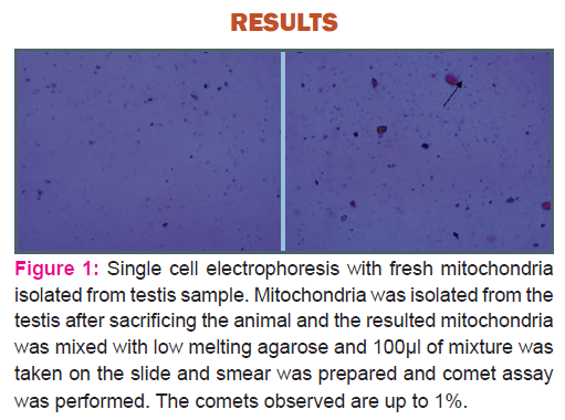
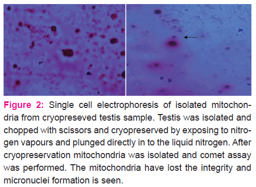
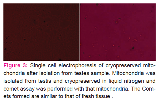
Comet assay of fresh and cryopreserved testis sample mitochondria has given the conclusion that cryopreservation may result in distortion of shape, micronuclei formation which may result low motility of spermatozoa therefore leading to infertility. Incase of cryopreserved mitochondria after isolation showed less distortion and absence of micronuclei and also comets formed was similar to fresh sample.
In fresh sample the comets was up to acceptable extent than compared to the cryopreserved mitochondria testes samples.
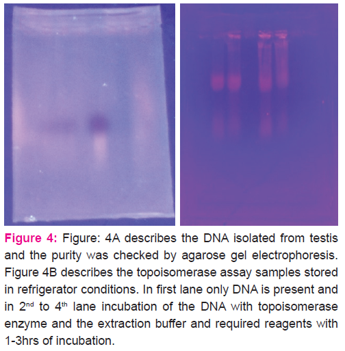
In the DNA isolated from the fresh tissue DNA fragments was not found where as in refrigerated samples treated with topoisomerase showed supercoiled structures and also the fragmented molecules which migrated to the longer distance in the gel. With increase in time period the intensity is also increased representing the cleavage.
Calculations:
Cell Counts - Each square of the haemocytometer, with cover-slip in place, represents a total volume of
0.1 mm3 or 10-4 cm3. Since 1 cm3 is equivalent to approximately 1 ml, the subsequent cell concentration per ml (and the total number of cells) will be determined using the following calculations:
Cells Per mL = the average count per squarex dilution factorx 104 (count 10 squares) If the average count per square is 15 cellsx 4x 104 = 6x 10⁵ cells/ml.
Total Cells = cells per mlx the original volume of fluid from which cell sample was removed.
6x 10⁵ (cells/ml)x 10 ml (original volume) = 6x 10⁶ total cells.
Cell Viability (%) = total viable cells (unstained) total cells (stained and unstained) x 100. If the average count per square of unstained (viable) cells is 0, the total viable cells = [0x 4x 104] viable cells/mlx 10 ml (original volume) = 0viable cells. Cell viability (%) = 0 (viable cells) 6x 10⁶ (total cells)x 100 = 0% viability.
From the cell proliferation assay it was clear that the drug incubation for 30min lead to 0% viability
Discussion:
Cell proliferation assay, topoisomerase assay and comet assay was used in the study for assessing the genome integrity. Comet assay was performed by so many people but for mitochondria is a new thing. Mitochondria isolated from the cryopreserved tissue showed micronuclei formation which indicates that DNA was fragmented and from fresh tissue revealed less by comets formation. Cell proliferation assay revealed that treatment with drug for 30min resulted in 0% viability. So, the drug was not safe and it should be confirmed with in vivo experiments also, in order to come to a conclusion.
Conclusion:
The drug metosartan resulted in formation of micronuclei in mitochondrial genome and also decreased viability by trypan blue, topoisomerase assay has revealed that with incubation the DNA was fragmented resulting in the bands as supercoiled and fragmented DNA.
Acknowledgement:
Authors acknowledge the immense help received from the scholars whose articles are cited and included in references of this manuscript. The authors are also grateful to authors / editors / publishers of all those articles, journals and books from where the literature for this article has been reviewed and discussed.
Conflict of interest:
No conflict of interest
Source of funding:
UGC- CSIR JRF
References:
1. Peggy L Olive and Judit P Banáth .The comet assay: a method to measure DNA damage in individual cells VOL.1 NO.1 | 2006 | 23-29.
2. Patrick Chène, Joëlle Rudloff, Joseph Schoepfer, Pascal Furet, Peter Meier, Zhiyan Qian, Jean- Marc Schlaeppi, Rita Schmitz and Thomas Radimerski. Catalytic inhibition of topoisomerase II by a novel rationally designed ATP-competitive purine analogue BMC Chemical Biology.
3. Nurcan Cetinkaya , Demet Ercin , Sümer Özvatan , Yakup Erel . Quantification of applied dose in irradiated citrus fruits by DNA Comet Assay together with image analysis. Food Chemistry
192 (2016) 370-373.
4. John R. Hofstetter, Aiwu Zhang, Aimee R. Mayeda,* Tim Guscar, John I. Nurnberger, Jr., and Debomoy K. Lahiri1Genomic DNA from Mice: A Comparison of Recovery Methods and Tissue Sources BIOCHEMICAL AND MOLECULAR MEDICINE 62, 197-202 (1997).
5. John L. Nitiss, Eroica Soans, Anna Rogojina, Aman Seth, and Margarita Mishina Molecular Pharmacology Department St. Jude Children's Research Hospital, Memphis,Tennessee Topoisomerase Assays. Curr Protoc Pharmacol. 2012 June ; CHAPTER: Unit3.3. doi:10.1002/0471141755.ph0303s57.
6. Cell Viability and Proliferation Assays By: Article Based on, Bio Files v6 n5, 17-21.
7. Sandra Amaral, Paula Mota, Ana Sofia Rodrigues, Luís Martins, Paulo J. Oliveira, João Ramalho-Santos Testicular aging involves mitochondrial dysfunction as well as an increase in UCP2 levels and proton leak
|
 IJCRR
IJCRR 


 This work is licensed under a Creative Commons Attribution-NonCommercial 4.0 International License


