IJCRR - 8(14), July, 2016
Pages: 01-08
Date of Publication: 21-Jul-2016
Print Article
Download XML Download PDF
HISTOPATHOLOGICAL STUDY OF SPINAL TUMOURS
Author: Jobanputra G. P., Parikh U. R., Goswami H. M.
Category: Healthcare
Abstract:Background: Spinal tumours are tumours that can occur within or adjacent to the spinal cord. Primary spinal cord tumours account for 2 to 4 percent of all primary central nervous system(CNS) tumours, one third of which are located in the intramedullary compartment.
Objective: To study the incidence of spinal tumour at Tertiary Care Teaching Hospital and to study the morphological and clinicoradiological correlation and relative incidence of various spinal tumours among different age groups and sex.
Methods: In the Present Study, all operated cases; exicsed bipsies and resected specimens are taken into consideration. After processing detail microscopic examination was carreied out.
Results: The peak age of incidence of spinal tumours was between 21-40 years of age, with the rmale:female ratio 1.57:1.Benign tumours (89%) are more common than malignant tumours (11%).Spinal tumours more commonly located in intradural (86%) than extradural(14%) locations. Malignant tumours mostly located on extradural locations(7%) than intradural locations(4%). Spinal tumours are more commonly located in thoracic region. Schwannoma(31%) is the most common spinal tumour followed by meningioma(24%), astrocytoma (11%), neurofibroma (8%) and ependymoma (8%). Hemangioma and lipoma are relatively less common.
Conclusion: The study can contribute to epidemiologic knowledge of Spinal cord tumours.
Keywords: Spinal Cord Tumours (SCT), Histopathology, Central Nervous System (CNS)
Full Text:
INTRODUCTION
Spinal tumours are tumours that can occur within or adjacent to the spinal cord. They are considered to be intraaxial in location and can be either primary or metastatic. Primary spinal cord tumours account for 2 to 4 percent of all primary central nervous system(CNS) tumours, one third of which are located in the intramedullary compartment.1 In this study spinal tumours are considered as spinal cord tumours. Spinal cord tumours can be classified according to their anatomic location2,3,4,5 Intramedullary — Intramedullary tumours arise within the spinal cord itself. Most primary intramedullary tumours are either ependymomas or astrocytomas.Low grade tumours are usually benign and high grade tumours are malignant. WHO grade I and II are considered as benign while grade III and IV are malignant. Metastases are being recognized with increasing frequency, primarily because of improvements in imaging modalities6 Intradural-extramedullary — Tumours arising within the dura but outside the actual spinal cord are termed intraduralextramedullary. The most common tumours in this group are meningiomas and nerve sheath tumours(23). Benign Meningioma are WHO grade I and grow slowly. In contrast atypical (WHO grade II) and anaplastic (WHO grade III) form can be aggressive. Extradural- Extradural tumours are usually metastatic and most often arise in the vertebral bodies. Metastasis mostly comes from breast, lung and prostate. Metastatic lesions can cause spinal cord compression either by epidural growth that results in extrinsic spinal cord or cauda equina compression or less frequently by intradural invasion7 Primary spinal tumours commonly occurs in young adults and commonly present with following symptoms.
• Back or neck pain.
• Pain that does not improve with rest and worse at night
• Pain accompanied by neurologic symptoms such as numbness or weakness of arms or legs or change in bowel or bladder routine
Spinal tumours can be treated with medications, surgery, radiation, or a combination of treatments. Spinal tumour oncology is a rapidly evolving and exciting field. Advances are being made through integration of systemic basic laboratory and clinical research. It is hoped that these advances will eventually culminate in safer and more effective treatment for the spinal tumour.8 Because of the major advances in diagnosis, multi-modality therapy, surgery, development of rational use of combination chemotherapy and improved supportive care, the cure rate in spinal tumours has increased tremendously.
AIMS AND OBJECTIVES
1. To study the incidence of spinal tumour at Tertiary Care Teaching Hospital, Gujarat
2. To study the morphological and clinicoradiological correlation of spinal tumours
3. To study the relative incidence of various spinal tumours among different age groups and sex.
4. To compare the data and other investigations with similar studies.
MATERIALS AND METHODS
Biopsies and whole tumours specimens were taken from admitted patients in different wards of our institute. A detailed history of each patient regarding age, sex, chief complaints were collected. Along with these radiological investigations in the form of CT and MRI findings were also collected in detail. All the surgically resected specimens were fixed in the 10% neutral buffered formalin for 24 hours. The received bony parts were decalcified with the help of the HNO3. After proper fixation and/ or decalcification gross examination is carried out. Thorough gross examination of each specimen for its size, shape, and consistency was performed. From received surgical specimens representative areas of tissue were taken and submitted to routine tissue processing and paraffin embedding. Hematoxylin and Eosin staining was performed in all cases. After staining thorough macroscopic examination was performed and diagnosis is made. Detail analysis of results are carried out.
RESULTS AND OBSERVATION
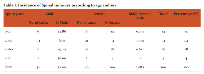
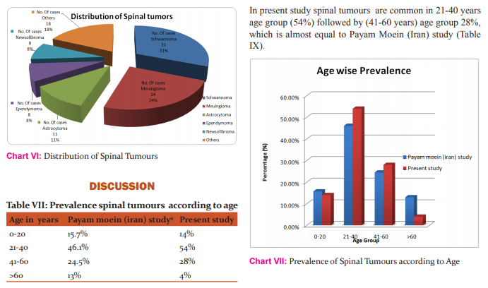
The present study was carried out in one of the tertiary care teaching hospital, Gujarat from April-2011 to November-2013. A total of 100 cases were studied in detail. Analysis of the study was collected. Table I and Chart I show age wise incidence of spinal tumours. It was found that spinal tumor was most common during 21-40 years, i.e. 54%. It also shows sex wise incidence of spinal tumour. It indicates that overall spinal tumours are common in male as compare to female, the male: female ratio is about 1.08:1. But due to unknown reason, during the third and forth decade it is much higher in male with the ratio of 1.57:1; and the incidence is equal after 60 years of age.
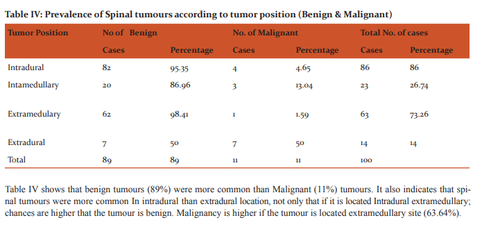
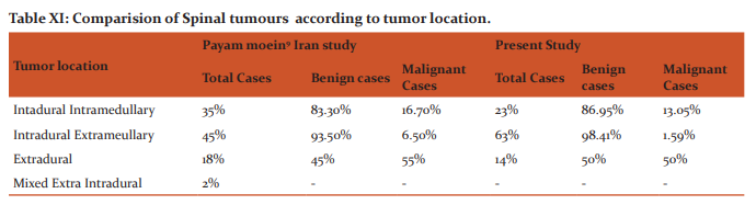
In present study most common location of spinal tumours is Intradural extramedullary 63%. Followed by Intradural Intramedullary 23% which is quite comparable with earlier study (Table XI). Malignant tumours were observed more frequently in extradural(50%) compared to intradural locations(14.64%) which is comparable with earlier study
Present study suggest that other tumours were more common in inextradural location11 cases. Cases of hemangioma, hemangioblastoma, angioma, osteochondroma, NHL and Rare case of metastatic SCC were found in present as well as moein study and comparable with that. Cases of plasmacytoma, chordoma, neuroblastoma, medulloblastoma which were not found in present study but present moein study. The difference is due to geographical difference.
SUMMARY
A histopathological study of spinal tumours was undertaken at Tertiary Care Teaching Hospitalfrom April-2011 to November-2013.
• It was found that spinal tumor was most common during 3nd and 4th decade of life.
• It was found that overall spinal tumours are common in male as compare to female.
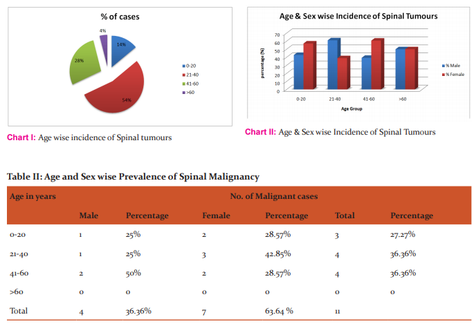
• Spinal Malignancy were more common in Female than Male.
• Benign tumours were more common than Malignant tumours.
• Spinal tumours were more common in intradural than extradural location,
• In present study most common location of spinal tumours is intradural extramedullary
CONCLUSION
A histopathological study of spinal tumours was undertaken at Tertiary Care Teaching Hospital, Gujarat to know the occurrence of different types of spinal tumours and was correlated with other studies. A total of 100 cases were studied from April 2011 to November 2013after routine tissue processing and H and E staining. The findings are as follows.
• The peak age of incidence of spinal tumours was between 21-40 years of age.
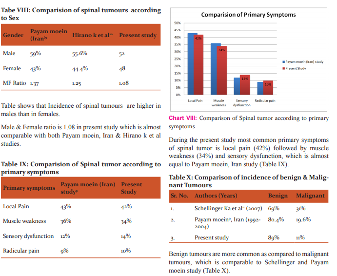
• Spinal tumours are more common in males(52%) than females(48%), with the rmale:female ratio 1.57:1.
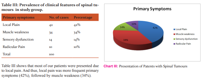
• Local pain was the commonest mode of presentation.
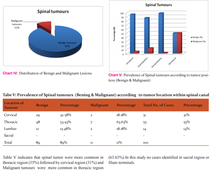
• Benign tumours (89%) are more common than malignant tumours (11%).
• Spinal tumours more commonly located in intradural (86%) than extradural(14%) locations. Malignant tumours mostly located on extradural locations(7%) than intradural locations(4%).
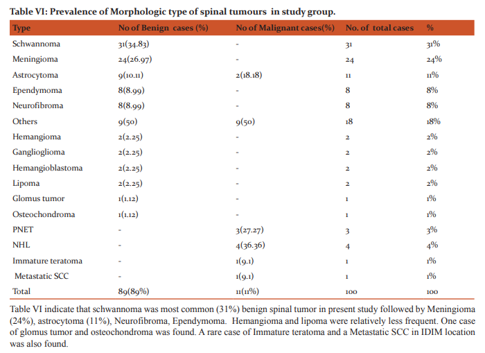
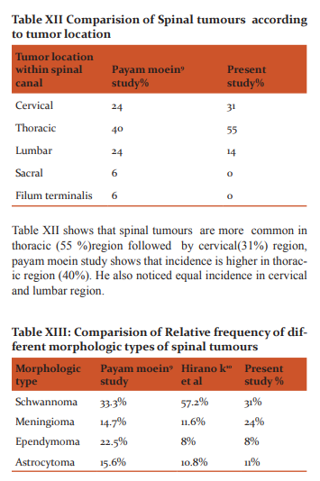
• Within spinal cord spinal tumours are more commonly located in thoracic (55%)region followed by cervical region(31%). • Malignant tumours were more common in thoracic region (63.63%)
• Among spinal tumours, schwannoma(31%) is the most common followed by meningioma(24%), astrocytoma (11%), neurofibroma (8%) and ependymoma (8%). Hemangioma and lipoma are relatively less common.
ACKNOWLEDGEMENT
The author acknowledges the help received from Dr.HansaGoswami, MD.PATH, Professor and Head, Department of Pathology for teaching me the scientific approach of the subject and its subtle aspects. I am also thankful to Dr.Urvi Parikh, MD.PATH, Assistant Professor Pathology Dept., B.J. Medical College, Ahmedabad for motivating me for doing the work meticulously and her kind co-operation. I would like to give my special thanks to all the technicians of Histopathology Section, Pathology Dept., B. J. Medical College, Ahmedabad for helping me while conducting the present study. Last but not least Author acknowledge the immense help received from the scholars whose articles are cited and included in references of this manuscript. The author is also grateful to authors / editors / publishers of all those articles, journals and books from where the literature for this article has been reviewed and discussed.
NOTE: The present study tumours was undertaken at Tertiary Care Teaching Hospital, Gujarat to know the occurrence of different types of spinal tumours. In the Present Study, all operated cases; excised bipsies and resected specimens are taken into consideration. Biopsies and whole tumours specimens were taken from admitted patients in different wards of our institute, which are with prior consent of the patients. Ethical committee clearance has not been required as confidentiality of patient’s details not been published.
References:
1. Chapter in book: Juan Rosai. Chapter-18. In : Rosai and Ackerman’s Surgical Pathology, Tenth Edition, Volume I, Testis; Mosby; Elsevier; 2004: 1412-1457.
2. Kleihues P, Louis DN, Scheithauer BW, Rorke LB, Reifenberger G, Burger PC, et al. The WHO classification of tumours of the nervous system. J Neuropathol Exp Neurol 2002;61:215-25.
3. Louis DN, Ohgaki H, Wiestler OD, Cavenee WK. Classification of Tumours of the Nervous System, IARC Press, Lyon, France 2007.
4. Lucein J. Rubinstein: Tumours of the central nervous system, 2nd series, fascicle 6, 1972.
5. Minimum dataset for the histopathological reporting of tumours of CNS; 10th Sept. 2003
6. Shrivastava RK, Epstein FJ, Perin NI, et al. Intramedullary spinal cord tumours in patients older than 50 years of age: management and outcome analysis. J Neurosurg Spine 2005; 2:249.
7. Descriptive epidemiology of malignant and nonmalignant primary spinal cord, spinal meninges, and cauda equina tumours , United States, 2004-2007. Cancer 118:4220.
8. The WHO Classification of Tumours of Central Nervous Systerm; David N. Louis, Hiroko Ohgaki, Otmar D. Wiestler, Webster K. Cavenee; International Agency for Research on Cancer (IARC) 69008 Lyon, France, Heidelberg November 17-18, 2006.
9. Moein P, Behnamfar O, Khalighinejad N, Farajzadegan Z, Fard SA, Razavi M et al. A 12-year epidemiologic study on primary spinal cord tumours in Isfahan, Iran. J Res Med Sci 2013;18:17- 21.
10. Hirano k,et al.Eur spine J. 2012. Oct;21(10):2019-26. doi:10.1007/s00586-012-2345-5.Epub 2012 May 12.
11. Schellinger KA, Propp JM, Villano JL, McCarthy BJ. Descriptive epidemiology of primary spinal cord tumours . J Neurooncol 2008;87:173-9.
|
 IJCRR
IJCRR 


 This work is licensed under a Creative Commons Attribution-NonCommercial 4.0 International License


