IJCRR - 3(2), February, 2011
Pages: 37-41
Print Article
Download XML Download PDF
STUDY OF DEGENERATIVE CHANGES IN UTERINE LEIOMYOMAS
Author: Ramesh B.H, Shashikala P, Kavita G.U, Doddikoppad, Chandrasekhar H.R
Category: Healthcare
Abstract:Leiomyomas are the commonest tumors in female genital tract and in the body as a whole. These
benign tumors of smooth muscles occur in 20-30% females in the reproductive age group and
tend to be symptomatic. A study of 314 uterine leiomyomas revealed some form of degeneration
in 45.79 % of specimens. Hyaline degeneration occurred most frequently, accounting for 94.11%
of all types of degeneration. Cystic change was observed in 3.5 % of cases. Calcification
occurred in 5.15 % of cases, Red degeneration in 0.74% of cases whereas sarcomatous change
was not encountered at all.
Full Text:
INTRODUCTION
Uterine leiomyomas, commonly referred to as fibroids are the most common benign tumor of the female genital tract. However, their true prevalence is probably underestimated, as the incidence at histology is more than double the clinical incidence.1 Regardless of their generally benign euplastic character, uterine fibroids are responsible for significant morbidity in a large segment of the female population. Although the cause or causes of fibroids are unknown, the scientific literature now contains a sizeable body of information pertaining to the epidemiology, genetics, hormonal aspects, and molecular biology of these tumors. 2 We have made an attempt to study the various degenerative changes that occur in leiomyomas.
AIM AND OBJECTIVES
To know the prevalence and various types of degenerative changes encountered in leiomyomas.
MATERIAL AND METHODS
This is a prospective study of degenerative changes encountered in leiomyomas over a period of two years at a tertiary hospital in Davangere, which is located in the central part of Karnataka. A total of 1830 specimens comprising of hysterectomies (1827) and myomectomies (3) were submitted to gross and microscopic examination. Of these, 314 specimens had leiomyomas which were included for the study. Specimens were fixed in 10% formalin and studied in detail to know the size, site, consistency, appearance and secondary changes. Apart from taking routine sections, particular attention was paid to the sampling of areas showing softening and or discoloration. Tissues were processed routinely and 4-5 µ thick sections were taken from paraffin blocks. Routine sections stained with hematoxylin and eosin were thoroughly studied microscopically to identify the various secondary changes like hyaline degeneration, cystic degeneration, calcification, red degeneration, myxomatous, mucoid, necrotic change, fatty degenerations and sarcomatous changes.
RESULTS
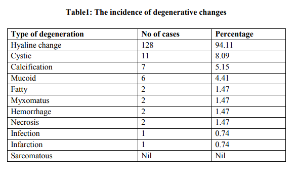
Of the 314 specimens of leiomyoma studied, associated degenerative changes were seen in 136 leiomyomas (45.79%). Hyaline change, the commonest form of degeneration, was seen in 128 (94.11%) of the 136 leiomyomas, whereas the remaining (5.88%) were associated with other degenerative changes. On histologic examination, typical hyaline change is characterized by diffuse homogenous glassy pink structure with marked acellularity. Grossly the mean size of these leiomyomas was 4.8cms. Cystic change (characterized by formation of cystic spaces of varying sizes but without an epithelial lining), which is an invariable accompaniment of hyaline change was observed in 11 cases (8.09%). Eight (5.88%) of these, were also associated with hyaline change. In five (4.11%) leiomyomas, a large part of the tumor was cystic containing colorless or straw colored fluid. Six (4.41%) leiomyomas showed mucoid degeneration, which also resulted in cystic change. Out of 7(5.15%) leiomyomas with calcareous degeneration, 4 were detected grossly and 3 showed microscopic foci of purplish amorphous lake produced by haematoxylin. These gave a gritty feeling while sectioning the tumor. Completely calcified leiomyoma was observed in one (0.74%) case, so called womb stone. Fatty change was observed in two cases (1.47%) Leiomyoma with hemorrhage was observed in 2 cases (1.47%), which included a case of red degeneration that occurred in absence of pregnancy. Myxoid change and Necrosis were also found in 2 cases (1.47%) each. Macroscopically leiomyoma with infection was not evident, but was detected microscopically in one case (0.74%). Leiomyoma with infarction was discernible in one (0.74%) case. However, there was no case of sarcomatous degeneration in the present study. Variants of leiomyoma constituted 0.96% of total 314 cases, which included one case each of cellular, epitheloid and symplastic leiomyomas.

DISCUSSION
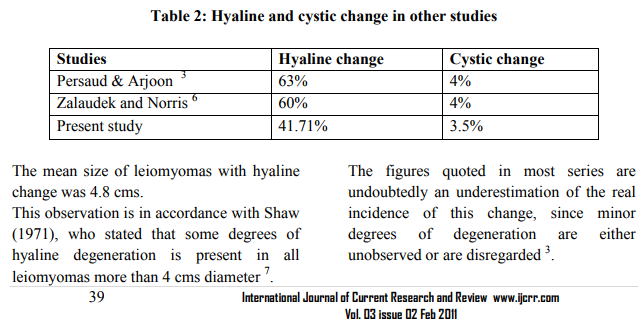
Degenerative changes in leiomyomas are considered to be due to inadequate blood supply, and the type of degenerative change seems to depend on the degree and rapidity of onset of the vascular insufficiency 3, 4 . Degenerative changes in various studies varies from 65% to 100% 3,5 . Hyaline change: It is generally accepted that hyaline change is the commonest form of degeneration seen in leiomyomas3, 5, 6. In the present study also it was the commonest form of degeneration encountered.
he incidence of leiomyomas with calcification was 2.22% which is consistent with other studies by Reddy and Malathy (1963, 2.5%), Torpin et al (1942, 2.4%) 3, 5. We report a completely calcified leiomyoma (so called Womb stone) in one case. Fatty degeneration in leiomyomas is very uncommon and its pathogenesis is somewhat controversial3, 4, 8. Starry (1925) believes that it is due to lipomatosis of the stroma of the uterine tumor, whereas Willis (1958) states that the adipose tissue is formed by metaplasia7 .Fatty change was observed in 0.64% of cases, similar to studies by Persaud and Arjoon (1970) and Reddy and Malathy (1963,) who reported low incidence of fatty change3,5. It should be differentiated from uterine lipoma, a rare tumor of obscure histogenesis3 . Jeffcoate (1967) considered myxomatous degeneration in leiomyomas to be rare, while Persaud and Arjoon (1970) reported higher incidence of 12%3 . Myxoid change was observed in 2 cases (0.64%) in our study. Myomas with mucoid degeneration (1.91%) also result in cystic change in the study. Persaud and Arjoon3 (1970) reported higher incidence of 5.36%. Leiomyoma with hemorrhage was reported in 2 cases in contrast to Norris and Zaloudek (1981)6 observation of 11% . One case of red degeneration (0.74%) showed hemorrhage with hyaline change microscopically, and was not associated with pregnancy in our study. Rosario pinto 1968, (1.2%), Persaud and Arjoon1970 (3.3%), and Reddy and Malathy 1963 (2.5%), found low incidence of red degeneration in patients without pregnancy 3, 5, 6. Faulkner 1947 estimated the frequency of red degeneration in leiomyomas to be 7-8%. Boyd reported that among 38 examples of red degeneration operated upon, 29% were associated with pregnancy 3 . Sarcomatous change in leiomyomas is rare. Corscaden and Singh indicated in their study that the true incidence of sarcoma developing within uterine leiomyomas is not more than 0.13%, and probably as low as 0.04%. Various other studies have also reported low incidence of sarcomatous change 3, 8. However, there was no case of sarcomatous degeneration in our study.
CONCLUSION
Leiomyomas are the most common benign tumors of the uterus. Hyaline change is the commonest degenerative change encountered and also coexists with other degenerative changes. Since leiomyomas are known to occur even in young reproductive age group, proper understanding, knowledge, intervention as well as medical advice regarding the tumor can help in preventing depression and anxiety related to the disease in many patients.
References:
REFERENCES
1. Okolo S .Incidence, etiology and epidemiology of uterine fibroids, best practice and research. Clinical obstetrics and gynecology . 2008 ;22 (4): 571–88.
2. Gordon P, Andersen FJ and Dixon D. Etiology and Pathogenesis of Uterine Leiomyomas,A Review Journal article byEnvironmental Health Perspectives. 2003; Vol. 111.
3. Persaud V and Arjoon P.D. Uterine Leiomyoma, Incidence of Degenerative change and a correlation of associated symptoms. Obst. and Gynec.1970; 35 (3):432-36.
4. Haines M and Taylor C.W. Adenomyosis and Fibromyoma, In Gynecological Pathology, 2nd edition. Ebinburg London, Churchill Livingstone. 1975;158-218
5. Pinto R,.Uterine fibromyomas. J. of Obstet. and Gynaecol. Of India, 1968;(18) 101-07.
6. Zaloudek ,C and Norris HJ . Mesenchymal Tumours of uterus In Blaunstein’s Pathology of Female Genital Tract. Editor R.J.Kurmann. IInd edition. New York, Springer-verlag, 1987;374-02
7. Padubidri VG and Daftary S.N. New Growths of uterus In Shaw’s text book of Gynaecology, Howkins and Browne.10th Edition. New Delhi, Churchill Livingstone.1989; 399-427.
8. Masani M.M.: New growths of cervix and uterus In A text book of Gynaecology. 6th Edition. Bombay, Popular Prakashan. 1971;313-417.
|


 This work is licensed under a Creative Commons Attribution-NonCommercial 4.0 International License