IJCRR - 3(10), October, 2011
Pages: 55-59
Print Article
Download XML Download PDF
BILATERAL VARIANT INNERVATION OF ANTERIOR COMPARTMENT MUSCLES OF UPPER ARM WITH
ABSENCE OF MUSCULO CUTANEOUS NERVE - A CASE REPORT
Author: Sunitha Vinnakota, Jayasree Neelee
Category: Healthcare
Abstract:During routine dissection for medical undergraduates, in a cadaver of around 50 years, musculo cutaneous
nerve was absent in both upper arms. Median nerve after its formation fused with lateral cord and
supplied all the muscles in upper arm and one long twig after supplying brachialis continued as lateral
cutaneous nerve of forearm. Awareness of anatomical variations of peripheral nerves is important in
repair of traumatic injuries and treatment of compression syndromes of these nerves.
Keywords: median nerve, musculo cutaneous nerve and coraco brachialis muscle.
Full Text:
INTRODUCTION
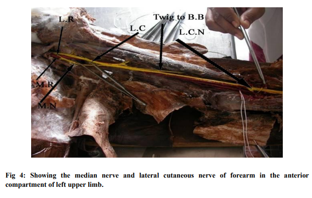
Variations in the formation and branching pattern of brachial plexus are well documented 1,2,. . Some of these variations include pre fixed and post fixed type of brachial plexus. The median nerve is formed by union of one root from lateral cord (C5, 6and7) and one root from medial cord (C8 and T1), where as musculocutaneous nerve (C5, 6 and7) arises from lateral cord of brachial plexus3 . The musculocutaneous nerve is derived as a terminal branch of lateral cord pierces the coracobrachialis and runs laterally downwards between biceps and brachialis to reach the lateral side of arm and continues as lateral cutaneous nerve of forearm. According to Tountas4 and Bergaman, musculocutaneous nerve arises from lateral cord in 90.5%, from lateral and posterior cords in 4%, from median nerve in 2% and has 2 separate bundles from medial and lateral cords in 1.4%. Case report: During routine dissection for medical undergraduates, musculocutaneous nerve was absent in the anterior compartments of both upper arms and median nerve after its formation fused with lateral cord and supplied all the muscles in upper arm and one long twig after supplying brachialis continued as lateral cutaneous nerve of forearm in approximately 50 year old male cadaver. Right side: Median nerve was formed from lateral and medial roots of lateral and medial cords infront of 3rd part of axillary artery. Median nerve was joined by lateral cord (See fig 1) 8cm below its formation. This combined nerve runs for a distance of 2cm and then twigs to biceps, coracobrachialis and one long twig to brachialis were noticed. The long twig after supplying brachialis continued as lateral cutaneous nerve of forearm. (See fig2) Left side: 5cm below its formation median nerve was joined by the lateral cord. Coracobrachialis was innervated by a small twig directly from the lateral cord and not from the median nerve as observed on right side. (See fig 3) The combined nerve runs downward for 2cm and gave twigs to biceps and brachialis. The long twig after supplying brachilis continued as lateral cutaneous nerve of forearm (See fig 4). Further course and distribution of median nerve was normal. No variation was noted in the vasculature of both upperlimbs
DISCUSSION
The variations in the formation of median nerve sited in the literature include formation of median nerve by 4 roots, one from medial cord and other three from the lateral cord5 . Arora6 and Dhingra (2005) reported a case in which the median nerve had 3 roots and musculocutaneous nerve was absent. Variations such as passing through a bony canal7 and abnormal communications with the musculocutaneous nerve have been recorded8and9. The reported variations of musculocutaneous include its total absence10 and communication with median nerve at various levels8,9. The musculo cutaneous, not piercing coraco brachialis is also known11. Venierator12 and Anagnostopoulous (1998) also described 3 different types of communication between musculocutaneous and median nerve in relation to coracobrachialis. Type I is communication proximal to muscle, where as type II is distal to muscle. Neither the nerve nor its communicating branch pierced the muscle is type III. In type V of Le Minor13 (1992) classification musculocutaneous nerve is absent and the entire fibers of musculocutaneous pass through lateral root and fibers to the muscles supplied by musculocutaneous nerve branch out directly from median nerve. The presence of communication between median and musculo cutaneous nerves may be attributed to andom factors influencing the mechanism of formation of limb muscles and peripheral nerves during embryonic life. Significant variations in nerve patterns may be a result of altered signaling between mesenchymal cells and neuronal growth cones 14 or circulatory factors at the time of fusion of brachial plexus cords15. The existing variations of the present case such as absence of musculo cutaneous nerve, derivation of all motor branches from median nerve directly falls into the type V of variations described by Le minor13 and it also falls into the type III of Venirator12. As per Arora6 and Dhingra the present case can be described as the median nerve with 3 roots and absence of musculo cutaneous nerve. Anatomical variations of peripheral nerves are important clinically and surgically. Precise knowledge of variations in median and musculo cutaneous nerves may prove valuable in traumatology of the arm, as well as in plastic and reconstructive repair operations.
. 2001; 80: 99- 101. 6. Arora L, Dhingra R (2005)- Absence of musculo cutaneous nerve and accessory head of biceps brachii- a case report, Indian Journal of paeditric surgery vol 38 issue 2, pp: 144-146. 7. Kazuki K, Egi T, Okada M, Takoka K. Anatomic variation – a bony canal for the median nerve at the distal humerus; a case report. J Hand Surg. (AM). 2004 ; 29: 953 - 958. 8. Loukas M, Aqueelah H. Musculocutaneous and median nerve connections within, proximal and distal to the coracobrachialis muscle. Folia Morphol. (Warsz). 2005; 84: 101- 108. 9. Prasada Rao PV, Chaudhary SC. Communication of the musculocutaneous nerve with the median nerve. East Air. Med. J.2000; 77: 498 – 503. 10. Gumshurun E, Adiguzel E. A Variation of the brachial plexus characterized by the absence of the musculocutaneous nerve: a case report. Surg Radiol. Anat. 2000;22:83- 85. 11. Nakatani T, Mizukami S, Tanaka S. Three cases of the musculocutaneous nerve not perforating the coracobrachialis muscle. Kalbogaku Zasshi. 1997; 72: 191-194. 12. Venierator D, Anagnostopoulous (1998) – classification of communications between musculo cutaneous and median nerves. Clinical Anatomy 11 (5), pp: 327-331. 13. Le minor JM. A rare variation of median & musculo cutaneous nerves in man. Arch Anat Histol Embryol 1990; 73: 33-42. 14.Sanes DH, Ruh TA, Harris MA. Development of the nervous system, New York, Academic press. 2010; 189-197. 15. Kosugi K, Motria, T; Yamashita, H (1986). Branching pattern of Musculo cutaneous nerve, 1 case possessing normal Biceps brachi, Ji Keakai Medical journal 33: pp 63- 71) Abbreviations:
Abbreviations:
1. MR: Medial root of Median nerve.
2. LR: Lateral root of Median nerve.
3. LC: Lateral cord of Brachial plexus.
4. MN: Median nerve.
5. LCN: Lateral cutaneous nerve of forearm.
6. C.B: Coracobrachialis muscle.
7. B.B: Biceps brachii muscle.


References:
REFERENCES
1. Ken AT (1918): The brachial plexus of nerves in man. The variation in its formation and branches. American Journal of Anatomy 23: 285-395 (s).
2. Linell EA (1921). The distribution of nerves in Upper limb with reference to variabilities and their clinical significance. Journal of Anatomy 55: 79-112 (s).
3. Snell S. Richard 1995- Clinical anatomy for medical students: 5th ed Little Brown and Company, USA pp: 393-398.
4. Tountas C Bergaman R- Anatomic variation of upper extremity- Churchill Livingstone, 1993 pp: 223-224.
5. Uzun A, Seelig LL Jr. A variation in the formation of the median nerve: communicating branch between the musculocutaneous and median nerves in man. Folia Morphol. (Warsz). 2001; 80: 99- 101.
6. Arora L, Dhingra R (2005)- Absence of musculo cutaneous nerve and accessory head of biceps brachii- a case report, Indian Journal of paeditric surgery vol 38 issue 2, pp: 144-146.
7. Kazuki K, Egi T, Okada M, Takoka K. Anatomic variation – a bony canal for the median nerve at the distal humerus; a case report. J Hand Surg. (AM). 2004 ; 29: 953 - 958.
8. Loukas M, Aqueelah H. Musculocutaneous and median nerve connections within, proximal and distal to the coracobrachialis muscle. Folia Morphol. (Warsz). 2005; 84: 101- 108.
9. Prasada Rao PV, Chaudhary SC. Communication of the musculocutaneous nerve with the median nerve. East Air. Med. J.2000; 77: 498 – 503.
10. Gumshurun E, Adiguzel E. A Variation of the brachial plexus characterized by the absence of the musculocutaneous nerve: a case report. Surg Radiol. Anat. 2000;22:83- 85.
11. Nakatani T, Mizukami S, Tanaka S. Three cases of the musculocutaneous nerve not perforating the coracobrachialis muscle. Kalbogaku Zasshi. 1997; 72: 191-194.
12. Venierator D, Anagnostopoulous (1998) – classification of communications between musculo cutaneous and median nerves. Clinical Anatomy 11 (5), pp: 327-331.
13. Le minor JM. A rare variation of median and musculo cutaneous nerves in man. Arch Anat Histol Embryol 1990; 73: 33-42.
14.Sanes DH, Ruh TA, Harris MA. Development of the nervous system, New York, Academic press. 2010; 189-197. 15. Kosugi K, Motria, T; Yamashita, H (1986). Branching pattern of Musculo cutaneous nerve, 1 case possessing normal Biceps brachi, Ji Keakai Medical journal 33: pp 63- 71
|


 This work is licensed under a Creative Commons Attribution-NonCommercial 4.0 International License